Ultrasound imaging apparatus and method thereof

Inactive Publication Date: 2006-06-01
LI PAI CHI
View PDF18 Cites 22 Cited by
  • Summary
  • Abstract
  • Description
  • Claims
  • Application Information

AI Technical Summary

Benefits of technology

[0009] Another aspect of the invention is to provide an ultrasound imaging apparatus and method, which uses an ultrasound transducer array for transmitting and receiving ultrasound signals, without rotating the array or the object, to obtain the B-mode image, the sound velocities, and the attenuation coefficients. The invention applies the B-mode image of the object for assisting in estimating the sound velocities and the attenuation coefficients.
[0010] Another aspect of the invention is to provide an ultrasound imaging apparatus and method, which applies the information extracted from the B-mode image of the object to efficiently improve the estimation accuracy of the sound velocities and the attenuation coefficients without using other imaging apparatus.
[0011] An ultrasound imaging apparatus and method achieving the above-mentioned aspects comprises a transducer array, a positioning device for the array, a reflector, a multiplexer, a transmitter / receiver, an image reconstruction and signal processing apparatus, and a monitor. The transducer array and the reflector are located on two sides of an object. Every channel of the transducer array can independently transmit and receive signal, and the reflector is used to reflect the transmitted signal from any channel. Therefore, the system can collect the echoes from the reflector for all transmit / receive combinations, and then extract from the echoes the time-of-flight data and the attenuation data necessary for estimating the sound velocities and the attenuation coefficients, respectively. When estimating the sound velocities and the attenuation coefficients of different tissues in an object, the B-mode image is applied for providing the segmentation information of the object to improve the estimation accuracy. The invention can be used to obtain the ultrasound B-mode image, the sound velocities, and the attenuation coefficients of an object, and can be extended to obtaining other parameters, such as elastic modulus, with a single apparatus.

Problems solved by technology

Although the sound velocities and the attenuation coefficients can be obtained using the conventional imaging apparatus 1, the B-mode imaging that can be used to obtain the backscattering information is not available with this setup.
Thus, the conventional apparatus and method is improvable.

Method used


Image

  • Ultrasound imaging apparatus and method thereof
  • Ultrasound imaging apparatus and method thereof
  • Ultrasound imaging apparatus and method thereof

Examples


Embodiment Construction

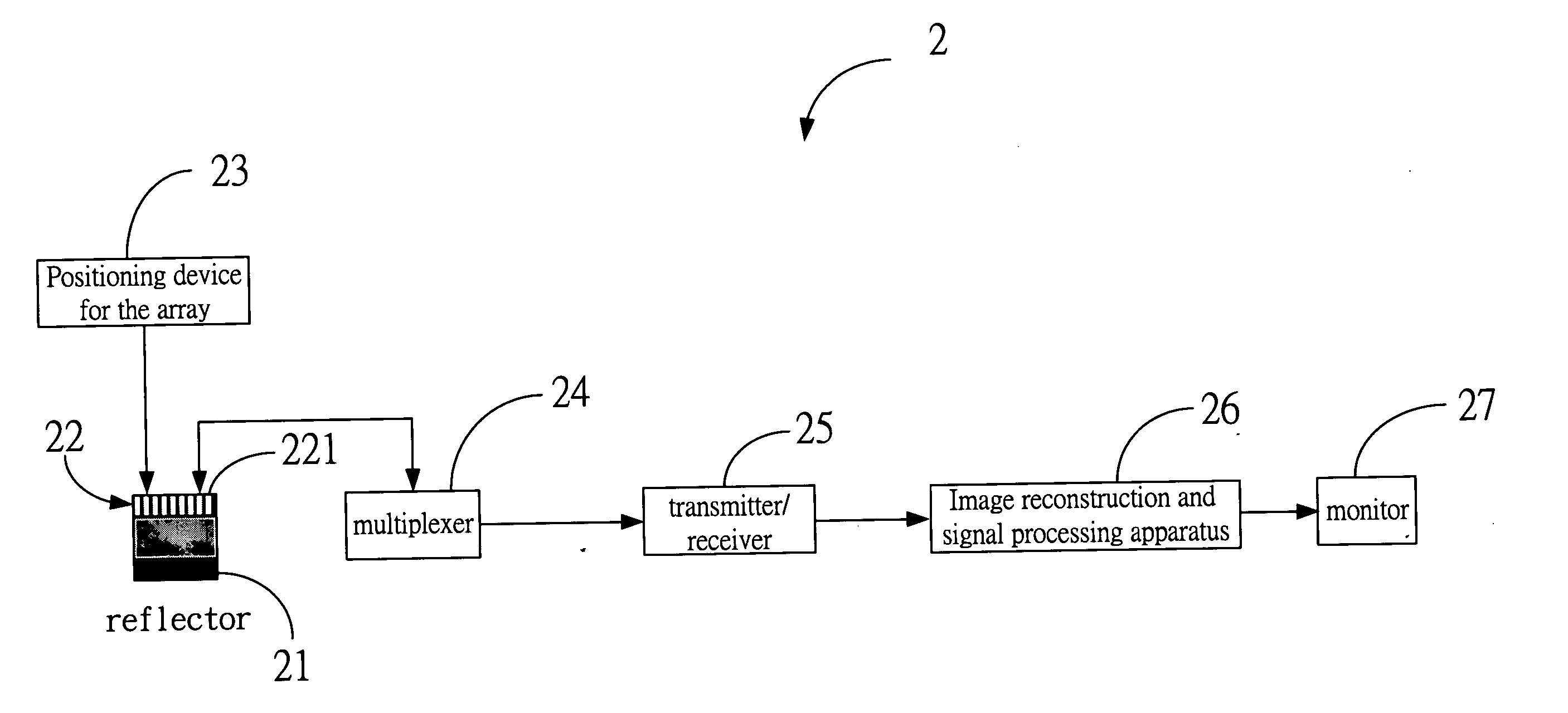
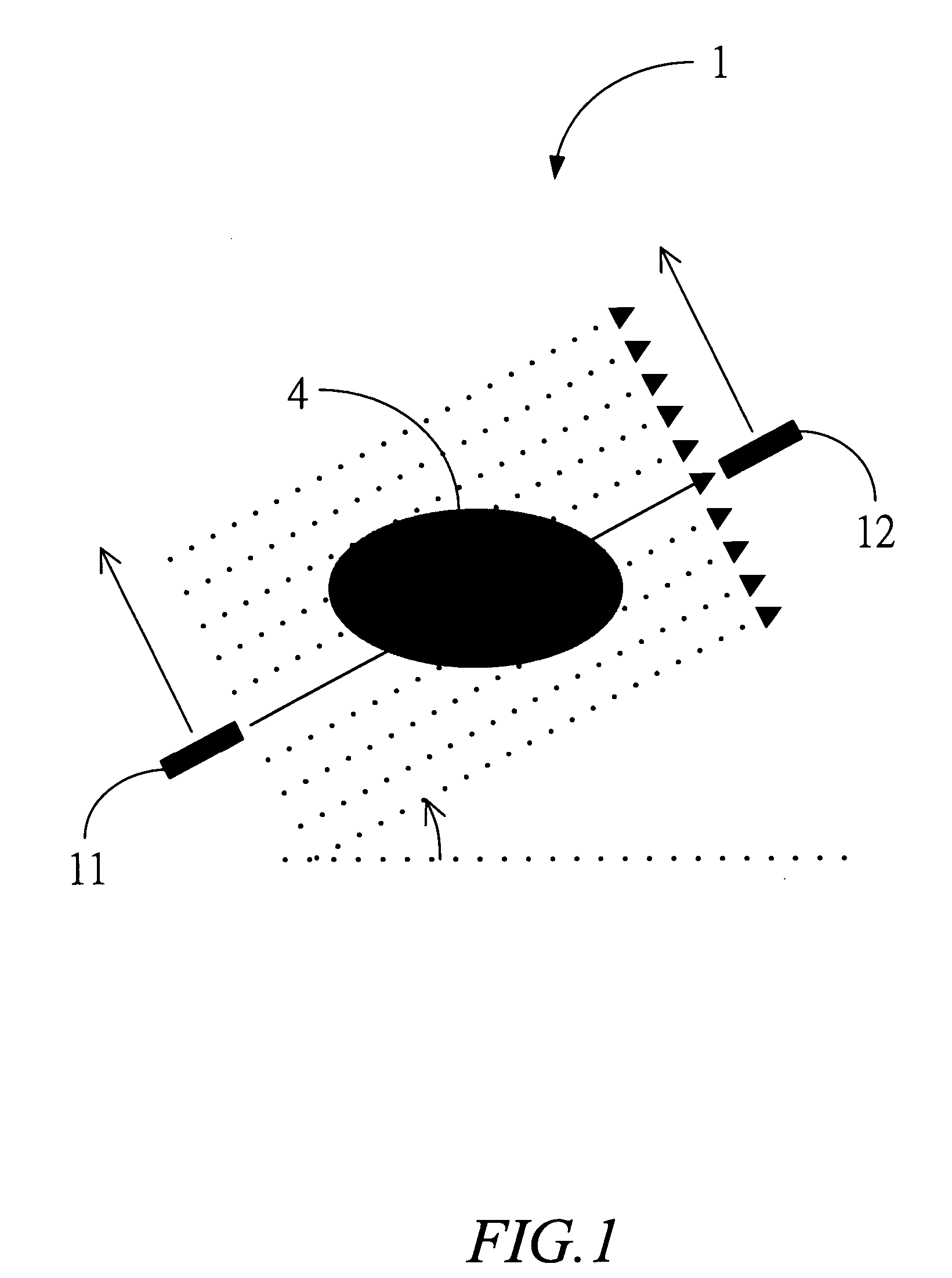
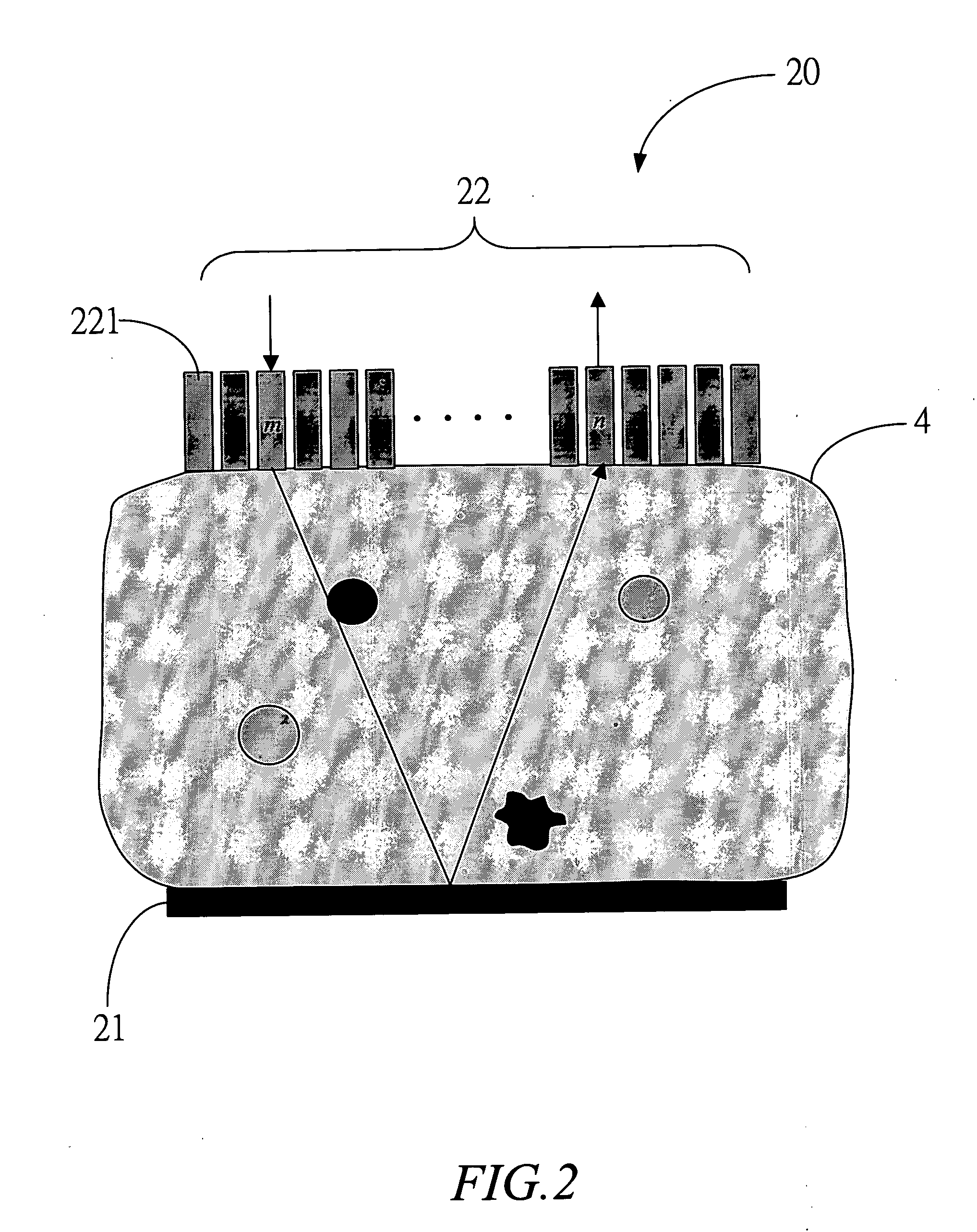
[0017]FIG. 2 is a diagram of imaging equipment 20 of the invention. Different from the conventional computed tomography apparatus, the imaging equipment 20 applies a reflector 21 reflecting the transmitted signal from every channel 221 of a transducer array 22. It enables the system to get the echoes from the reflector for all transmit / receive combinations, and then to reconstruct the sound velocities and the attenuation coefficients of different tissues of an image object 4 using the echoes.

[0018] Comparing with the conventional method, the invention does not need to rotate the ultrasound transducers, and is compatible with the conventional B-mode imaging apparatus. Despite the advantages, the sound velocities and the attenuation coefficients cannot be accurately estimated with the conventional computed tomography reconstruction methods due to the fact that that measurement angles span less than 180 degrees (i.e., incomplete data). The invention applies proper reconstruction const...


PUM


Abstract

An ultrasound imaging apparatus and method comprises a transducer array, a positioning device for the array, a reflector, a multiplexer, a transmitter / receiver, an image reconstruction and signal processing apparatus, and a monitor. The transducer array and the reflector are located on two sides of an object which are opposite to each other. Every channel of the transducer array can independently transmit and receive signal, and the reflector is used to reflect the transmitted signal from any channel. Thus, the system can collect the echoes from the reflector for all transmit / receive combinations, and then extract from the echoes the time-of-flight data and the attenuation data necessary for estimating the sound velocities and the attenuation coefficients, respectively. When estimating the sound velocities and the attenuation coefficients of different tissues in an object, the B-mode image is applied to provide the segmentation information of the object in order to improve the estimation accuracy. The invention can be used to obtain the ultrasound B-mode image, the sound velocities, and the attenuation coefficients of an object, and can be extended to obtaining other parameters, such as elastic modulus, with a single apparatus.

Description

BACKGROUND OF THE INVENTION [0001] 1. Field of the Invention [0002] The present invention relates to an ultrasound imaging system, and, in particular, to an ultrasound imaging apparatus and method thereof for obtaining the ultrasound B-mode image, the sound velocities, the attenuation coefficients, and the elastic properties of an object, which can be used for tissue characterization (e.g., to detect the breast tumor). [0003] 2. Description of the Prior Art [0004] Ultrasound computed tomography is a standard approach to reconstructing the sound velocities and the attenuation coefficients [1]˜[3]. As shown in FIG. 1, two ultrasound transducers 11 and 12 are located next to an object, and opposite to each other. Given a location in a specific angle, the transmit transducer 11 transmits an ultrasonic signal, and the receive transducer 12 receives the signal to complete a single measurement. Then the transducers 11 and 12 are moved transversely together in a short distance to next locat...

Claims


Application Information

Patent Timeline
IPC IPC(8): A61B8/14
CPCA61B8/0825G01S7/52042G01S7/5206G01S15/8993
Inventor LI, PAI-CHIHUANG, SHENG-WEN
Owner LI PAI CHI
Why Eureka
Try Eureka