
Method for determining organ-dependent parameters for image post-processing and image processing device

a technology of image post-processing and organ-dependent parameters, which is applied in the field of method for determining organ-dependent parameters of image post-processing and image processing devices, can solve the problems of affecting workflow, demanding and difficult, and complex methods that require more and more parameters

Inactive Publication Date: 2006-06-22
SIEMENS HEALTHCARE GMBH
View PDF6 Cites 19 Cited by
  • Summary
  • Abstract
  • Description
  • Claims
  • Application Information

AI Technical Summary

Benefits of technology

[0012] In doing this, the method in accordance with the invention is particularly advantageous in enabling the automatic acquisition of the optimal postprocessing parameters, and the automatic postprocessing. Unlike the prior art, the sole starting basis for the automatic determination of the parameters is the preprocessed raw image of the examination area, as a function of which the determination of the comparison image and determination of the parameters is carried out. The doctor or medical assistant is no longer required to determine manually and subjectively the required organ program with predefined image processing parameters, and possible sources of error associated with this are likewise excluded. The entire procedural sequence can be structured to be significantly faster, since corrections necessitated by errors are no longer required because the image postprocessing is always carried out on the basis of optimal postprocessing parameters determined beforehand, thus ensuring that in respect of image quality and image content the image which can be output is optimized by the processing. So optimal image processing is possible without the need for current information about the organ or the examination area, the patient, the exposure etc., because the sole basis for the selection is the preprocessed raw image data, and hence the content of the preprocessed image.

Problems solved by technology

Common to the image processing algorithms in these cases is the fact that the complex methods require more and more parameters to be selected for optimal image processing.
This can sometimes be very demanding and difficult.
With the variations in patient parameters, e.g. age, gender, weight, either additional organ programs must be provided or alternatively additional selection steps must be offered, which can have a negative effect on the workflow.
In general there is a high risk of incorrect settings.
In sum, for the doctor the manual and subjective selection method which has been the practice until now, with the multiplicity of individual different organ programs, is awkward, tedious and very easy to make mistakes with.

Method used


Image

  • Method for determining organ-dependent parameters for image post-processing and image processing device
  • Method for determining organ-dependent parameters for image post-processing and image processing device

Examples


Embodiment Construction

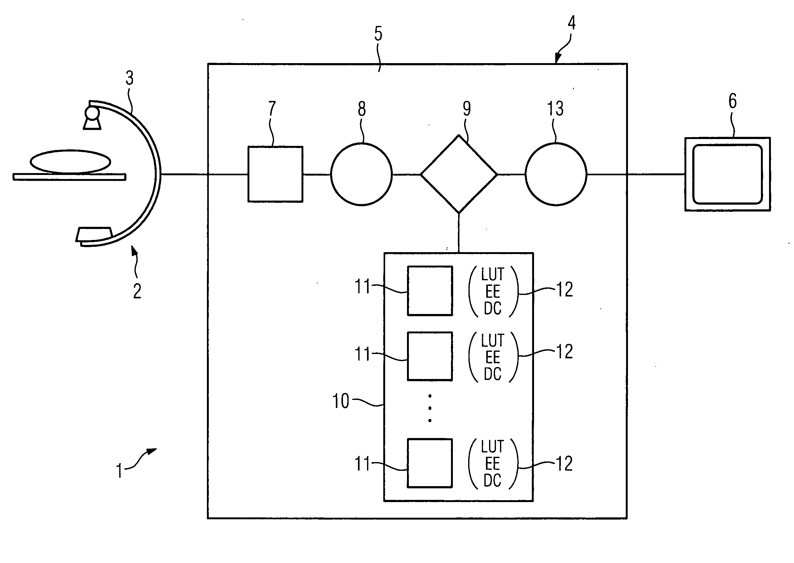
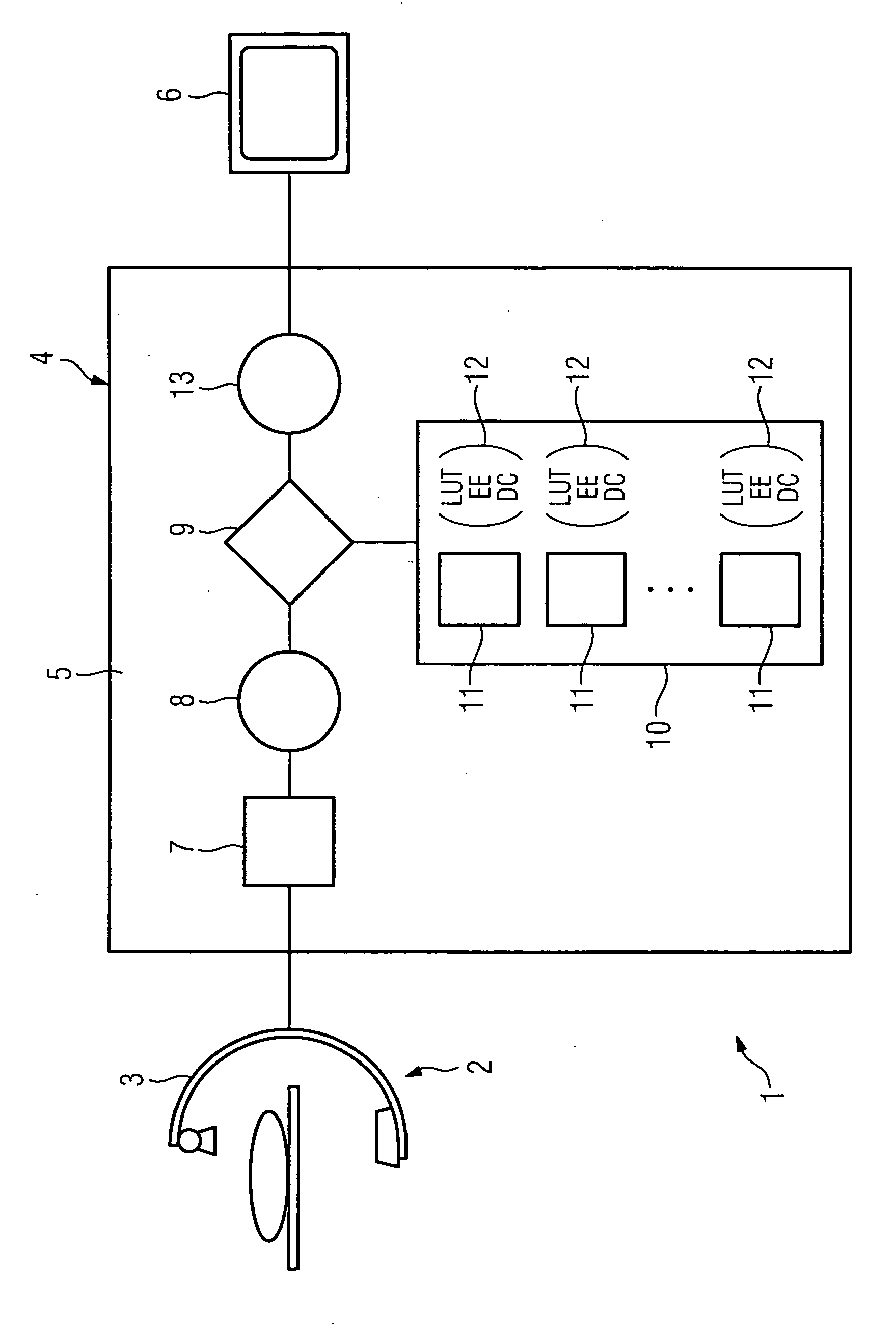
[0024] The FIGURE shows an image recording device 1 incorporating an image recording facility 2 in the form of an X-ray device 3. The digital raw image data which is recorded is passed to an image processing device 4 in accordance with the invention, which incorporates a computing device 5, which performs the relevant image processing steps and generates from the raw image data passed to it a postprocessed image which can be output, and which is output on a monitor 6.

[0025] As described, the raw image data 7 is first passed from the X-ray device 3 to the image processing device 4. The image processing device 4 or the computing device 5, as applicable, has a facility or a device 8 for image preprocessing. As part of this image preprocessing, the raw image data supplied is preprocessed in respect, for example, of the offset, the gain and possible defects etc. as part of the normal pre-processing.

[0026] The preprocessed image or preprocessed image data, as applicable, is then passed ...


PUM


Abstract

Method for determining organ-dependent parameters for image post-processing and image processing device The invention relates to a method for the automatic determination of one or more organ-dependent parameters for image post-processing, having the following steps: recording of a raw image and the execution of image preprocessing on the raw image data in order to generate a preprocessed image, determination of an organ-specific comparison image, stored in a database, which has been optimized by postprocessing using organ-specific postprocessing parameters, and acquisition of the postprocessing parameters for the comparison image, and postprocessing of the preprocessed raw image, using the postprocessing parameters of the comparison image.

Description

CROSS REFERENCE TO RELATED APPLICATIONS [0001] This application claims priority to the German application No. 10 2004 060 868.7, filed Dec. 17, 2004 which is incorporated by reference herein in its entirety. FIELD OF INVENTION [0002] The invention relates to a method for the automatic determination of one or more organ-dependent parameters for image post-processing, together with an image processing device for carrying out the method. BACKGROUND OF INVENTION [0003] Digital imaging methods play an ever more important role in medical diagnosis. Whereas digital techniques have been used from the beginning in such diagnostic methods as, for example, computer tomography, magnetic resonance, ultrasound and nuclear medical methods, a transition to digital imaging is currently taking place on a large scale in “conventional” X-ray methods such as radiography, mammography, angiography or cardiology, in particular with the assistance of solid state radiation detectors or planar detectors. [000...

Claims


Application Information

Patent Timeline
IPC IPC(8): G06K9/00
CPCG06T2207/30004G06T5/00A61B6/03A61B6/5294G06T2207/20084G06T2207/20081G06T5/60
Inventor SPAHN, MARTIN
Owner SIEMENS HEALTHCARE GMBH
Learn More