
Feature-based editing for electrocardiography

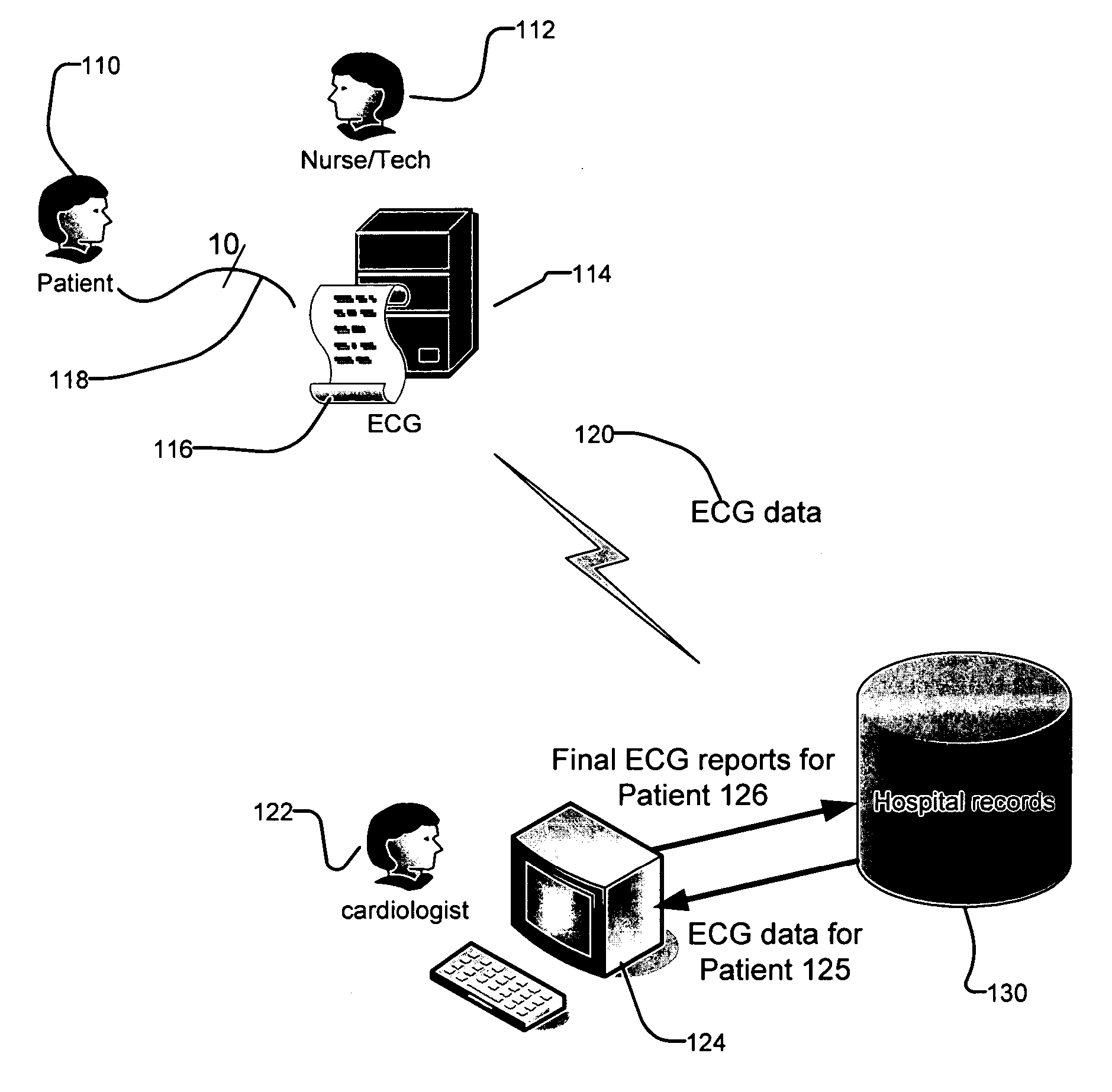
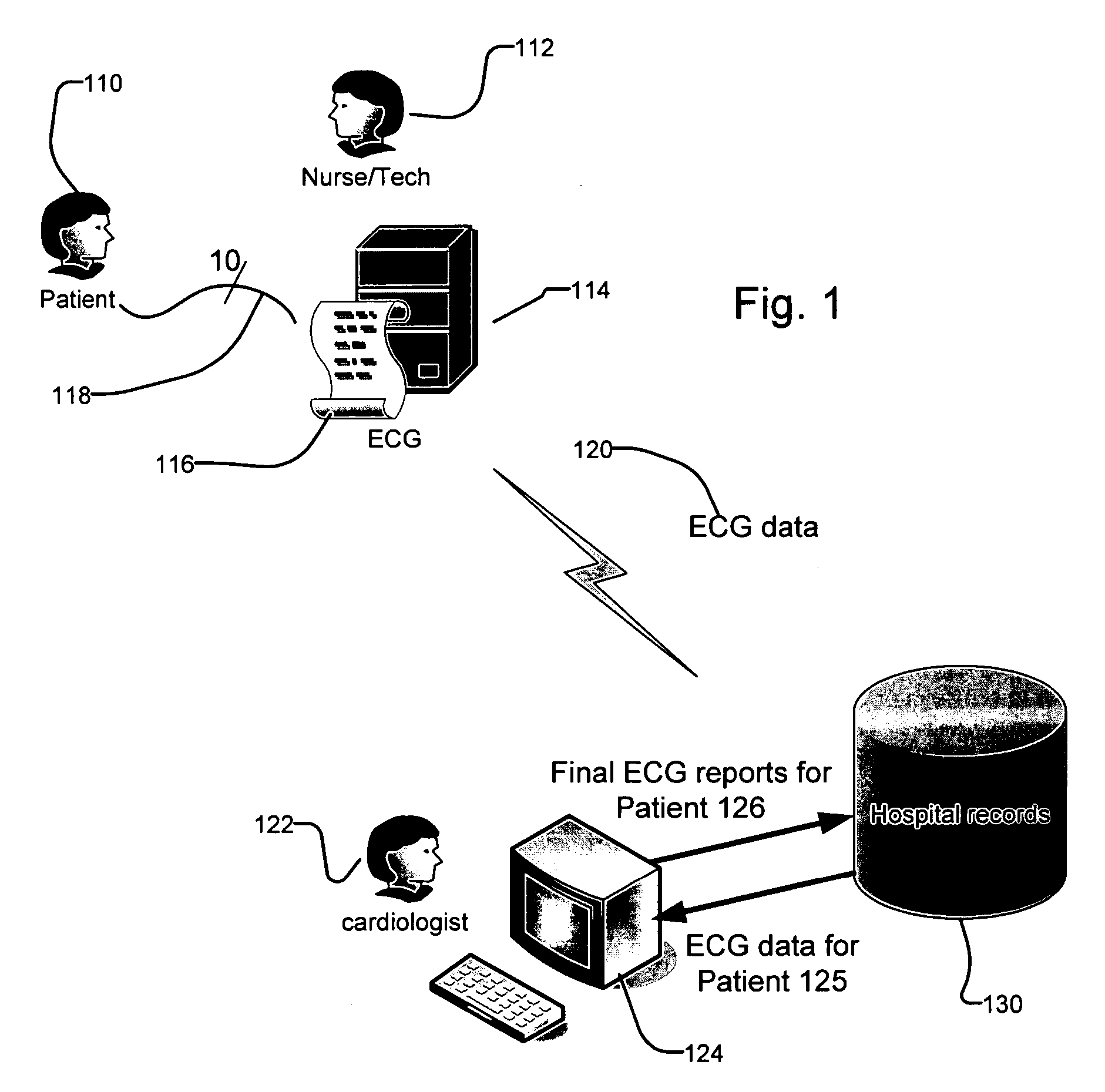
a feature-based editing and electrocardiography technology, applied in the field of electrocardiography diagnosis, can solve the problems of inability to accurately determine the low-level, the error is commonly made by the computer in making measurements, and the non-trivial number of machine-generated ecg interpretations will be incorrect, so as to facilitate analysis and generation of the final, reduce inadvertent errors and/or inconsistencies, and improve the accuracy of machine-generated ecg data interpretation.

Inactive Publication Date: 2006-07-20
AGFA HEALTHCARE INC
View PDF7 Cites 48 Cited by
  • Summary
  • Abstract
  • Description
  • Claims
  • Application Information

AI Technical Summary

Benefits of technology

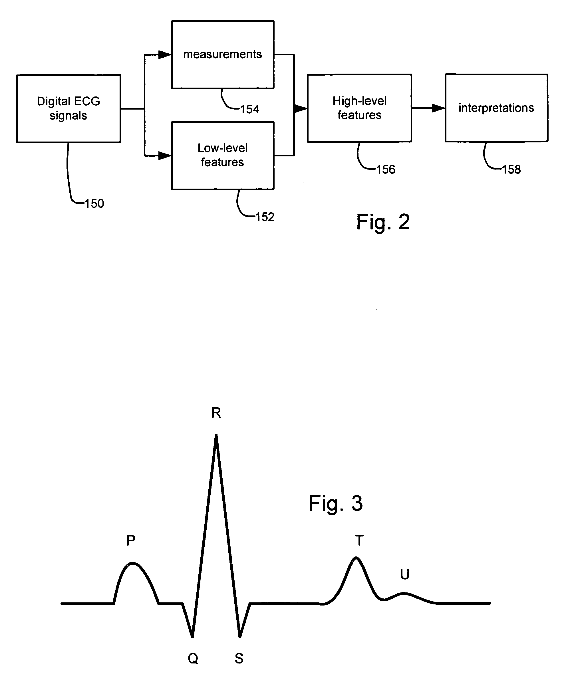
[0007] Even though algorithms for computer-based ECG interpretation are generally quite accurate, errors are commonly made by the computer in making measurements and determining the low-level and high-level features in the first place. When these are made available for review and correction, the cardiologist can interact at a higher level of sophistication, acting more as a domain expert and less like a clerk or secretary. In addition, all of the internal checks for mutually exclusive conditions can remain in place (done during the final generation step of the interpretation process), thereby greatly reducing inadvertent errors and / or inconsistencies.
[0008] The present invention is directed to a method and system for generating electrocardiogram reports. It allows for the editing of features in the electrocardiogram interpretation process. This improves the accuracy of machine interpretation of the ECG data thereby facilitating the analysis and generation of the final report by the physician.

Problems solved by technology

Despite this trend, however, a nontrivial number of these machine-generated ECG interpretations will be incorrect.
And, these incorrect ECG interpretations often represent a frustration to the physician / cardiologic, since the physician will be forced to function in a clerical role using text-editing tools to correct the machine-generated interpretations in the process of drafting the patient's ECG report.
Even though algorithms for computer-based ECG interpretation are generally quite accurate, errors are commonly made by the computer in making measurements and determining the low-level and high-level features in the first place.

Method used


Image

  • Feature-based editing for electrocardiography
  • Feature-based editing for electrocardiography
  • Feature-based editing for electrocardiography

Examples


example 1

[0043] The duration of one portion of the ECG wave, called the QRS complex, is used to determine if there is an interruption in the flow of electricity in the heart's conduction system. The QRS duration is a feature that directly leads to specific diagnoses on the ECG report. If the QRS duration feature is calculated to be greater than 120 milliseconds (and the QRS configuration matches a certain pattern), the condition is referred to as Left Bundle Branch Block, or “LBBB.” This means that the electricity is not conducting properly through the Left Bundle of the conducting system.

[0044] In the presence of LBBB on the ECG report, it is not possible to make any definitive statements about other clinical conditions such as anterior myocardial infarction (“AMI”) or left ventricular hypertrophy (“LVH”).

[0045] It is not uncommon for a previous ECG report for a patient to show a QRS duration of 118 milliseconds (below the cutoff for LBBB), along with an AMI and LVH. When the next ECG on ...

example 2

[0048] P-waves are very tiny, and hard for the computer to distinguish on an ECG, especially if the patient is moving or other noise is present. The computer might also incorrectly think that P-waves are present, getting confused by the presence of noise. In this latter case, the reading might be “Sinus rhythm” and “1st degree AV block.” A measurement of the PR Interval will be reported (the time duration between the occurrence of the P-wave and the occurrence of the QRS complex).

[0049] In a statement-based editing environment, the cardiologist might change the reading of “sinus rhythm” to “atrial fibrillation.” If the reading is left this way, which is very common, there is a conflict. “1st degree AV block” means that the PR interval is longer than 200 milliseconds, but there is no PR interval if the is no P-wave.

[0050] The correct next step is for the cardiologist with convention editing systems is to remove measurement of the PR interval, and delete the line that says “1st degr...


PUM


Abstract

A method and system for generating electrocardiogram reports allows for the editing of features in the electrocardiogram interpretation process. This improves the accuracy of machine interpretation of the ECG data thereby facilitating the analysis and generation of the final report by the physician. The present method and system are most useful in host-based ECG interpretation systems where the physician accesses the ECG data at a workstation including a machine interpretation that is generated typically at the ECG cart. The physician is then provided with the ability to modify the features in the ECG data and generate a new host-based interpretation based on the original ECG data and the features specified by the physician.

Description

RELATED APPLICATIONS [0001] This application claims the benefit under 35 USC 119(e) of U.S. Provisional Application No. 60 / 644,888, filed on Jan. 18, 2005, which is incorporated herein by reference in its entirety.BACKGROUND OF THE INVENTION [0002] Electrocardiography is a technology for the detection and diagnosis of cardiac conditions. An electrocardiograph is a medical device capable of recording the potential differences generated by the electrical activity of the heart. An electrocardiogram (ECG or EKG) is produced by the electrocardiograph. It typically comprises the ECG wave data that describes the heart's electrical activity as a function of time. [0003] The heart's electrical activity is detected by sensing electrical potentials via a series of electrode leads that are placed on the patient at defined locations on the patient's chest and limbs. Systems with ten (10) separate ECG leads and digital data capture / storage are typical. During electrocardiography, the detected ele...

Claims


Application Information

Patent Timeline
IPC IPC(8): A61B5/04
CPCA61B5/044A61B5/0452A61B5/7435A61B5/7445A61B5/339A61B5/349
Inventor ELION, JONATHAN L.
Owner AGFA HEALTHCARE INC
Learn More