
Medical image output system, medical image transferring apparatus and program

Inactive Publication Date: 2006-08-17
KONICA MINOLTA MEDICAL & GRAPHICS INC
View PDF2 Cites 2 Cited by
  • Summary
  • Abstract
  • Description
  • Claims
  • Application Information

AI Technical Summary

Benefits of technology

[0017] Moreover, when the medical image forming apparatus expands the medical image data to perform the image formation, the medical image data which the medical image transferring apparatus transfers to the medical image forming apparatus is the data before the expansion. Consequently, the communication capacity of the data between the medical image transferring apparatus and the medical image forming apparatus can be reduced.
[0023] Accordingly, the medical image data includes the radiography image produced by radiographing the subject and the patient information. Consequently, because the radiography images and the patient information are drawn on the sheet-like recording media output by the plurality of medical image forming apparatus, when the plurality of sheet-like recording media is transmitted in the sate of being superposed on one another, each of the radiography images and the patient information is in the same size, and do not produce any shifts. Consequently, for example, whether the sheet-like recording medium of another patient is mixed among the plurality of sheet-like recording media output from different medical image forming apparatus or not can be easily confirmed by superposing the sheet-like recording media on one another.

Problems solved by technology

For example, when a doctor performs a diagnosis using recording media severally output from a plurality of output apparatus, a diagnostic mistake may be caused from the difference in each size of the medical image of each recording medium.
Hence, even if recording media are superposed on one another as disclosed in Patent Document 1, a character of each recording medium is shifted from one another, and it is difficult to confirm the mixing of a film of another patient.

Method used


Image

  • Medical image output system, medical image transferring apparatus and program
  • Medical image output system, medical image transferring apparatus and program
  • Medical image output system, medical image transferring apparatus and program

Examples


first embodiment

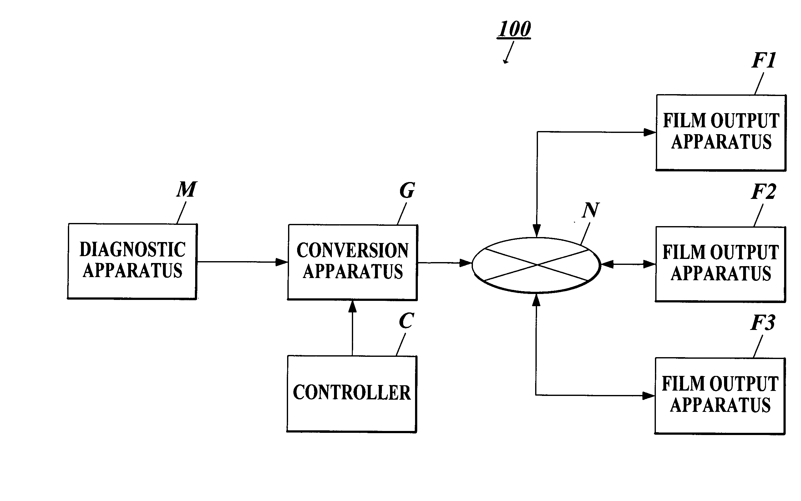
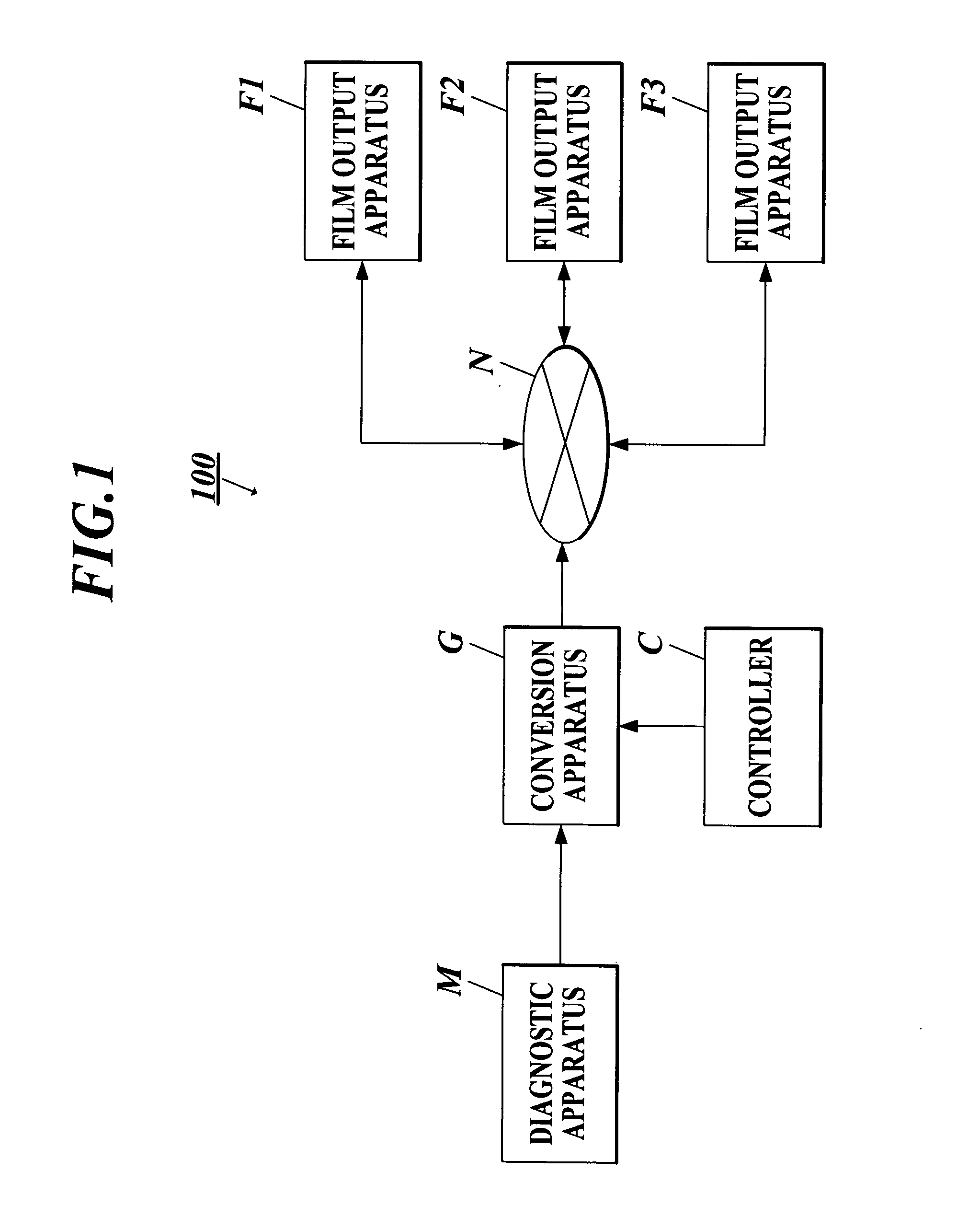
[0046] First, a medical image output system in the case where a medical image transferring apparatus according to the present invention is composed of a conversion apparatus and a controller and a medical image forming apparatus is applied to a film output apparatus is described in detail by referring to FIGS. 1-9.

[0047]FIG. 1 is a diagram showing an example of the system configuration of a medical image output system 100. According to the diagram, the medical image output system 100 is composed of a conversion apparatus G transferring a medical image output from a diagnostic apparatus M in conformity with an instruction of a controller C, and film output apparatus F1, F2 and F3, which are connected to one another through a predetermined communication network N.

[0048] A user determines whether to perform the film output of the data of a medical image (hereinafter referred to as “medical image data”) generated by the radiography with the diagnostic apparatus M or not by using the co...

second embodiment

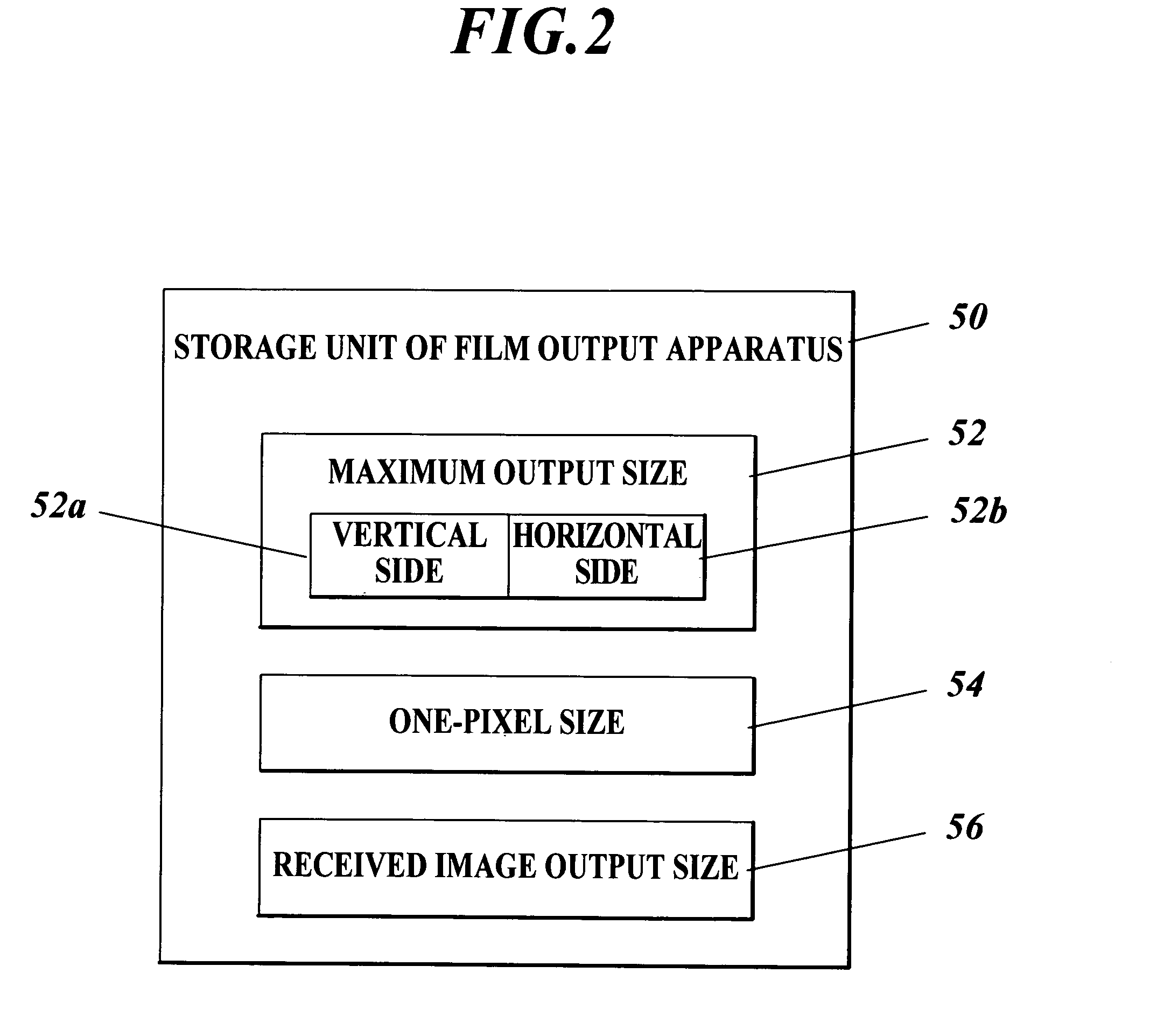
[0097]FIG. 10B is a diagram showing an example of the data configuration of the memory 13 of the According to the diagram, the memory 13 stores the controller input information 130, the output apparatus setting information table 132, the image output size 134, the patient information setting items 136, the patient information 138, the one-frame image region size 140, a received image size 144, a transmitted image size 146, an expansion or contraction rate 148, and an actual size template data 149.

[0098] The received image size 144 is an image size (number of pixels) of medical image data received from the diagnostic apparatus M, i.e. the medical image data stored in the medical image table 32. For example, the CPU 11 obtains the received image size 144 from the header information included in the data at the time of the reception of the medical image data, or calculates the received image size 144 from the data capacity of the data.

[0099] The transmitted image size 146 is an image ...


PUM


Abstract

A medical image output system includes: a medical image transferring apparatus and plural medical image forming apparatus. Each medical image forming apparatus includes a storage unit for storing image formable region information. The transferring apparatus includes: an obtaining unit for obtaining the stored image formable region information; a determination unit for determining an image formation size based on the obtained image formable region information; a unit for expanding or contracting the received medical image data according to the determined image formation size; and a transfer unit for transferring the expanded or contracted image data to the medical image forming apparatus.

Description

BACKGROUND OF THE INVENTION [0001] 1. Field of the Invention [0002] The present invention relates to a medical image output system provided with a medical image transferring apparatus transferring received medical image data and a plurality of medical image forming apparatus each outputting a sheet-like recording medium having an image formable region in which the image formation of the medical image based on the transferred medical image data is performed, and the like. [0003] 2. Related Art [0004] There has been conventionally known a medical image output system outputting a medical image formed (drawn) on a sheet-like recording medium such as a film, recording paper or the like. The medical image is one having been produced by radiography with various medical image generating apparatus (hereinafter referred to as “diagnostic apparatus”) of calculated tomography (CT), calculated radiography (CR), magnetic resonance imaging (MRI), a mammography apparatus, an ultrasonic diagnostic a...

Claims


Application Information

Patent Timeline
IPC IPC(8): G06F7/00A61B5/00A61B5/055A61B8/00G16H10/60G16H30/20G16H30/40
CPCG06F19/321H04N1/32133H04N2201/3242H04N2201/3271A61B5/0013G16H30/20G16H30/40
Inventor YAMASAKI, TOSHIO
Owner KONICA MINOLTA MEDICAL & GRAPHICS INC
Learn More