
Temporary catheter for biopsy site tissue fixation

a tissue fixation and temporary catheter technology, applied in the field of medical treatment devices, can solve the problem of high cost of prior catheters

Inactive Publication Date: 2006-09-14
HOLOGIC INC
View PDF99 Cites 74 Cited by
  • Summary
  • Abstract
  • Description
  • Claims
  • Application Information

AI Technical Summary

Benefits of technology

[0012] Additionally, the catheter device and method of using the device may include application of a vacuum within the body cavity that is effective to draw tissue surrounding the body cavity towards the surface of the cavity filling member of the catheter device, which is placed within the body cavity.

Problems solved by technology

The prior catheters are expensive and a catheter used only to hold the shape of the biopsy cavity is disposed of afterwards.

Method used


Image

  • Temporary catheter for biopsy site tissue fixation
  • Temporary catheter for biopsy site tissue fixation
  • Temporary catheter for biopsy site tissue fixation

Examples


Embodiment Construction

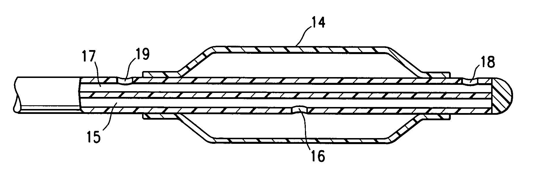
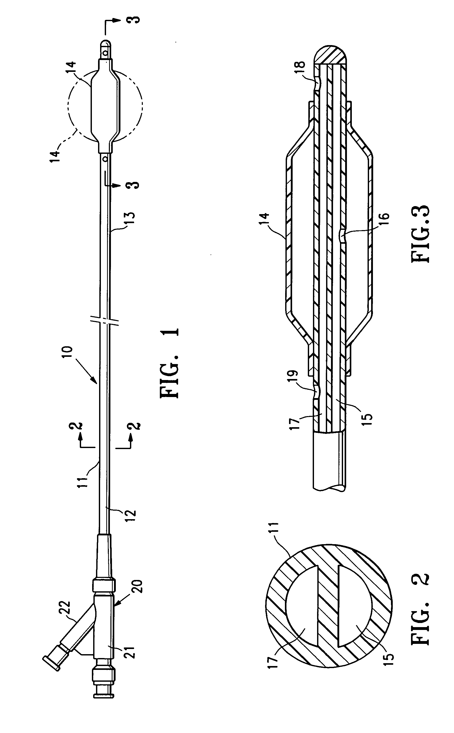
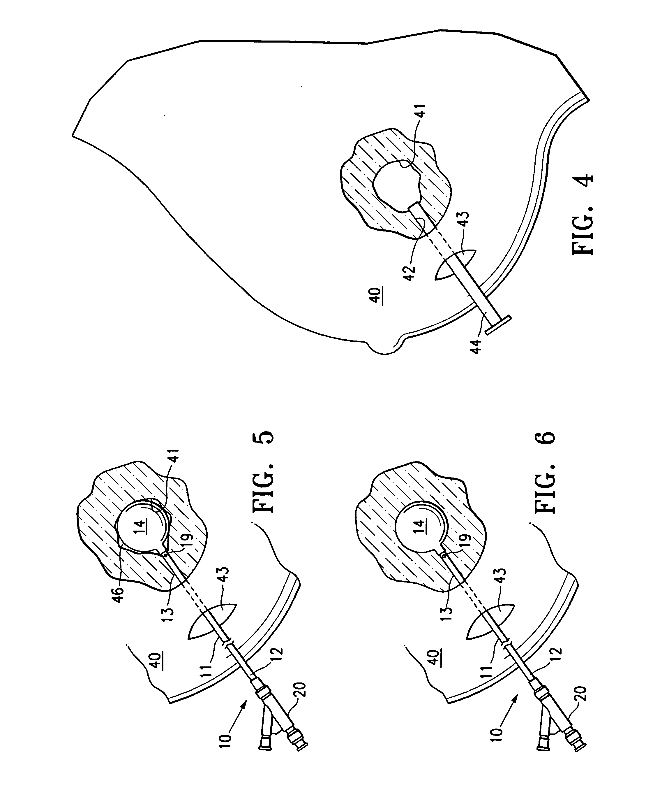
[0027] The present invention is directed to catheter devices and methods of using such devices for temporarily maintaining access to an intracorporeal cavity in a targeted tissue region within in a patient's body, such as a biopsy site or a cavity left after removal of a tissue specimen. The catheter device embodying features of the invention has an elongated shaft with a proximal shaft portion that is either detachable or foldable or coilable to facilitate deployment of the proximal end of the catheter within the tissue surrounding the biopsy site. After tissue has been removed from the targeted tissue region, the cavity filling member on the distal end of the catheter is inserted through an opening in the patient's skin and advanced through a passageway in the patient to the body cavity where the cavity filling member is deployed. The proximal shaft section of the catheter is folded or coiled and placed in a subcutaneous location and the opening in the patient's skin is closed or ...


PUM


Abstract

Devices and methods are provided for temporarily maintaining access to a body cavity in a targeted tissue region within a patient's body. One embodiment of the catheter device includes an elongated shaft having a proximal shaft section which is flexible enough to be folded or coiled into a configuration for deployment within the patient. An alternate embodiment includes a catheter device having one or more detachable proximal shaft sections and having at least one one-way valve to restrict fluid flow of inflation fluid to flow to the balloon. After deployment of the catheter device completely within the patient, the opening through which the catheter device is deployed is closed, e.g. by sutures, adhesives and the like to minimize infection at the site. Within a few days or weeks after the tissue has been evaluated for cancer, the temporary catheter device may be removed from the patient. If cancer or pre-cancer cells are found in the specimen removed from the cavity, then a radiation balloon catheter or other irradiation device can be inserted into the patient to irradiate tissue surrounding the biopsy cavity to ensure that cancer cells within the tissue surrounding the cavity are killed.

Description

RELATED APPLICATIONS [0001] This application is a continuation-in-part of application Ser. No. 11 / 105,856 filed Apr. 14, 2005 which is a continuation of application Ser. No. 10 / 849,410 filed May 19, 2004, now U.S. Pat. No. 6,955,641 which is a continuation of application Ser. No. 10 / 290,002 filed Nov. 6, 2002, now U.S. Pat. No. 6,923,754. All of these applications are incorporated herein in their entireties by reference.FIELD OF THE INVENTION [0002] This invention generally relates to medical treatment devices and methods of using such devices. In particular, the invention is directed to devices and methods for temporarily maintaining access to a cavity in a targeted tissue region, such as a biopsy site from which cancerous, pre-cancerous or other tissue has been removed, to provide subsequent treatments to the tissue surrounding the cavity. BACKGROUND OF THE INVENTION [0003] In diagnosing and treating certain medical conditions, it is often desirable to perform a biopsy, in which a...

Claims


Application Information

Patent Timeline
IPC IPC(8): A61N5/00A61F2/958
CPCA61B10/0041A61B17/0218A61B19/24A61B2017/00557A61B2017/00796A61B2019/5408A61M25/10A61M29/02A61M2025/1095A61N5/1015A61M29/00A61B90/02A61B2090/3908
Inventor LUBOCK, PAULJONES, MICHAEL L.
Owner HOLOGIC INC
Learn More