Detector head position correction for hybrid SPECT/CT imaging apparatus

a technology of hybrid spect/ct imaging and detector head position correction, which is applied in the field of hybrid nuclear medical imaging, can solve the problems of head deflection and alignment, inaccuracy in image registration of images produced, and differences in resolution between ct images and spect images, and achieve the effect of more accurate spect/ct image registration

a technology of hybrid spect/ct imaging and detector head position correction, which is applied in the field of hybrid nuclear medical imaging, can solve the problems of head deflection and alignment, inaccuracy in image registration of images produced, and differences in resolution between ct images and spect images, and achieve the effect of more accurate spect/ct image registration

US20060214097A1Active Publication Date: 2006-09-28SIEMENS MEDICAL SOLUTIONS USA INC

Image

  • Detector head position correction for hybrid SPECT/CT imaging apparatus
  • Detector head position correction for hybrid SPECT/CT imaging apparatus
  • Detector head position correction for hybrid SPECT/CT imaging apparatus

Examples


Embodiment Construction

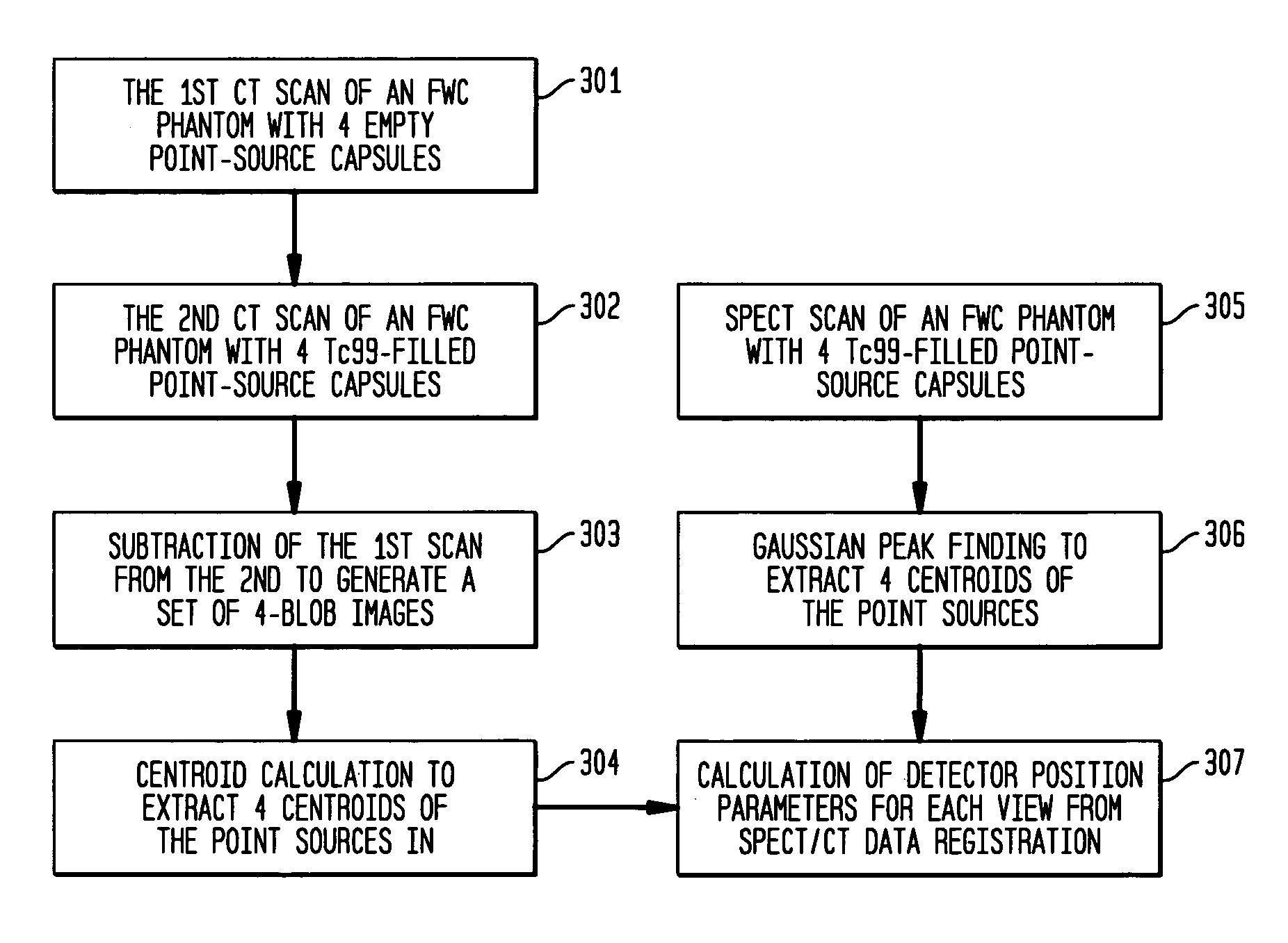
[0028] According to the present invention, a test phantom is provided with a number of radioisotope point sources, and is imaged with both CT and SPECT modalities. For the CT test data, two images are taken of the phantom: one with blank point sources and another with loaded point sources. The difference between these two images will then represent only the point source locations in three-dimensional space. The three-dimensional space is defined by a fixed coordinate system.

[0029] The phantom with loaded point sources is then subjected to SPECT imaging over an entire range of projection view angles. For each view angle, centroids are calculated for the point sources, using a coordinate system that is attached to the detector heads, and thus is moving with respect to the fixed coordinate system.

[0030] These two sets of test data then are used to calculate detector head position correction parameters for each different view angle of the SPECT procedure. The detector correction param...


PUM


Abstract

A system and method provide for more accurate SPECT / CT image registration. CT data is utilized to establish a global spatial coordinate system of a common test phantom. The common test phantom is then used to obtain a set of point source nuclear images. Three-dimensional CT point source data is mapped to a two-dimensional image plane of corresponding point source data, to obtain a pair of intersecting projection cones that are used to obtain a set of detector head position correction parameters to correct detector head positioning in the CT coordinate system when obtaining SPECT projection images of the same object.

Description

BACKGROUND OF THE INVENTION [0001] 1. Field of the Invention [0002] This invention relates generally to hybrid nuclear medical imaging, wherein nuclear medical images are co-registered or fused with images of the same region obtained with a different imaging modality, such as Computerized Tomography (CT), Magnetic Resonance Imaging (MRI) and Ultrasound (US). More specifically, this invention relates to improvement in image registration accuracy between nuclear medical images and images obtained with a different imaging modality such as CT, where both modalities are obtained with a single hybrid imaging apparatus. [0003] 2. Background and Prior Art [0004] In nuclear imaging, a patient is injected with or swallows a radioactive isotope which has an affinity for a particular organ, structure or tissue of the body. Gamma rays are then emitted from the body part of interest, are collimated by a collimator so that only gamma photons traveling in a direction perpendicular to the surface of...

Claims


Application Information

Patent Timeline
28 Sep 2006
Publication
US20060214097A1
IPC
G01T1/166
CPC
G01T1/1611; G01T1/1612; G01T1/1648; G01T1/169; G01T7/005
Inventors
WANG, SHARON XIAORONG; FANG, JOSEPH