Image processing apparatus and method, photographing system, controlling method for the system, and computer-readable memory
- Summary
- Abstract
- Description
- Claims
- Application Information
AI Technical Summary
Benefits of technology
Problems solved by technology
Method used
Image
Examples
first embodiment
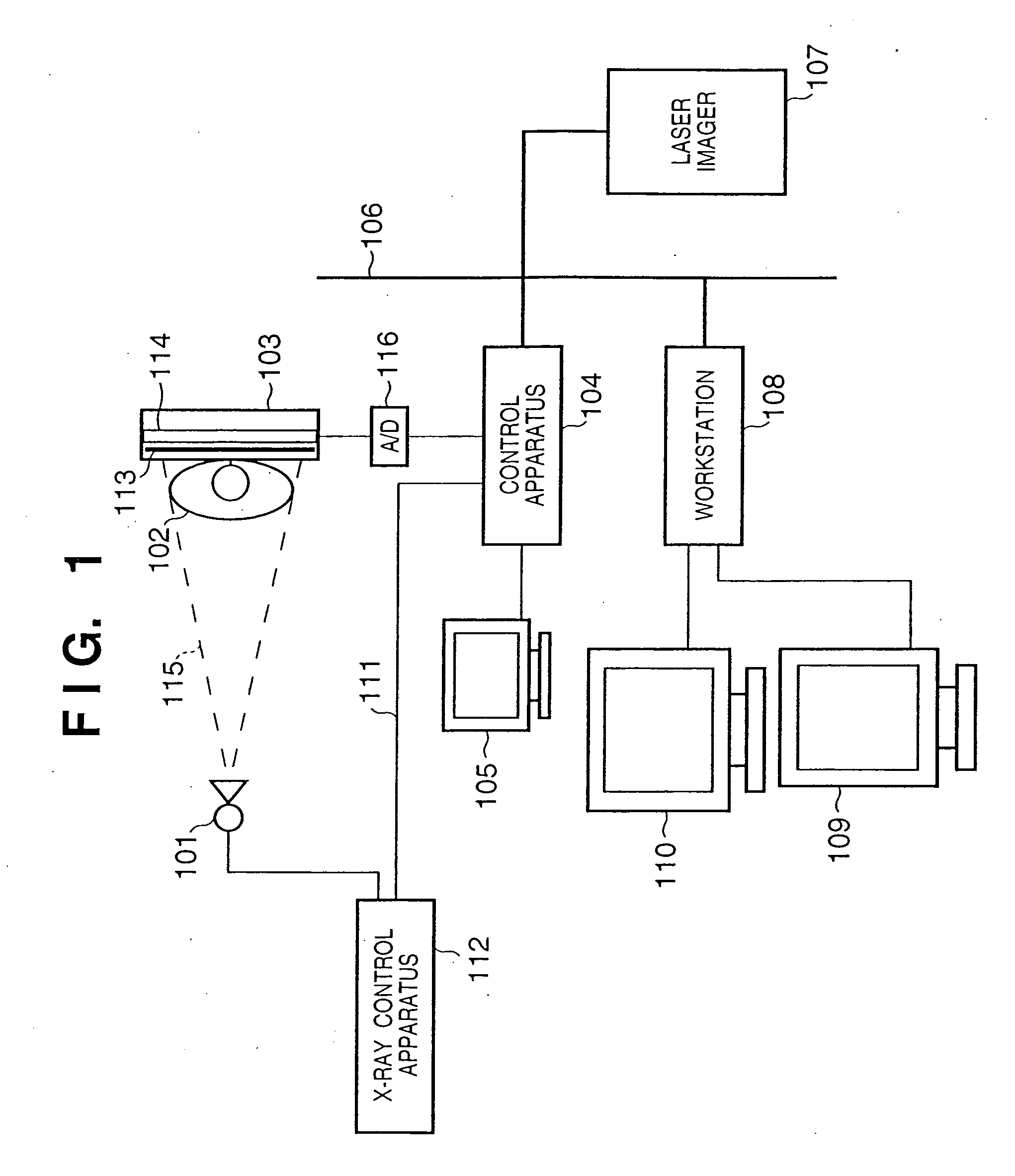
[0053]FIG. 1 is a view showing the schematic arrangement of a digital X-ray photographing system according to the first embodiment of the present invention.
[0054] Referring to FIG. 1, an X-ray source 101 generates X-rays. An X-ray control apparatus 112 controls the tube voltage or tube current of a radiation tube for generating ray. X-rays 115 generated by the X-ray source 101 pass through a person 102 to be examined as a diagnosis object and enter an X-ray detection apparatus 103. At this time, the X-rays 115 pass through the person 102 to be examined and become incident on a phosphor 113 for converting the X-rays 115 into visible light. The X-rays 115 that have passed through the person 102 to be examined contain image information with different transmission amounts according to the sizes or shapes of bones and viscera or the presence / absence of a morbid portion in the person 102 to be examined. The X-rays 115 are converted into visible light by the phosphor 113 and enter an X-ra...
second embodiment
[0076] In the first embodiment, the observation area setting information is set by the observation area setting section 302 using the X-ray irradiation stop information obtained from the X-ray control apparatus 112. However, this is not always necessary. For example, as shown in FIG. 15, on the basis of digital image data acquired by an image input section 801, an irradiation field recognition section 802 automatically acquires an area where an X-ray detection apparatus 103 is irradiated with X-rays, and determines the area as an observation area. The processing described in the first embodiment is executed for this observation area. As the irradiation field recognition section 802, for example, a method disclosed in Japanese Patent Application No. 10-243020 proposed by the present applicant can be applied.
[0077] The image input section 801, an image processing section 804, output mode input section 807, output medium size input section 803, output determination section 805, and im...
third embodiment
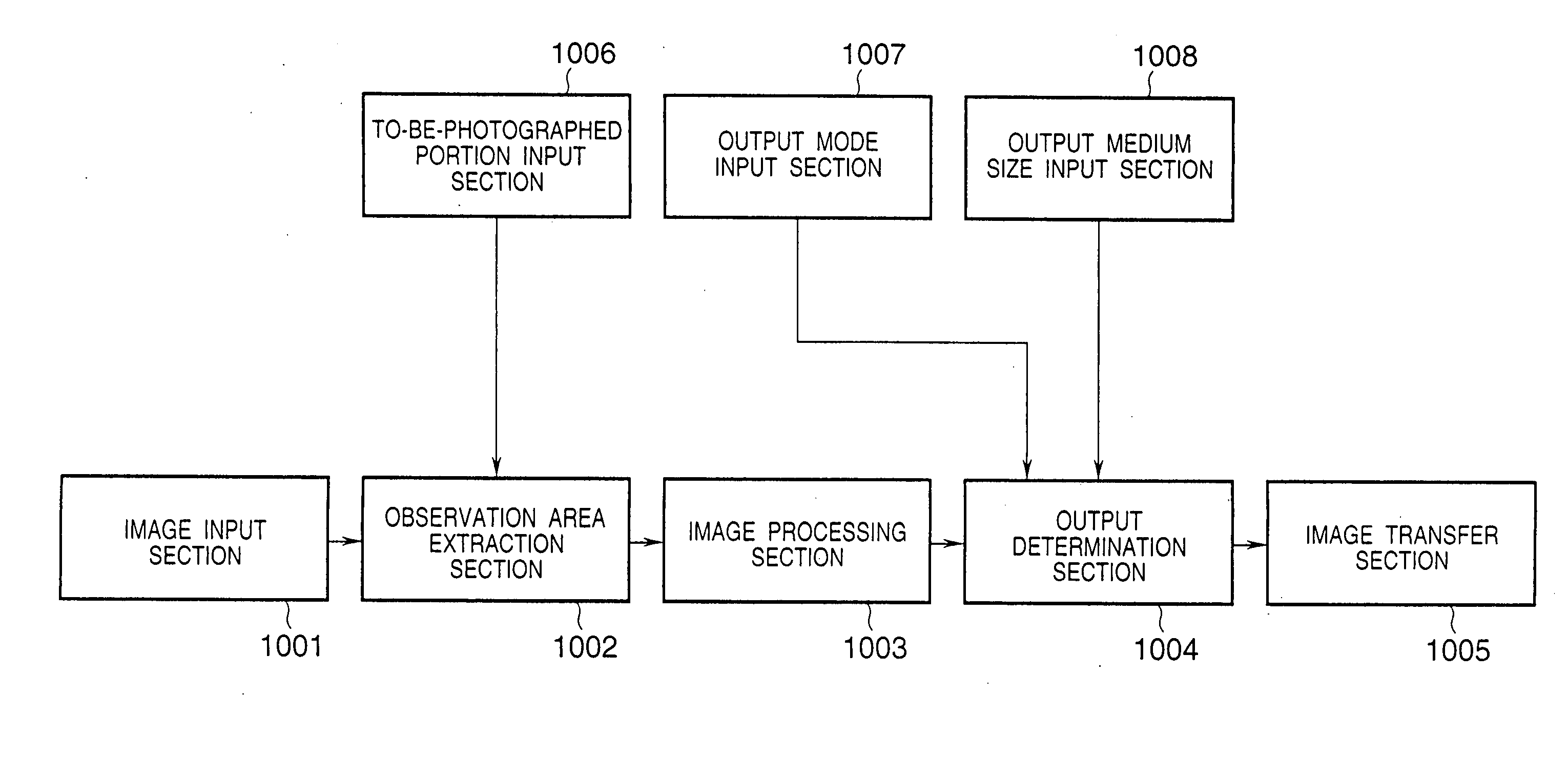
[0079] In the first embodiment, the observation area setting information is set by the observation area setting section 302 using the X-ray irradiation stop information obtained from the X-ray control apparatus 112. However, this is not always necessary. For example, as shown in FIG. 18, observation area setting information may be obtained by setting portion information input from a to-be-photographed portion input section 1006. An observation area extraction section 1002 automatically extracts the basic observation area for the doctor from that portion. An observation area is set on the basis of the basic observation area, thereby obtaining the observation area setting information. FIGS. 16 and 17 show this example. FIG. 16 shows the front image of a chest. FIG. 17 shows the side image of the chest. In both cases, an area (an area indicated by a thin dotted line) including the area of lungs is the basic observation area for the doctor. This basic observation area is extracted by th...
PUM
 Abstract
Description
Claims
Application Information
 - R&D
- Intellectual Property
- Life Sciences
- Materials
- Tech Scout
- Unparalleled Data Quality
- Higher Quality Content
- 60% Fewer Hallucinations
Browse by: Latest US Patents, China's latest patents, Technical Efficacy Thesaurus, Application Domain, Technology Topic, Popular Technical Reports.
© 2025 PatSnap. All rights reserved.Legal|Privacy policy|Modern Slavery Act Transparency Statement|Sitemap|About US| Contact US: help@patsnap.com


