
3d-originated cardiac roadmapping

a technology of cardiac roadmapping and original cardiac image, which is applied in the field of 3d-originated cardiac roadmapping, can solve problems such as confusion and inaccuracy, and achieve the effect of improving the accuracy of information, without any additional burden

Inactive Publication Date: 2013-05-09
KONINKLIJKE PHILIPS ELECTRONICS NV
View PDF7 Cites 23 Cited by
  • Summary
  • Abstract
  • Description
  • Claims
  • Application Information

AI Technical Summary

Benefits of technology

The technical effect of this patent text is to improve the accuracy of information provided to users as navigation information without adding any additional burden to the patient, such as additional X-ray dose.

Problems solved by technology

However, using 2D image data for cardiac roadmapping leads to inaccuracy and confusions.

Method used


Image

  • 3d-originated cardiac roadmapping
  • 3d-originated cardiac roadmapping
  • 3d-originated cardiac roadmapping

Examples


Embodiment Construction

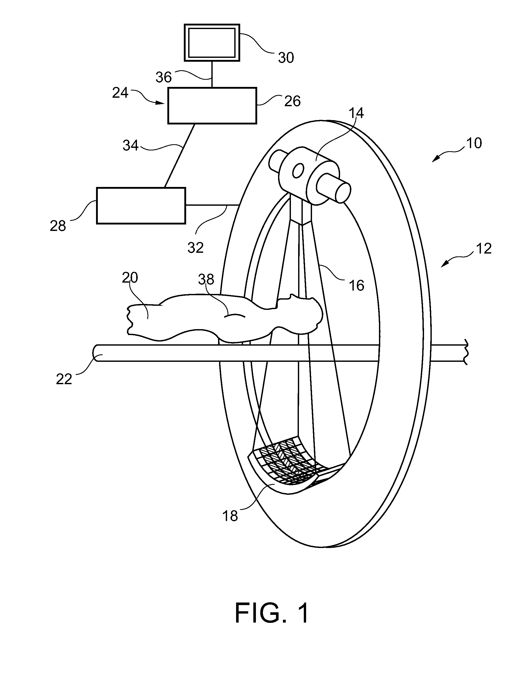
[0038]FIG. 1 schematically shows a medical imaging system 10, for the use in a catheterization laboratory, for example. The medical imaging system 10 for examination of an object of interest comprises X-ray image acquisition means 12. The X-ray image acquisition means 12 are provided with a source of X-ray radiation 14 to generate X-ray radiation, indicated by an X-ray beam 16. Further, an X-ray image detection module 18 is located opposite the source of X-ray radiation 12 such that, for example, during a radiation procedure, an object, for example a patient 20, can be located between the source of X-ray radiation 14 and the detection module 18. Further, a table 22 is provided to receive the object to be examined.

[0039]Further, the medical imaging system 10 comprises a device 24 for 3D-originated cardiac roadmapping. The device 24 comprises a processing unit 26, an interface unit 28, and a display 30.

[0040]The interface unit 28 is adapted to provide 3D+t image data of a vascular str...


PUM


Abstract

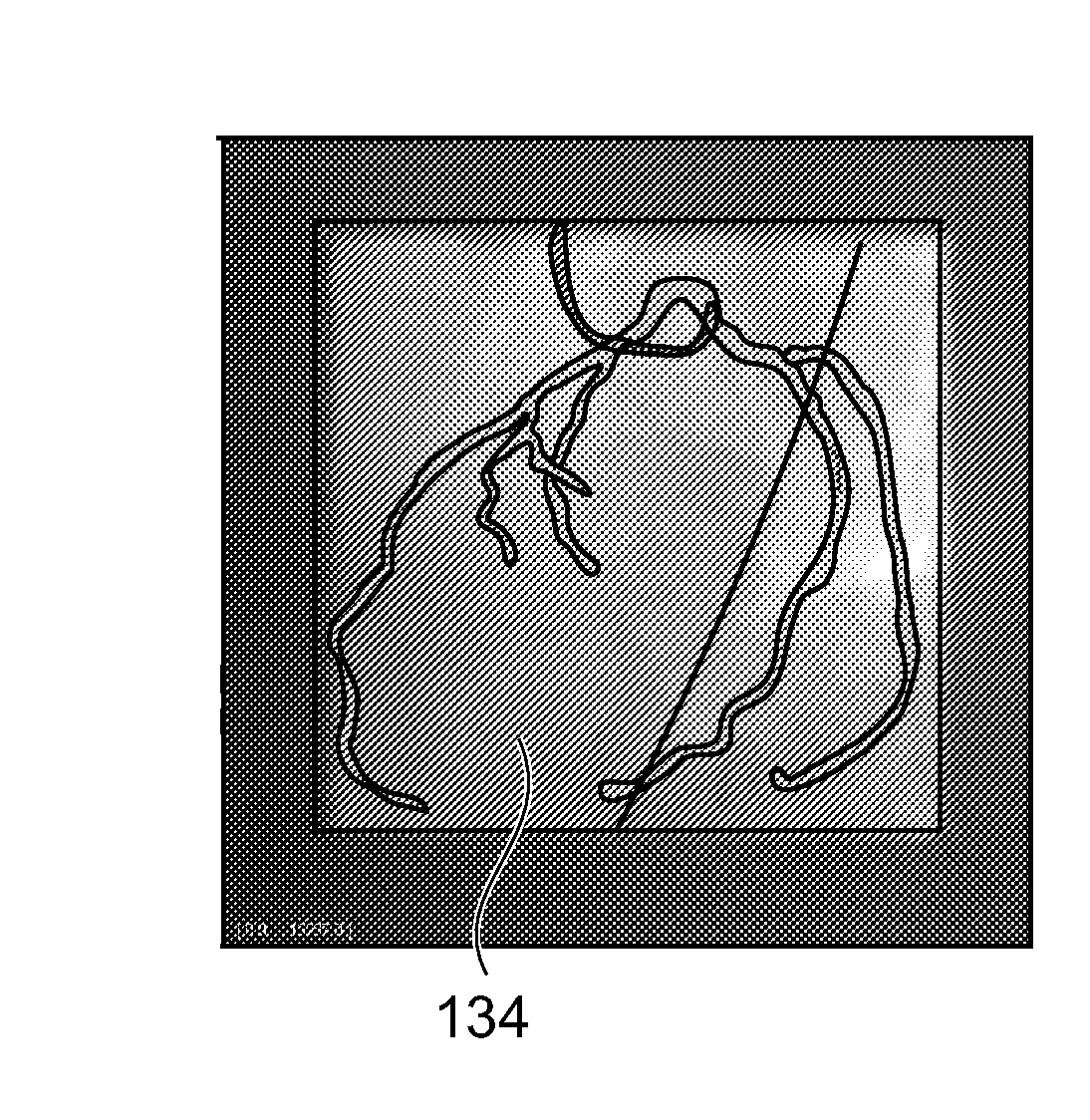
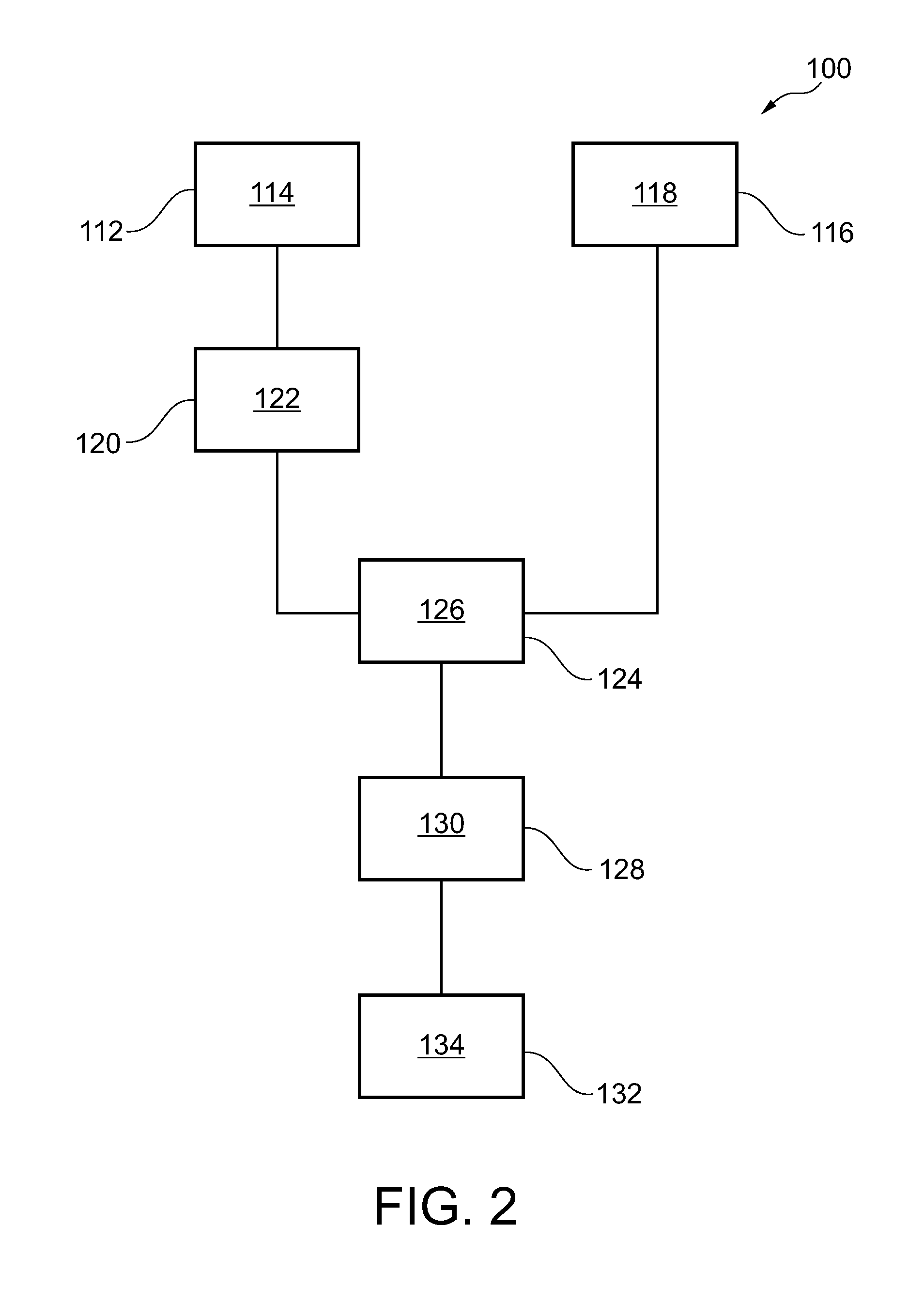
The present invention relates to 3D-originated cardiac roadmapping. In order to improve the accuracy of the information provided to the user as navigation information, without any additional burden to the patient such as additional X-ray dose, a method is described comprising the steps of a) providing 3D+t image data of a vascular structure of an object; b) acquiring two-dimensional image data of the object, which object comprises the vascular structure, the 2D image data comprising at least one 2D image; c) projecting the vascular structure, thereby generating a plurality of mask images on the basis of a 3D+t image data; d) registering the at least one 2D image with one of the plurality of the mask images, wherein the registration comprises finding the maximum of a similarity factor between the mask images and the at least one 2D image; e) generating a combination of the at least one 2D image and a projection of the vascular structure on the basis of the 3D+t image data according to the registration; and f) displaying the combination as a guiding vessel tree projection. Thus improved cardiac roadmapping in form of a topographical roadmapping is provided since the roadmapping is based on three-dimensional data.

Description

FIELD OF THE INVENTION[0001]The present invention relates to a device and a method for 3D-originated cardiac roadmapping as well as to a medical imaging system for examination of an object of interest, a computer program element, and a computer readable medium.BACKGROUND OF THE INVENTION[0002]Navigation systems are known to provide navigating information, for example to a cardiologist, a physician or other clinical staff for example, or to provide the user with information about an object of interest, for example a patient. Such navigation information is in particular needed during catheter interventional procedures, such as PTCA (Percutaneous Transluminal Coronary Angioplasty), for example to treat cardiac stenoses. In WO 2008 / 104921, cardiac roadmapping is performed on the basis of at least one of a global correlation determination of a first image sequence of the object of interest and a second image sequence of the object of interest and a correlation determination of a first im...

Claims


Application Information

Patent Timeline
IPC IPC(8): A61B6/02
CPCA61B6/022A61B6/12A61B6/4441A61B6/463A61B6/487A61B6/504A61B6/5223A61B6/5235A61B2090/367A61B2090/364
Inventor FLORENT, RAOULAUVRAY, VINCENTGRASS, MICHAELSCHAEFER, DIRKSCHOONENBERG, GERT
Owner KONINKLIJKE PHILIPS ELECTRONICS NV
Learn More