
System and method for vascular segmentation by Monte-Carlo sampling

a vascular segmentation and monte-carlo technology, applied in the field of tubular structure segmentation, can solve the problems of not taking into account prior knowledge of the form of the vessel, morphological filters as well as geometric contours can be very sensitive to local minima, and existential approaches suffer from certain limitations

Inactive Publication Date: 2006-10-26
SIEMENS MEDICAL SOLUTIONS USA INC
View PDF11 Cites 16 Cited by
  • Summary
  • Abstract
  • Description
  • Claims
  • Application Information

AI Technical Summary

Benefits of technology

The patent describes a method for segmenting tubular structures in digital images using a particle-based approach. The method models the vessel as a probability space and uses a learning process to account for bifurcations and branchings. The method also takes into account the non-linearity of the vessel's appearance and structure through a statistical model. The final result is a probability density function that describes the vessel's centerline and appearance. The method can be used for both pathological and non-pathological cases and is robust against noise. The method also includes a training set of tubular cross sections and a principle component analysis to estimate the representation of the tubular cross section. The method can be used in various medical applications and is efficient in terms of computational resources.

Problems solved by technology

One can claim that existing approaches suffer from certain limitations.
Local operators, region growing techniques, morphological filters as well as geometric contours can be very sensitive to local minima and fail to take into account prior knowledge on the form of the vessel.
On the other hand, cylindrical models, parametric active contours and template matching techniques might not be well suited to account for the non-linearity of the vessel structure and require particular handling of branchings and bifurcations.
Tracking methods can often fail in the presence of missing and corrupted data.

Method used


Image

  • System and method for vascular segmentation by Monte-Carlo sampling
  • System and method for vascular segmentation by Monte-Carlo sampling
  • System and method for vascular segmentation by Monte-Carlo sampling

Examples


Embodiment Construction

[0033] Exemplary embodiments of the invention as described herein generally include systems and methods for particle-filter based propagation approaches for the segmentation of vascular structures in 3D volumes.

[0034] As used herein, the term “image” refers to multi-dimensional data composed of discrete image elements (e.g., pixels for 2-D images and voxels for 3-D images). The image may be, for example, a medical image of a subject collected by computer tomography, magnetic resonance imaging, ultrasound, or any other medical imaging system known to one of skill in the art. The image may also be provided from non-medical contexts, such as, for example, remote sensing systems, electron microscopy, etc. Although an image can be thought of as a function from R3 to R, the methods of the inventions are not limited to such images, and can be applied to images of any dimension, e.g. a 2-D picture or a 3-D volume. For a 2- or 3-dimensional image, the domain of the image is typically a 2- o...


PUM


Abstract

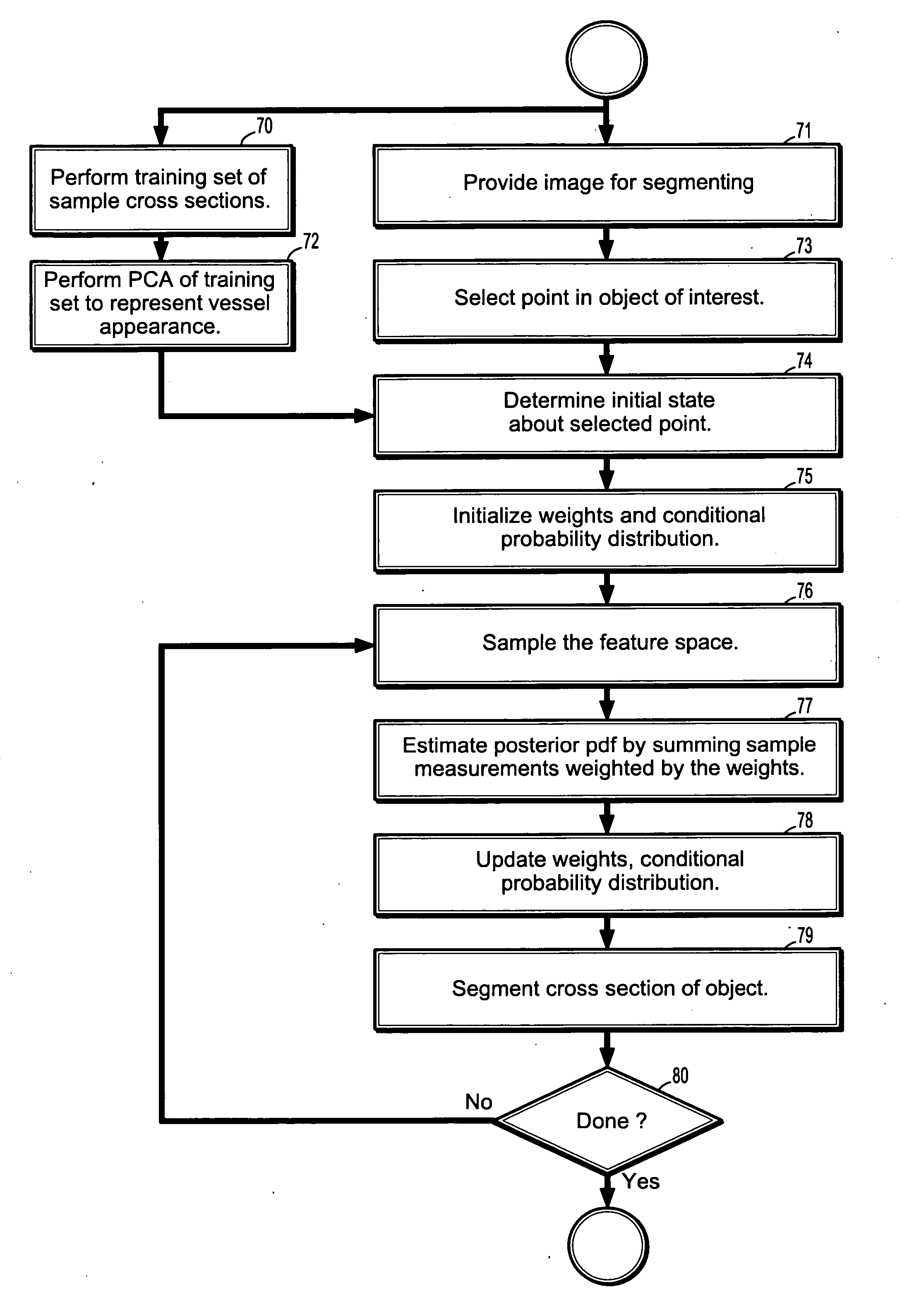
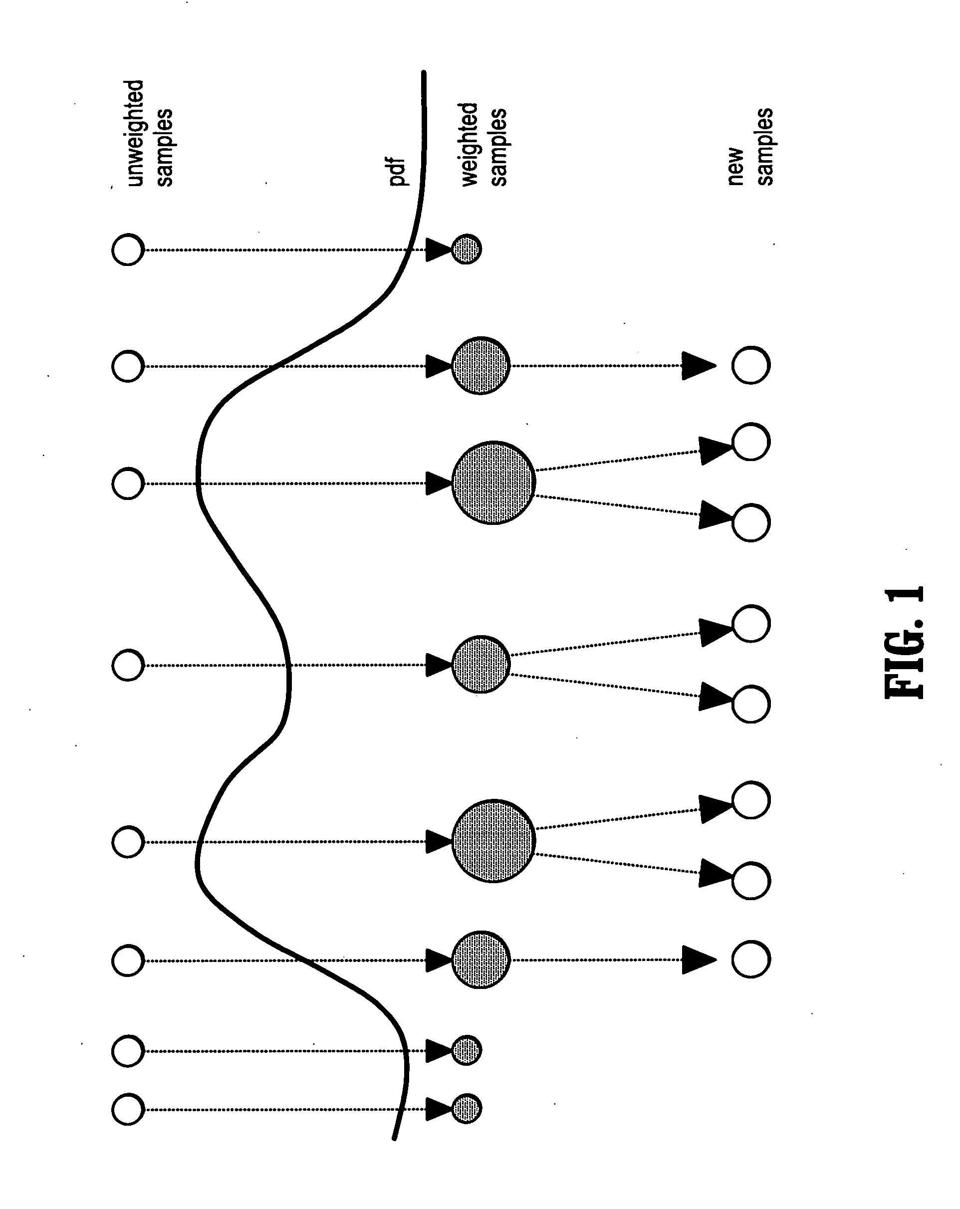
A method of segmenting tubular structures in digital images includes selecting a point in an image of a tubular object to be segmented, defining an initial state of the selected point, initializing measurement weights, a conditional probability distribution and a prior probability distribution of a feature space of the initial state, sampling the feature space from the prior probability distribution, estimating a posterior probability distribution by summing sample measurements weighted by the measurement weights, and segmenting a cross section of the tubular object from the posterior probability distribution.

Description

CROSS REFERENCE TO RELATED UNITED STATES APPLICATION [0001] This application claims priority from “Vascular Segmentation by Monte-Carlo Sampling, with Prior Appearance Model”, U.S. Provisional Application No. 60 / 664,627 of Florin, et al., filed Mar. 23, 2005, the contents of which are incorporated herein by reference,TECHNICAL FIELD [0002] This invention is directed to the segmentation of tubular structures in medical imaging and computer vision applications. DISCUSSION OF THE RELATED ART [0003] Quasi-Monte-Carlo methods are a dynamic field of study for Computer Vision, due to the increasing capacities of computers. Furthermore, for a class of segmentation problems for which ground-truth is widely available, statistical learning leads to faster and more robust results. [0004] Segmentation of tubular structures arises in numerous applications of computer vision. Medical imaging is an example where such need exists, in particular in vascular and retinal images. The coronary arteries p...

Claims


Application Information

Patent Timeline
Patent Type & Authority Applications(United States)
IPC IPC(8): G06K9/00G06K9/34G06T15/00G06V10/26
CPCG06K9/34G06K2009/00932G06K2209/05G06T7/0081G06T2207/30101G06T2207/10081G06T2207/20081G06T2207/20101G06T7/0087G06T7/11G06T7/143G06V40/14G06V10/26G06V2201/03
Inventor FLORIN, CHARLESWILLIAMS, JAMES P.PARAGIOS, NIKOLAOS
Owner SIEMENS MEDICAL SOLUTIONS USA INC
Learn More