Ultrasound image diagnosis apparatus and an apparatus and method for processing an image display

an ultrasonic image and diagnostic equipment technology, applied in ultrasonic/sonic/infrasonic diagnostics, instruments, applications, etc., can solve the problems of wasting time, wasting operator's time, and wasting resources, so as to achieve efficient operation and facilitate image diagnosis

Active Publication Date: 2006-10-26
TOSHIBA MEDICAL SYST CORP
View PDF10 Cites 6 Cited by
  • Summary
  • Abstract
  • Description
  • Claims
  • Application Information

AI Technical Summary

Benefits of technology

[0018] According to embodiments of the present invention, it becomes possible to rapidly search for and to display a patient's cardiac images of a diastolic end period and a systolic en

Problems solved by technology

However, the present inventors recognized these prior proposals have never intended to automatically display a dual image of a diastolic end period and a systolic end period at an optional heart cycle at all.
Thus, the conventional techniques have never disclosed nor suggested to solve the above-mentioned problems for displaying a pair of ultrasound images of a cardiac diastolic period and systolic period that are det

Method used


Image

  • Ultrasound image diagnosis apparatus and an apparatus and method for processing an image display
  • Ultrasound image diagnosis apparatus and an apparatus and method for processing an image display
  • Ultrasound image diagnosis apparatus and an apparatus and method for processing an image display

Examples


Embodiment Construction

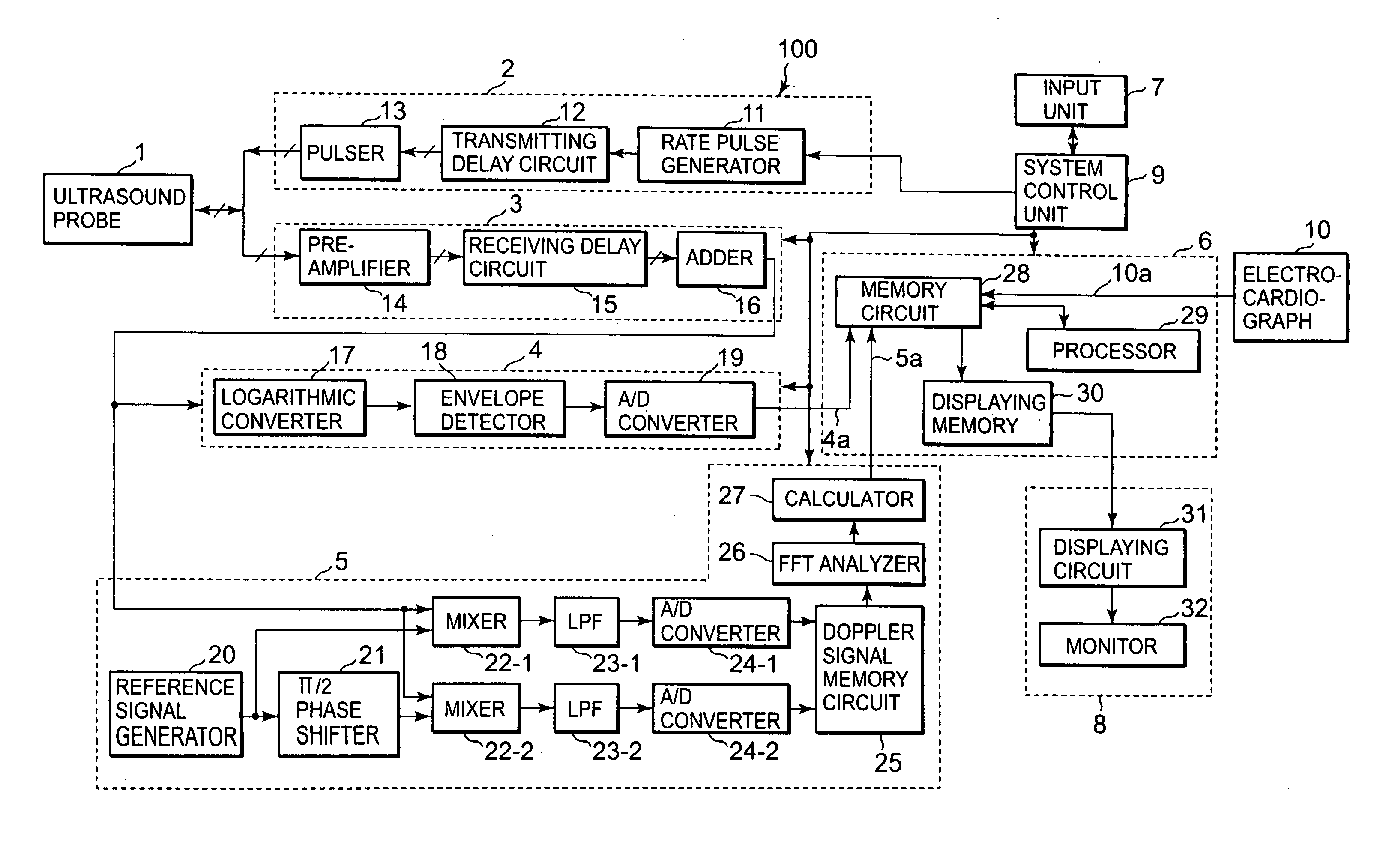
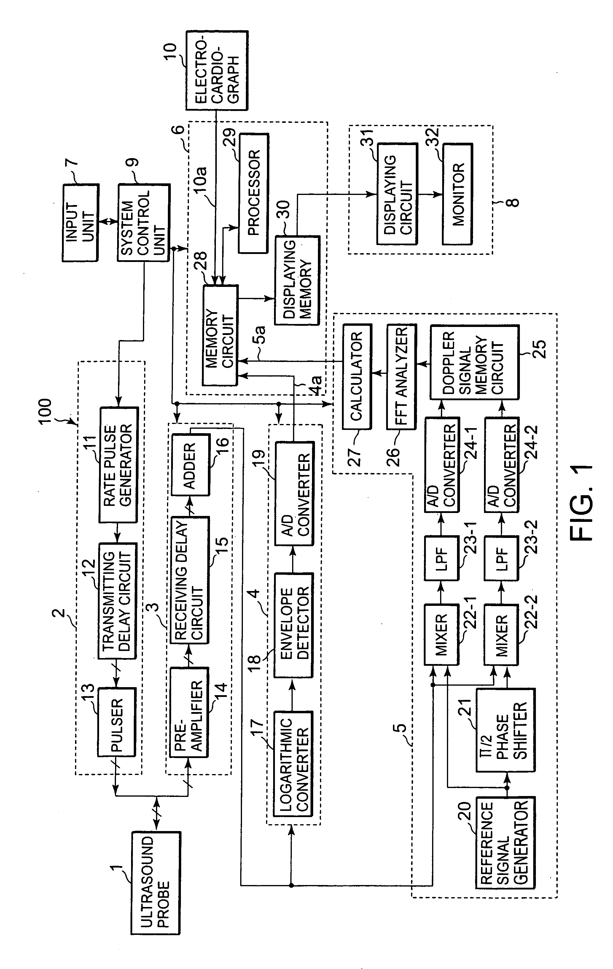
[0034] With reference to the drawings, the embodiments of an ultrasound image diagnosis apparatus and method consistent with the present invention are explained. FIG. 1 is a block diagram for illustrating an entire construction of an embodiment of the ultrasound diagnostic apparatus consistent with the present invention.

[0035] As illustrated in FIG. 1, the ultrasound image diagnosis apparatus 100 includes an ultrasound probe 1 for transmitting ultrasound to a patient body and receiving echo signals reflected from the patient body by being placed in contact with a patient body surface, an ultrasound transmitting unit 2 for transmitting ultrasound, an ultrasound receiving unit 3 for receiving echo signals as received signals, a B mode processing unit 4 and a Doppler mode processing unit 5 for respectively processing the received signals, an image processing unit 6 for executing image processing operations, an input unit 7 for inputting image selection data, a display unit 8 for displ...


PUM


Abstract

An ultrasound image diagnosis apparatus and an ultrasound image processing method that can automatically display a pair of a cardiac systolic end period image and a cardiac diastolic end period image based on living body signal data. A plurality of ultrasound images are collected from a patient and stored in a memory in correspondence with living body signal data, such as an ECG wave. When a freeze instruction is input during a reproduction of ultrasound images, a pair of images of a cardiac diastolic end period and a systolic end period is searched for going back from the freeze input time. A pair of the diastolic end period image and the systolic end period image is automatically displayed on a screen of the ultrasound image diagnosis apparatus.

Description

CROSS-REFERENCE TO RELATED APPLICATIONS [0001] This application claims priority from, and the benefit of, Japanese Patent Application No. 2005-114265, filed on Apr. 12, 2005, the contents of which are expressly incorporated herein by reference in their entirety. BACKGROUND OF THE INVENTION [0002] 1. Field of the Invention [0003] The present invention relates to an ultrasound image diagnosis apparatus and an apparatus and method for processing an image display thereof, and more particularly to an ultrasound image diagnosis apparatus and method that can automatically search for a cardiac diastolic end period and a cardiac systolic end period image, the searched cardiac diastolic end period image appearing just following the searched diastolic end period image, and to display such images in parallel on a screen. [0004] 2. Discussion of the Background [0005] An ultrasound image diagnosis apparatus transmits ultrasound through ultrasound transducers installed in an ultrasound probe to an...

Claims


Application Information

Patent Timeline
IPC IPC(8): A61B8/00A61B5/352
CPCA61B5/0456A61B8/463A61B8/00A61B5/352
Inventor OONUKI, MASATO
Owner TOSHIBA MEDICAL SYST CORP
Why Eureka
Try Eureka