
Apparatus and method for holding a transesophageal echocardiography probe

a transesophageal echocardiography and apparatus technology, applied in the field of medical apparatus and methods, can solve the problems of not providing a very user-friendly configuration, unable to provide a suitable device, and generally unstable supports

Inactive Publication Date: 2006-10-26
LOUBSER PAUL G
View PDF29 Cites 13 Cited by
  • Summary
  • Abstract
  • Description
  • Claims
  • Application Information

AI Technical Summary

Benefits of technology

[0041] The above and other features of the invention, including various novel details of construction and combinations of parts and method steps will now be more particularly described with reference to the accompanying drawings and pointed out in the claims. It will be understood that...

Problems solved by technology

One of the currently existing problems is the lack of a suitable device for stabilizing TEE probe 5A, 5B, 5C at the head of the operating table.
However, these supports are generally very unstable and do not provide a very user-unfriendly configuration.
However, the Kainuma device does not provide optimal stability to various sized TEE probe handles 25 and also requires a separate pole to support it.
None of the above-identified currently practiced techniques or devices is universally used inasmuch as each is believed to be either unsatisfactory or unpopular.
Damage to TEE Probe
This type of positioning is extremely unstable as TEE probe 5A, 5B, 5C may fall off the operating room table onto the floor and become damaged.
Such damage can be significant inasmuch as a TEE probe typically sells for about $14,000.
In addition, repeated falls may subject the electrical connections of TEE probe 5A, 5B, 5C to repeated flexion and / or stretching, which in turn may result in excess wear and tear.
In addition, excess wear and tear of the TEE probe may produce a break in the coverings of the TEE probe shaft or transducer tip, thereby exposing wires to the esophagus, which may serve as a conduit of electrical energy during cardioversion of the heart, and produce burns to the esophagus.
Improper positioning may also cause TEE probe 5A, 5B, 5C to fall off an operating room table and such a fall may actually tear open the tissue at the side of a patient's mouth.
Improperly positioning or unsecured use may cause TEE probe 5A, 5B, 5C to become twisted so that it is pressed on a patient's lip.
This prolonged contact with a lip can produce mucosal abrasions or blistering.
Dysrrhythmias or even rupture of the coronary sinus may occur without optimal TEE images.
Another problem that exists in the operating room is the maintenance of sterility and infection control.
All of the known, currently available devices do not address the issue of isolating or separating TEE probe handle 25 from any other structure.
Furthermore, currently available TEE probe holders are subject to cross contamination from a previously used TEE probe inasmuch as these holder devices each make direct contact with the contaminated TEE handles used by previous patients.

Method used


Image

  • Apparatus and method for holding a transesophageal echocardiography probe
  • Apparatus and method for holding a transesophageal echocardiography probe
  • Apparatus and method for holding a transesophageal echocardiography probe

Examples


Embodiment Construction

[0057] Referring to FIGS. 4, 5A, 5B, 6, 7A, 7B, 8, 9A, 9B, 12A-12C, 13A-13C, and 14A and 14B, and in a preferred embodiment of the present invention, there is shown a holder device 40 for supporting one of TEE probes 5A, 5B, 5C. For the sake of simplification, and unless otherwise specified hereinbelow, TEE probe 5A is identified and described but TEE probe 5B or TEE probe 5C may be used in place thereof.

[0058] Preferably, holder device 40 is positioned at the head of an operating room table 45 (FIG. 14A) or at the side of table 45 (FIG. 14B). Holder device 40 preferably holds TEE probe 5A in such a fashion that it does not stretch a patient's bodily structures, such as the lips, and allows the anesthesiologist to place TEE probe 5A in holder 40 when the examination is finished. Later, when a follow-up examination is needed, TEE probe 5A is lifted off holder 40 quite easily.

[0059] Holder 40 is preferably attached to the side or the top of operating room table 45 (FIGS. 14A and 14B...


PUM


Abstract

A support device for holding a transesophageal echocardiography probe is disclosed, the device comprising a liner component forming an interior portion and an exterior portion, the interior portion of the liner configured to receive the transesophageal echocardiography probe therein; a base component forming a support region having a support surface, the support region of the base component configured to receive the liner component therein; and an attachment component in connection with the base component, the attachment component configured to attach the base component to a support structure. A method for holding a transesophageal echocardiography probe is disclosed, the method comprising attaching an anchor portion to a support structure so as to attach a base portion to the support structure; and securing a handle of the transesophageal echocardiography probe and a liner in the base portion by positioning a cord of the transesophageal echocardiography probe through the slot of the base portion and positioning the handle into a top end of the base portion with a liner substantially surrounding the handle.

Description

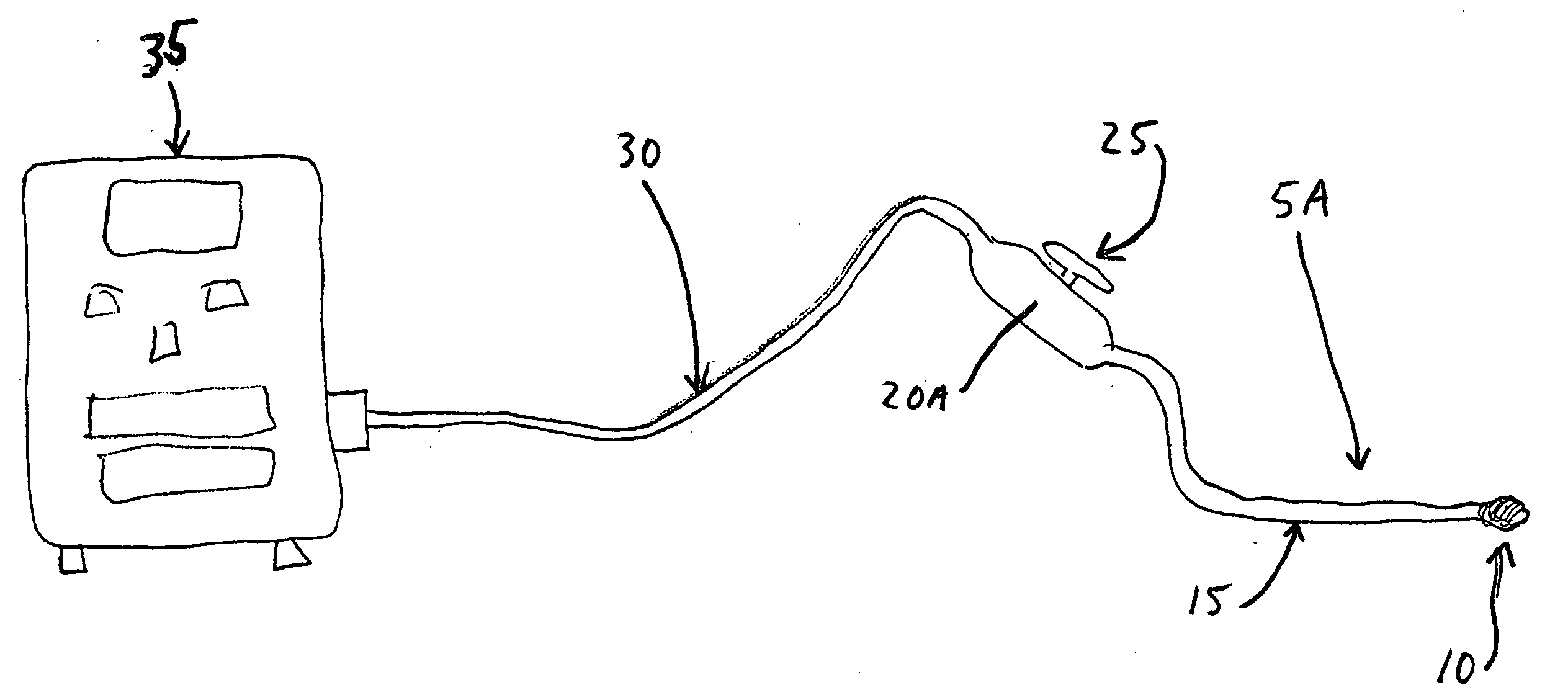
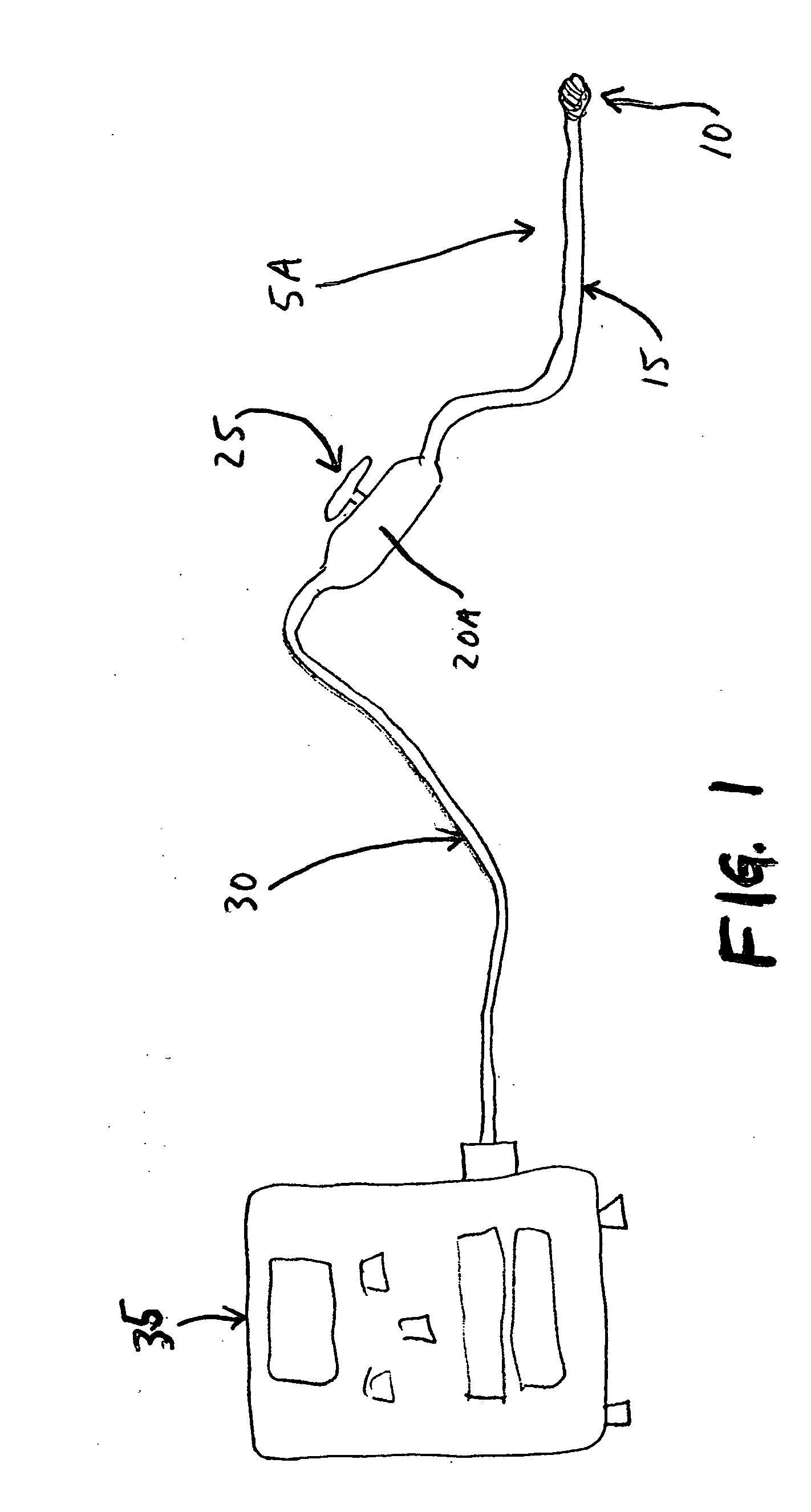
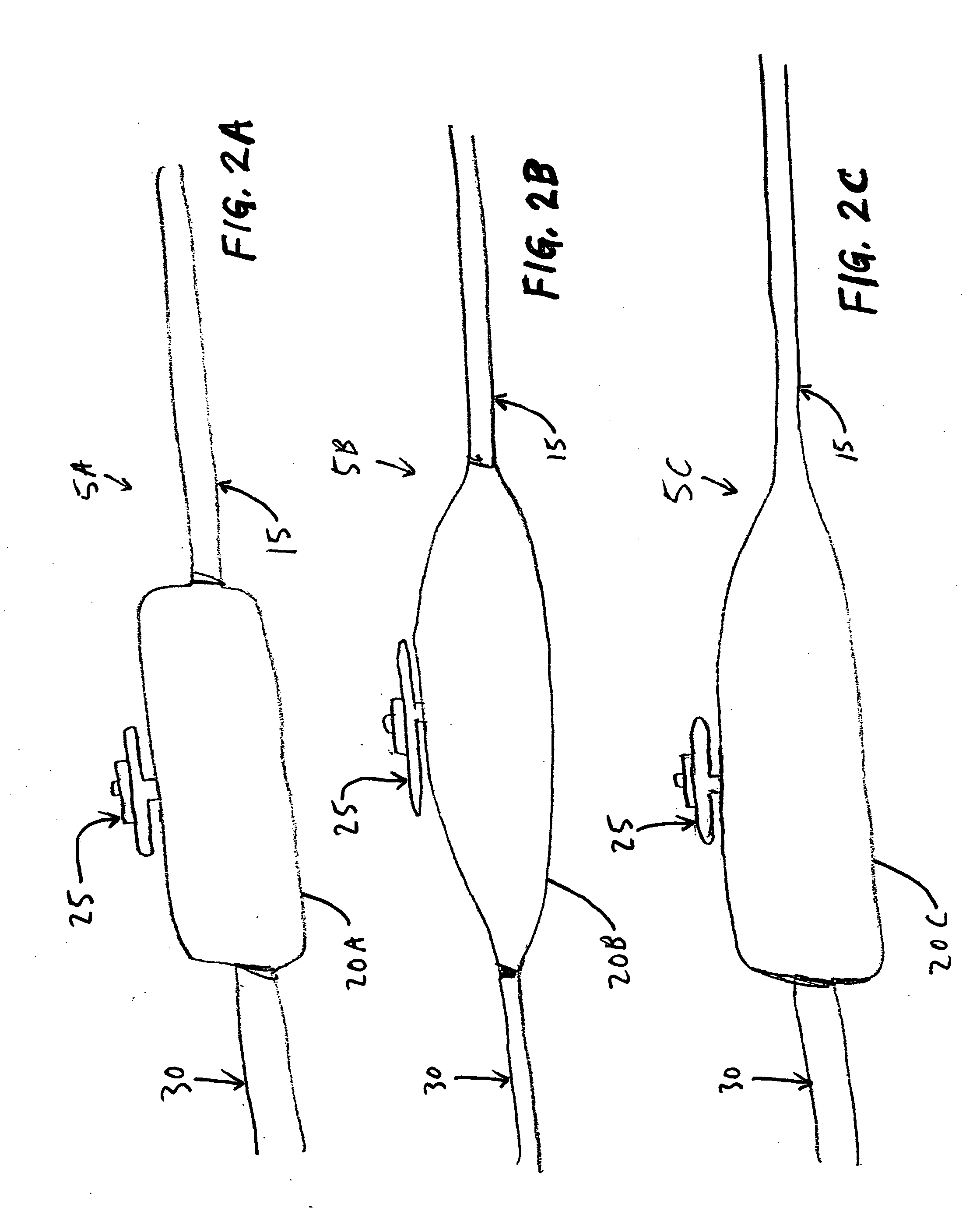
FIELD OF THE INVENTION [0001] This invention is related to medical apparatus and methods in general, and more particularly to apparatus and methods for holding a transesophageal echocardiography probe. BACKGROUND OF THE INVENTION [0002] Looking at FIGS. 1 and 2A-2C, transesophageal echocardiography (TEE) probes 5A, 5B, 5C typically include a distal tip 10 or transducer 10 which houses the ultrasound crystal technology and sensors. A long neck 15, typically about 3-4 feet long, attaches distal tip 10 to a proximal portion 20A, 20B, 20C or handle 20A, 20B, 20C. Proximal portion 20A, proximal portion 20B, and proximal portion 20C each have a different shape from one another. The specific shape of proximal portion 20A, 20B, 20C varies by the manufacturing company and is generally about 12 inches long, about 2-3 inches wide, and either tapered or rounded at each end. Handle 20A, 20B, 20C typically has a rounded knob 25 or gear 25 on one side. Knob 25 enables twisting or flexation of tip ...

Claims


Application Information

Patent Timeline
IPC IPC(8): A61B8/14
CPCA61B8/12A61B8/4209
Inventor LOUBSER, PAUL G.
Owner LOUBSER PAUL G
Learn More