
Catherter for measuring an intraventricular pressure and method of using same

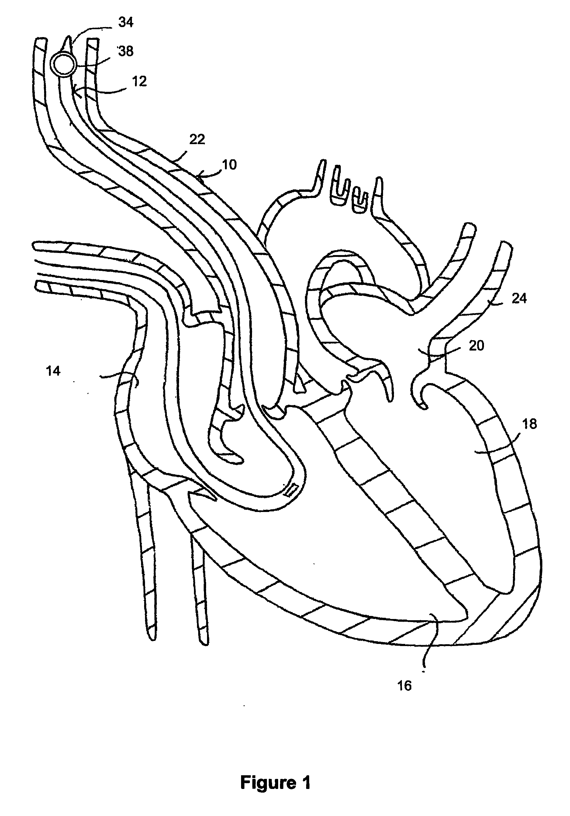
a catheter and intraventricular pressure technology, applied in the field of catheters for measuring intraventricular pressure, can solve the problems of pulmonary edema or cardiac malfunction, hemodynamic instability, and high cost of echocardiography, and achieve the effect of more cost-effectiveness

Inactive Publication Date: 2007-01-18
INST DE CARDIOLOGIE DE MONTREAL
View PDF5 Cites 87 Cited by
  • Summary
  • Abstract
  • Description
  • Claims
  • Application Information

AI Technical Summary

Benefits of technology

[0036] Advantageously, the device allows taking measurements of intraventricular pressure with minimal risks of injuries and other complications, such as arrhythmias, for the subject.
[0041] The method takes advantage of the common insertion of intracardiac catheters to add a functionality to this type of catheter to measure additional parameters that are of clinical importance. For example, the direct measurement of intraventricular pressure without the need to use echocardiography is simpler and more cost-effective.

Problems solved by technology

The major cause of death after cardiac surgery is hemodynamic instability.
When the heart experiences diastolic dysfunction, it requires a higher pressure to be filled, which in some cases leads to serious problem such as pulmonary edema or cardiac malfunction.
The latter manifests itself as hemodynamic instability that can lead to death.
Unfortunately, echocardiography is a highly specialized method that requires extensive knowledge in the interpretation of the data obtained through the technique.
Furthermore, echocardiography is a procedure that is not very suitable for monitoring a patient.
However, there are situations, for example in case of a right ventricular diastolic dysfunction, when this injection of volume is not beneficial and is even nocive.

Method used


Image

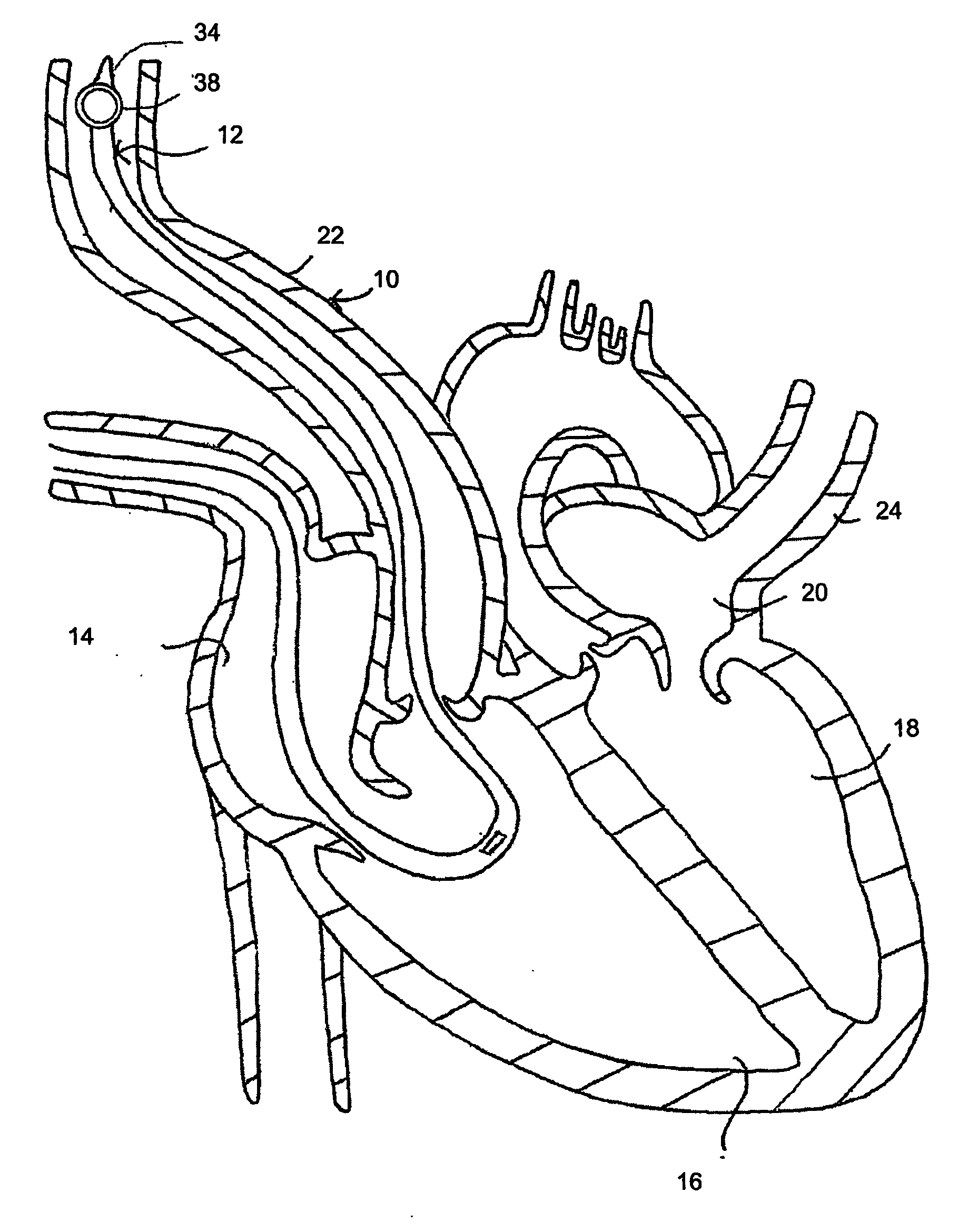
  • Catherter for measuring an intraventricular pressure and method of using same
  • Catherter for measuring an intraventricular pressure and method of using same
  • Catherter for measuring an intraventricular pressure and method of using same

Examples


examples 1

[0124]FIG. 5, shows the effect of administering 500 ml of a colloid (Pentaspan) in two patients presenting different right intraventricular pressure (RVP) waveforms. The first patient (left-hand side of FIG. 5), who responded to the administration of the administration of the colloid by increasing an ejection volume, presents a normal right intraventricular pressure waveform with a substantially constant measured pressure during the diastole. The second patient, who did not respond to the administration, presents an increasing right intraventricular pressure waveform that increases during the diastole.

example 2

[0125]FIG. 6 illustrates measurements taken in a 75 years-old man suffering from right ventricular outflow tract obstruction after coronary revascularization and aortic valve replacement. The procedure was complicated by difficult weaning from cardiopulmonary bypass requiring intra-aortic balloon counterpulsation after a second failed attempt of weaning from the cardiopulmonary bypass. Panels A and B illustrate a trans-gastric mid-papillary short-axis echographic view (respectively with an echographic image and a segmented model obtained from the echographic image) revealing a dilated and hypertrophied right ventricle (RV). Unexplained acute right heart failure was present without pulmonary hypertension. As shown in panel C, the pulmonary artery pressure (Ppa) was 34 / 22 mmHg and right atrial pressure 20 mmHg. However a significant systolic pressure gradient between the right intraventricular pressure (Prv) and the pulmonary artery was present. (LV: left ventricle, Pa; arterial press...

example 3

[0130]FIG. 9 shows a hemodynamic and transesophageal echocardiographic evaluation of a 46 years-old woman scheduled for aortic valve endocarditis. Despite a pulmonary artery pressure (Ppa) of 34 / 16 mmHg and pulmonary vascular resistance index (PVRI) at 286 dyn.s.cm-5 m-2, this patient had abnormal right intraventricular pressure (Pvr) diastolic filling waveform characterized by a rapid upstroke (Panel A illustrating the right intraventricular pressure waveform) and abnormal S / D ratio<1 in the pulmonary (panel B) and hepatic (panel C) venous flow obtained from Doppler imaging consistent with both left and right ventricular diastolic dysfunction. In addition a dilated right atrium (RA) and right ventricle (RV) were present without significant tricuspid regurgitation in a mid-oesophageal right ventricular view (panel D, which is a an echocardiographic image).

[0131] The mean arterial (MAP) to mean pulmonary artery pressure (MPAP) ratio was 65 / 23 or 2.8. Weaning from cardiopulmonary byp...


PUM


Abstract

A method for diagnosing a right ventricular dysfunction of a subject. The method includes measuring a right intraventricular pressure waveform in the subject over at least one cardiac cycle, extracting a ventricular parameter indicative of a right ventricular function from the measured right intraventricular pressure waveform, and establishing a diagnosis at least in part on a basis of the ventricular parameter.

Description

FIELD OF THE INVENTION [0001] The present invention relates to a catheter for measuring a pressure and a method of using same. Specifically, the present invention concerns a catheter for measuring an intraventricular pressure and a method of using same. BACKGROUND OF THE INVENTION [0002] The major cause of death after cardiac surgery is hemodynamic instability. There are specific factors that can predispose a patient to hemodynamic instability. These factors are related to the inability of the heart to relax and accept or receive blood, which is called diastolic dysfunction. When the heart experiences diastolic dysfunction, it requires a higher pressure to be filled, which in some cases leads to serious problem such as pulmonary edema or cardiac malfunction. The latter manifests itself as hemodynamic instability that can lead to death. [0003] There are several types and causes of hemodynamic instability that can occur alone or in combination1. A few are presented hereinbelow: [0004]...

Claims


Application Information

Patent Timeline
Patent Type & Authority Applications(United States)
IPC IPC(8): A61B5/02A61B5/0215
CPCA61B5/412A61B5/0215A61B5/02108
Inventor DENAULT, ANDRE
Owner INST DE CARDIOLOGIE DE MONTREAL
Learn More