Tissue connector apparatus and methods

Inactive Publication Date: 2007-02-01
GARDINER BARRY +3
View PDF99 Cites 74 Cited by
  • Summary
  • Abstract
  • Description
  • Claims
  • Application Information

AI Technical Summary

Benefits of technology

[0012] According to one aspect of the invention, a tissue connector assembly is provided and comprises a flexible member and a surgical clip which may be releasably coupled to the flexible member. With this construction, a needle may be coupled to

Problems solved by technology

Constructing an arterial anastomosis is technically challenging for a surgeon in open surgical procedures, and is almost a technical impossibility using minimally invasive techniques.
Many factors contribute to the difficulty of performing arterial replacement or bypass grafting.
If one of the tissues is affixed too close to its edge, the suture can rip through the tissue and impair both the tissue and the anastomosis.
Another factor is that, even after the tissues are properly aligned, it is difficult and time consuming to pass the needle through the tissues, form the knot in the suture material, and ensure that the suture material does not become tangled.
These difficulties are exacerbated by the small size of the artery and graft.
Another factor contributing to the difficulty of such procedures is the limited time available to complete the procedure.
The time the surgeon has to complete an arterial replacement or bypass graft is limited because there is no blood flowing through the artery while the procedure is being done.
If blood flow is not promptly restored, sometimes in as little as thirty minutes, the tissue the artery supplies may experience significant damage, or even death (tissue necrosis).
In addition, arterial replacement or bypass grafting is

Method used


Image

  • Tissue connector apparatus and methods
  • Tissue connector apparatus and methods
  • Tissue connector apparatus and methods

Examples


Embodiment Construction

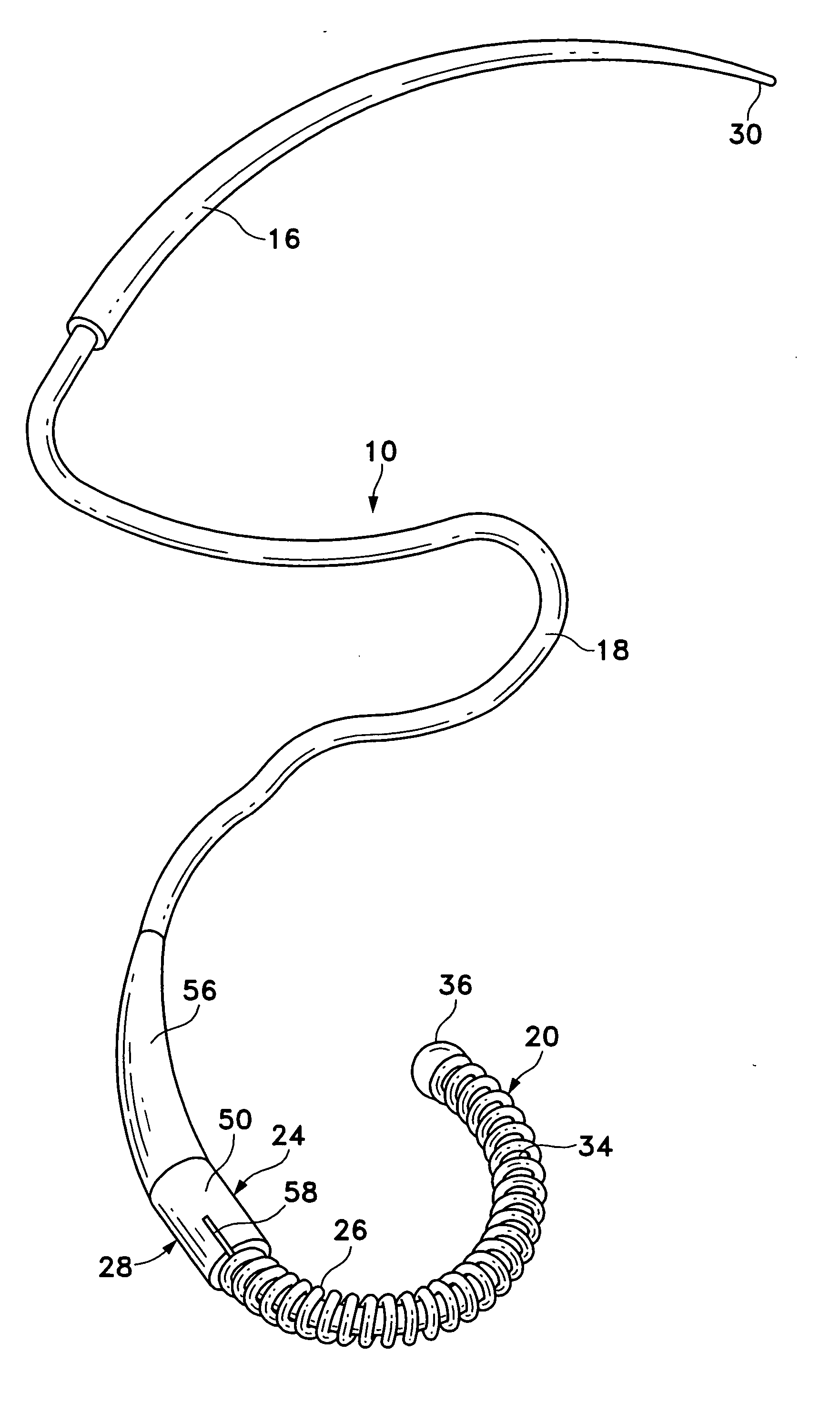
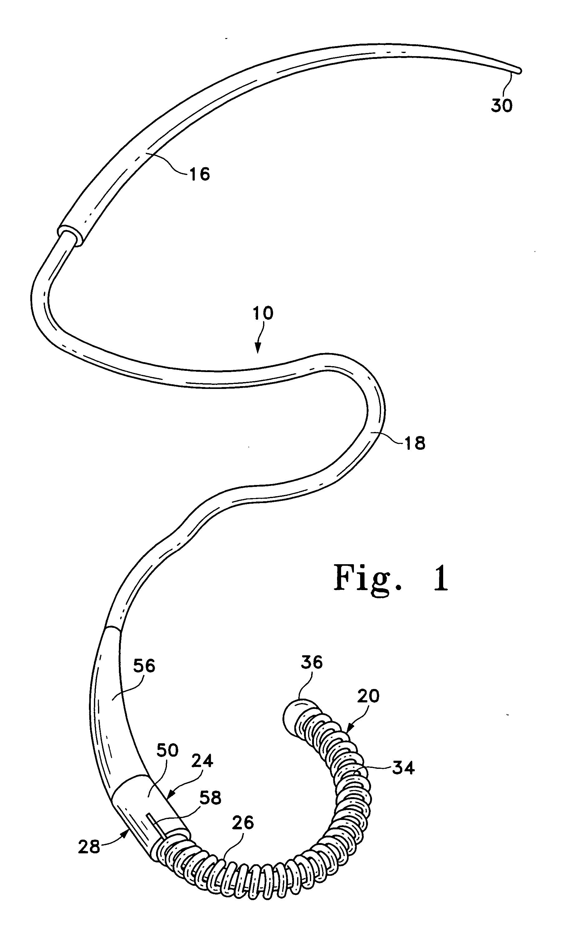
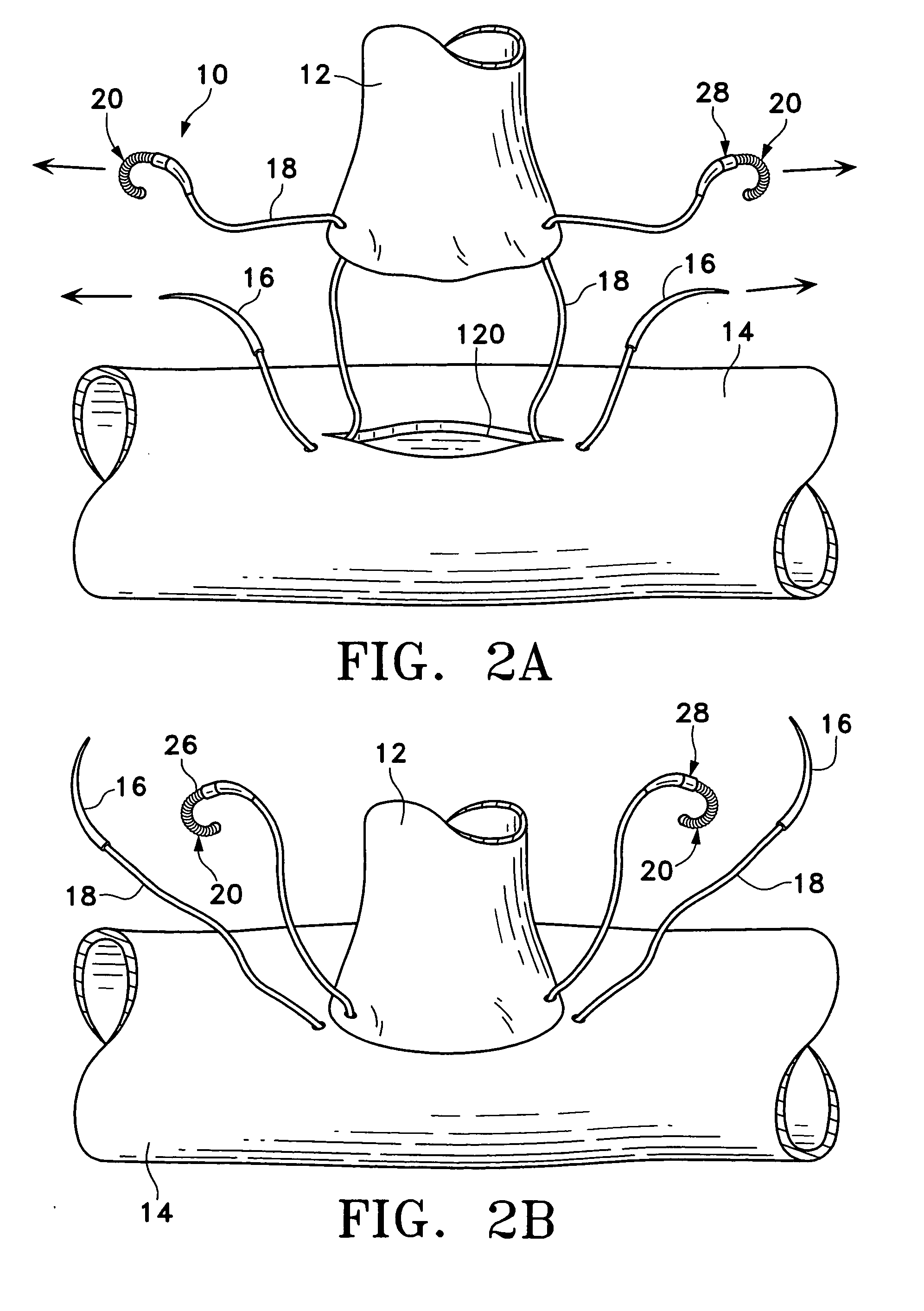
[0040] Referring now to the drawings, and first to FIG. 1, a tissue connector assembly constructed according to the principles of the present invention is shown and generally indicated with reference numeral 10. The tissue connector assembly 10 may be used to manipulate and align tissues, or tissue and prosthesis with respect to each other and thereafter connect the tissues or tissue and prosthesis together (FIGS. 2A-2G). As used herein, the term graft includes any of the following: homografts, xenografts, allografts, alloplastic materials, and combinations of the foregoing. The tissue connector assembly 10 may be used in vascular surgery to replace or bypass a diseased, occluded, or injured artery by connecting a graft vessel 12 to a coronary artery 14 or vein in an anastomosis, for example. The tissue connector assembly 10 may be used in open surgical procedures or in minimally invasive or endoscopic procedures for attaching tissue located in the chest, abdominal cavity, or retrop...


PUM


Abstract

A tissue connector assembly having a flexible member and a surgical clip releasably coupled to the flexible member. A needle may be secured to one end portion of the flexible member with the surgical clip coupled to the other end portion of the flexible member. A locking device may be used to couple the flexible member to the surgical clip. A method for connecting tissues is also disclosed. The method includes drawing tissue portions together with a clip assembly and securing the tissue portions together with the clip assembly.

Description

FIELD OF THE INVENTION [0001] The present invention relates to instruments and methods for connecting body tissues, or body tissue to prostheses. BACKGROUND OF THE INVENTION [0002] Minimally invasive surgery has allowed physicians to carry out many surgical procedures with less pain and disability than conventional, open surgery. In performing minimally invasive surgery, the surgeon makes a number of small incisions through the body wall to obtain access to the tissues requiring treatment. Typically, a trocar, which is a pointed, piercing device, is delivered into the body with a cannula. After the trocar pierces the abdominal or thoracic wall, it is removed and the cannula is left with one end in the body cavity, where the operation is to take place, and the other end opening to the outside. A cannula has a small inside diameter, typically 5-10 millimeters, and sometimes up to as much as 20 millimeters. A number of such cannulas are inserted for any given operation. [0003] A viewin...

Claims


Application Information

Patent Timeline
IPC IPC(8): A61B17/08A61B17/06A61B17/064A61B17/11
CPCA61B17/06A61B17/064A61B2017/06057A61B17/11A61B2017/06009A61B17/0644
Inventor GARDINER, BARRYSCHALLER, LAURENTGANDIONCO, ISIDRO MATIASNGUYEN, JOHN
Owner GARDINER BARRY
Why Eureka
Try Eureka