Automated fundus imaging system

a fundus imaging and automatic technology, applied in the field of electronic imaging apparatus for fundus imaging, can solve the problems of significant hurdles to widespread acceptance and usability of these devices, relative complexity of operation, and considerable effort expended in automating specific functions of these imaging apparatus, and achieve the effect of enhancing the sense of control over the imaging process

Inactive Publication Date: 2007-02-08
CARESTREAM HEALTH INC
View PDF13 Cites 88 Cited by
  • Summary
  • Abstract
  • Description
  • Claims
  • Application Information

AI Technical Summary

Benefits of technology

[0018] It is an advantage of the present invention that it can allow the patient to have an enhanced sense of control over the imaging process, without requiring operational expertise or training of the patient.
[0019] These and other objects, features, and adv

Problems solved by technology

While conventional fundus imaging apparatus require manual operation, there has been considerable effort expended toward automating specific functions of these imaging apparatus.
While these patents attest to continuous improvements in fundus camera design, there are still significant hurdles to widespread acceptance and usability of these devices.
Among disadvantages noted with current apparatus are relative complexity of operation.
One inherent problem relates to the operator learning curve.
While a number of these systems offer ease of operation, they still require some amount of practice.
However, only a small percentage of patients in the PCP environment wi

Method used


Image

  • Automated fundus imaging system
  • Automated fundus imaging system
  • Automated fundus imaging system

Examples


Embodiment Construction

[0047] The present description is directed in particular to elements forming part of, or cooperating more directly with, apparatus in accordance with the invention. It is to be understood that elements not specifically shown or described may take various forms well known to those skilled in the art.

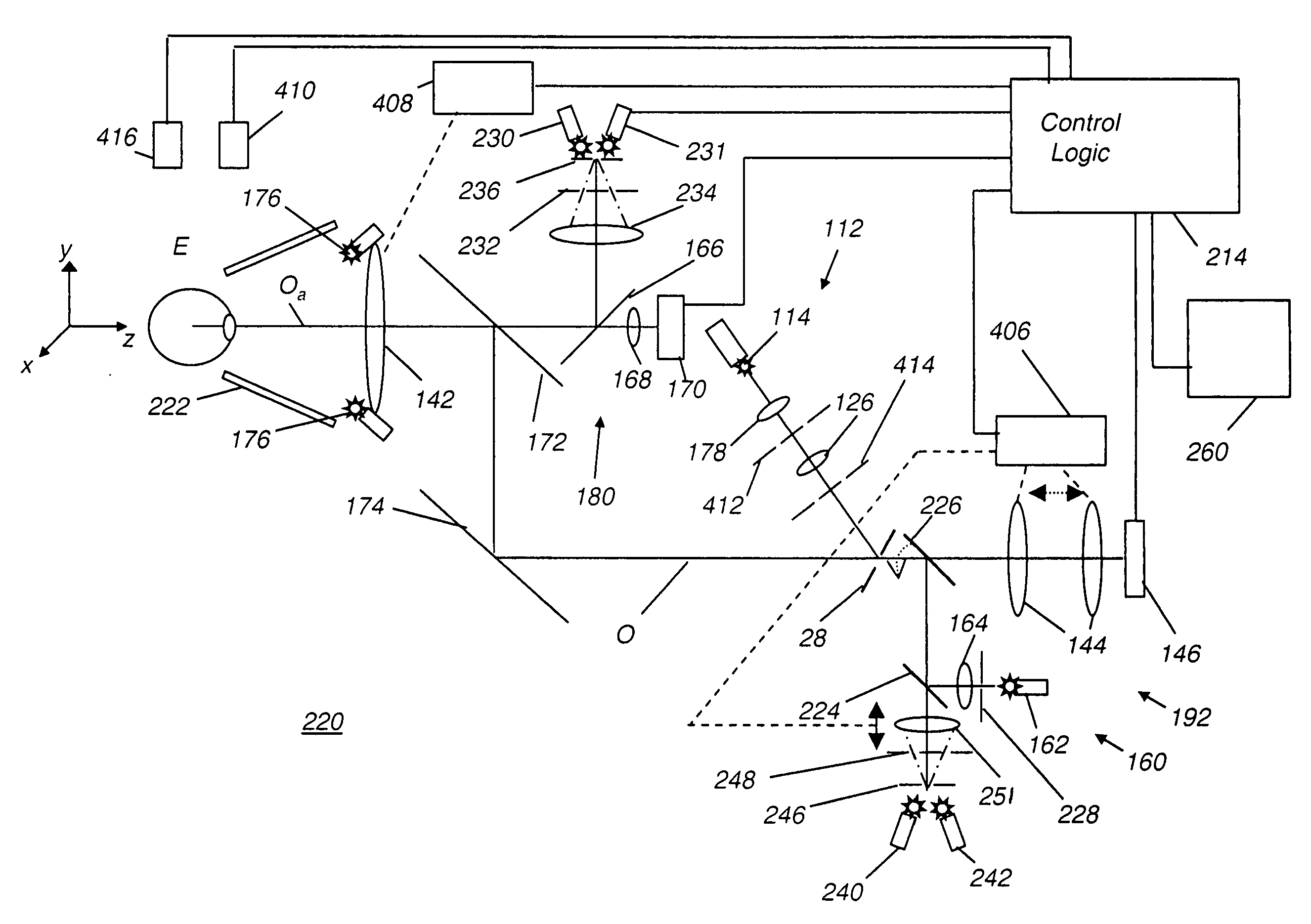
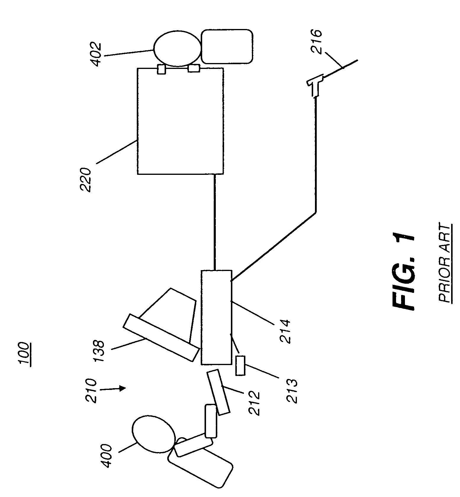
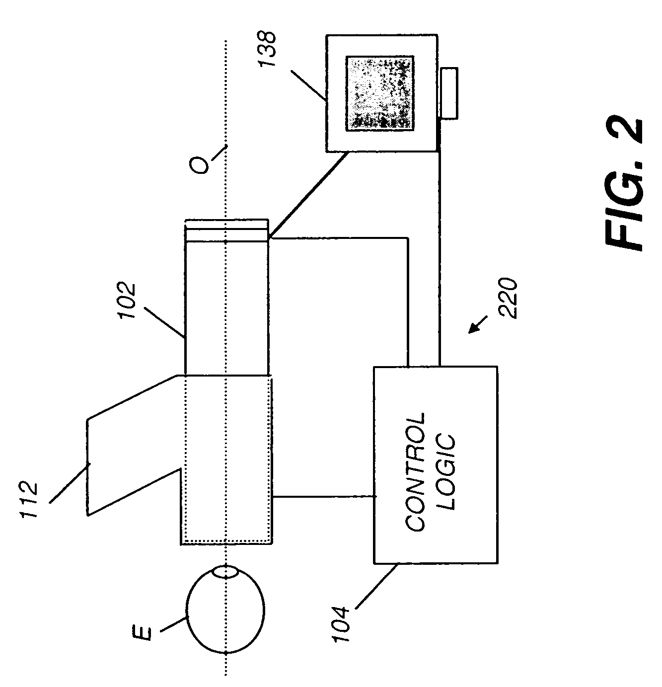
[0048] System Configuration Referring to FIG. 1, there is shown a conventional fundus imaging system 100 for obtaining an image from a patient 402. To support fundus imaging system 100, a control workstation 210 has a display 138, a keyboard 212, and a control logic processor 214 for providing control logic and interface functions for an operator 400. Imaging functions are provided by optical, electro-optical, and electronic components within a fundus imaging appliance 220. A network 216 allows communication between fundus imaging system 100 and processing and storage devices at other networked sites. Using network 216, for example, fundus images obtained by fundus imaging system 100 can...


PUM


Abstract

An apparatus for obtaining a retinal image of an eye has a control logic processor (214) for executing a sequence of operations for obtaining the image. A visual target (162) orients the eye of a patient when viewed. An indicator element (410) provides a signal that indicates that the patient is in position. A cornea focus detection section (450) indicates cornea focus, in cooperation with the control logic processor (214). An alignment actuator aligns the optical path according to a signal obtained from the cornea focus detection section (450). A retina focus detection section (452) detects retina focus in cooperation with the control logic processor (214). A focusing actuator (406) is controlled by instructions from the control logic processor (214) according to a signal obtained from the retina focus detection section (452). An image capture light source is energized by the control logic processor (214) for illuminating the retina.

Description

FIELD OF THE INVENTION [0001] This invention generally relates to electronic imaging apparatus for fundus imaging and more particularly relates to an improved fundus imaging apparatus for automated imaging without operator intervention. BACKGROUND OF THE INVENTION [0002] Fundus camera imaging is acknowledged to be an important diagnostic tool for detection of various conditions affecting the eye, including diabetic retinopathy and macular degeneration. Various embodiments of fundus imaging apparatus are disclosed, for example in U.S. Pat. Nos. 5,713,047 (Kohayakawa); 5,943,116 (Zeimer); 5,572,266 (Ohtsuka); 4,838,680 (Nunokawa); 6,546,198 (Ohtsuka); and 6,636,696 (Saito). [0003] While conventional fundus imaging apparatus require manual operation, there has been considerable effort expended toward automating specific functions of these imaging apparatus. For example, U.S. Pat. No. 6,296,358 (Cornsweet et al.) discloses an automated sequence that is executed for pupil alignment and f...

Claims


Application Information

Patent Timeline
IPC IPC(8): A61B3/14
CPCA61B3/14
Inventor LIANG, RONGGUANGTUCKER, DALEHAWVER, JEFFERYBAEK, SEUNG-HOABHYANKAR, VISHWASWEIL, RICHARDBLANDING, DOUGLASS
Owner CARESTREAM HEALTH INC
Why Eureka
Try Eureka