
System and method for automatic segmentation of vessels in breast MR sequences

Inactive Publication Date: 2007-02-15
SIEMENS MEDICAL SOLUTIONS USA INC
View PDF10 Cites 6 Cited by
  • Summary
  • Abstract
  • Description
  • Claims
  • Application Information

AI Technical Summary

Benefits of technology

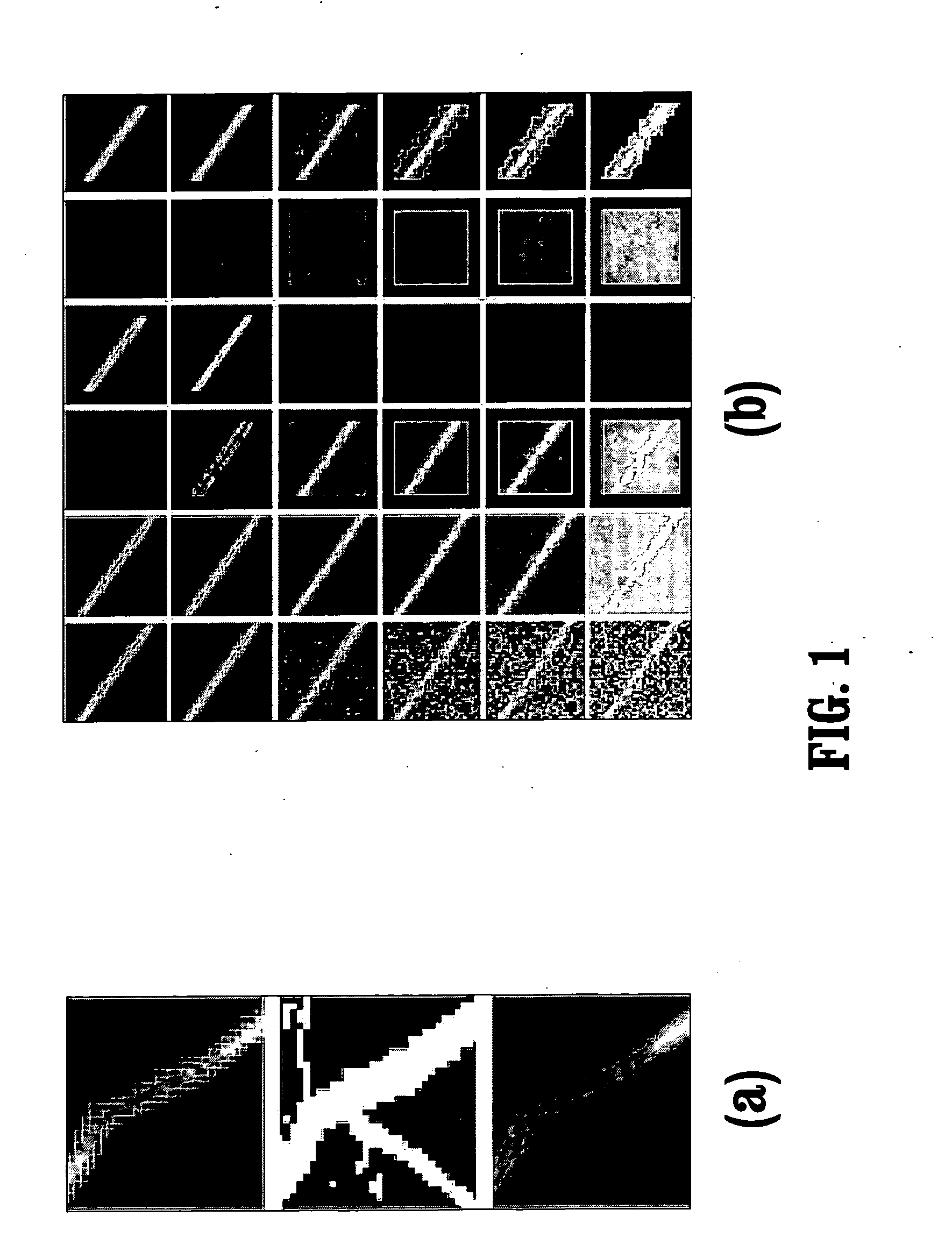
[0007] Exemplary embodiments of the invention as described herein generally include methods and systems for automatic detection of bright tubular structures and its performance for automatic segmentation of vessels in breast MR sequences based on geometrical moments for the extraction of tubular structures from images. A method according to an embodiment of the invention is based on the eigenvalues of the shape tensor, and reconciles not having to threshold the image with reliably recovering structure under very low signal to noise (SN) ratios. A method according to an embodiment of the invention does not rely on image derivatives of either first order, like methods based on the eigenvalues of the mean structure tensor, or second order, like methods based on the eigenvalues of the Hessian, and the smoothing of the output which is inherent to approaches based on the Hessian or structure tensor is avoided. A method according to an embodiment of the invention can execute quickly, needing only a few seconds per sequence. Testing results based motion-corrected breast MR sequences indicate that a method according to an embodiment of the invention reliably segments vessels while leaving lesions intact, and out-performs differential techniques both in sensitivity and localization precision and is less sensitive to scale selection parameters.

Problems solved by technology

A difficulty for such a system is the fact that, besides the lesions, a number of non-suspicious structures also enhance in the post-contrast image.
The problem with this is that the threshold is usually difficult to choose and might not allow detection of small vessels because a low threshold will cause smaller vessels, which tend to have lower intensities, to be merged with neighboring structures.
Another problem with thresholding is that the structure becomes “pixelized”, i.e. develops sharp edges that render the computation of its shape imprecise with respect to the true shape of the underlying structure.
However, in regions where the signal-to-noise (SN) ratio is low, it becomes difficult to establish a threshold on the eccentricity of a fitted ellipse to detect elongated structures.
The threshold was adjusted to capture both vessels, a task that is quite difficult to achieve automatically.

Method used


Image

  • System and method for automatic segmentation of vessels in breast MR sequences
  • System and method for automatic segmentation of vessels in breast MR sequences
  • System and method for automatic segmentation of vessels in breast MR sequences

Examples


Embodiment Construction

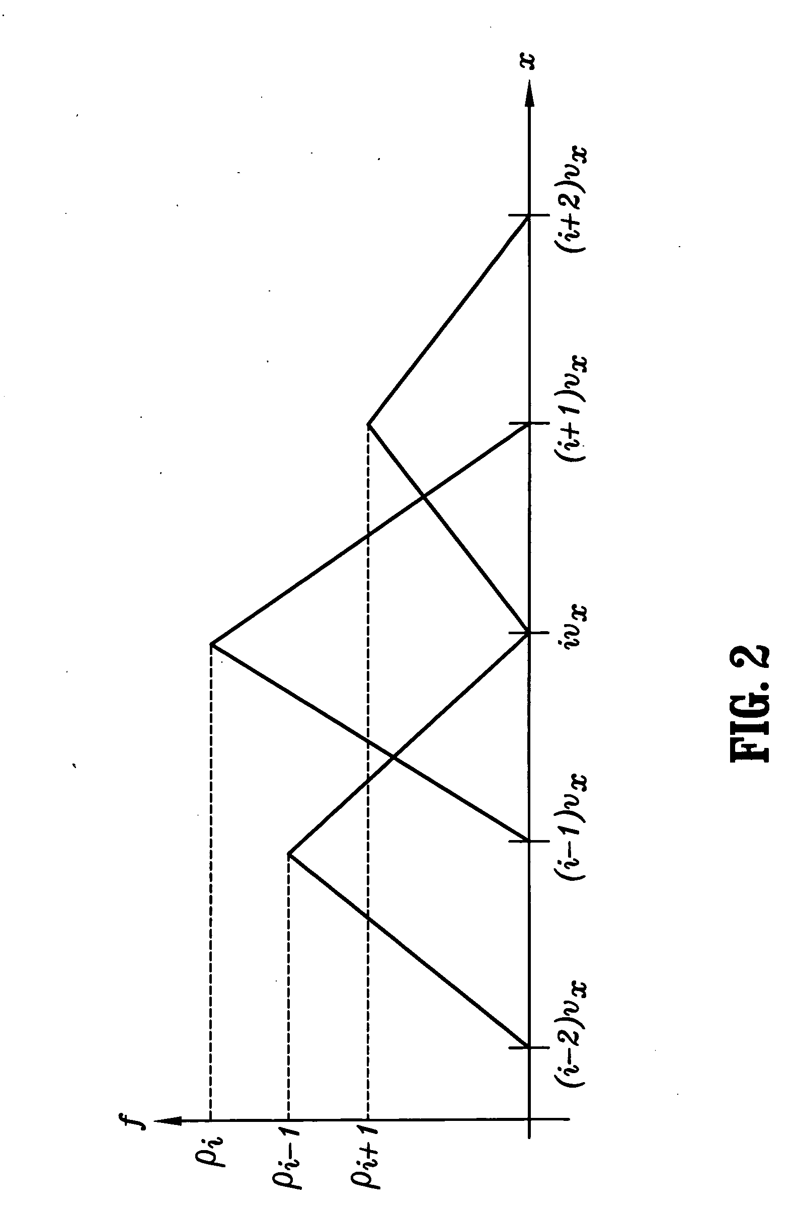
[0026] Exemplary embodiments of the invention as described herein generally include systems and methods for automatic detection of bright tubular structures and its performance for automatic segmentation of vessels in breast MR sequences. A method according to an embodiment of the invention is based on the eigenvalues of a shape tensor. It can be compared to methods based on the eigenvalues of the mean Hessian and those based on the eigenvalues of the mean structure tensor. The Hessian, being defined from the second-order derivatives, can be regarded as a structure descriptor of order two. Similarly, the structure tensor is a structure descriptor of order one. The shape tensor can be regarded as a structure descriptor of order zero.

[0027] As used herein, the term “image” refers to multi-dimensional data composed of discrete image elements (e.g., pixels for 2-D images and voxels for 3-D images). The image may be, for example, a medical image of a subject collected by computer tomogr...


PUM


Abstract

A method for segmenting digitized images includes providing a digitized image, selecting a point with a median enhancement greater than a predefined threshold, wherein a contrast enhancing agent was applied to the subject matter of said digitized image prior to acquisition of said image, defining a shape matrix for the selected point in said image from moments of the intensities in a window of points about said selected point, calculating eigenvalues of said shape matrix, determining an eccentricity of a structure underlying said point from said eigenvalues, and segmenting said image based on said eccentricity values, wherein the steps of defining a shape matrix, calculating eigenvalues of said shape matrix, and determining the eccentricity of the underlying structure are repeated for all points in said image.

Description

CROSS REFERENCE TO RELATED UNITED STATES APPLICATIONS [0001] This application claims priority from “Automatic segmentation of vessels in breast MR sequences as a false positive elimination technique for automatic lesion detection and segmentation using the shape tensor”, U.S. Provisional Application Ser. No. 60 / 764,122 of Hermosillo, et al., filed Feb. 1, 2006, and from “Method for automatic extraction of image structure based on the second order geometric moment”, U.S. Provisional Application Ser. No. 60 / 704,930 of Hermosillo, et al., filed Aug. 2, 2005, the contents of both of which are incorporated herein by reference.TECHNICAL FIELD [0002] This invention is directed to segmentation of digitized medical images. DISCUSSION OF THE RELATED ART [0003] Contrast enhanced MR sequences are a powerful diagnostic tool for the detection of lesions in breast. Typically, the diagnosis begins by identifying suspicious regions of enhancement in post contrast acquisitions with respect to a pre-c...

Claims


Application Information

Patent Timeline
IPC IPC(8): G06K9/00
CPCG06T7/0012G06T7/0081G06T2207/30101G06T2207/10088G06T2207/30068G06T7/606Y10S128/92G06T7/11G06T7/66
Inventor VALADEZ, GERARDO HERMOSILLOJIANG, XUGUANG
Owner SIEMENS MEDICAL SOLUTIONS USA INC
Learn More