Method for production of neuroblasts

a neuroblast and cell technology, applied in the field of cell populations derived from neurons, can solve the problems of delayed neuropharmacologicai studies in the cns, inability to establish cell lines from the central nervous system (cns) and neuronal tissues in the absence of immortalization, and adult neurons that do not survive well in vitro

Inactive Publication Date: 2007-03-08
RGT UNIV OF CALIFORNIA
View PDF9 Cites 0 Cited by
  • Summary
  • Abstract
  • Description
  • Claims
  • Application Information

AI Technical Summary

Problems solved by technology

Only a few neuronal cell types have been reported to divide in the adult brain and adult neurons do not survive well in vitro.
To date, even with all of the recent advances in neurobiology, genetics, immunology and molecular biology, no reliable procedure exists to establish cell lines from the central nervous system (CNS) and neuronal tissues in the absence of immortalization.
To date, neuropharmacologicai studies in the CNS have been delayed by the lack of cell systems needed to investigate potentially useful neuroactive compounds.
In live animals, the complexity of the brain makes it difficult to effectively measure which cellular receptors are being targeted by these compounds.
Additionally, the expense involved in live animal research and the current controversies stemming from animal rights movements have made in vivo animal studies less acceptable for initial research.
Primary cells from neuronal tissue are often used for CNS studies, however, long-term culture of primary neurons has not been achieveD.
In fact, the proliferation of neuronal cells has proven so elusive that it has become ingrained in the scientific community that neuronal cells do not proliferate in vitro.
As a consequence, fresh dissections must be performed for each study in order to obtain the necessary neuronal cell types, resulting in costly research with increased variability in-the experimental results.
In general, these tumor cell lines do not mimic the biology of the primary neurons from which they were originally established and, as a result, are not suitable for drug discovery screening programs.
However, it should be noted that these differentiated neurons were not cultured for lengthy periods of time nor was there any evidence that these cells could be frozen and then thawed and recultured.

Method used


Image

  • Method for production of neuroblasts
  • Method for production of neuroblasts
  • Method for production of neuroblasts

Examples


example 1

Materials and Methods

[0050] Materials: DMEM:F12 medium, N2 supplement and laminin were obtained from Gibco / BRL (Bethesda, Md.); polyornithine (PORN) was obtained from Sigma (St. Louis, Mo.). Recombinant bFGF was from Syntex / Synergen Consortium (Boulder, Colo.). Bovine bFGF was purchased from R&D, Minneapolis, Minn. NeuroTag™ green was obtained from Boehringer Mannheim, Indianapolis, Ind. Cell proliferation kit containing bromodeoxyuridine (BrdU), anti-BrdU antibody and streptavidin / Texas Red was purchased from Amersham, Arlington Heights, Ind. The antibodies used to determine the phenotypes of cells in culture were obtained from the following sources and used at the indicated dilutions: polyclonal rabbit anti-neurofilament 200 (NF) (1:500; Chemicon International, Temecula, Calif.), monoclonal anti-neuron specific enolase (NSE) (1:50; DAKO, Carpenteria, Calif.), monoclonal anti-glia fibrillary acidic protein (GFAP) (1:500-1:10,000; Amersham, Arlington Heights, III), monoclonal anti-...

example 2

Growth of Neurons In Vitro

[0062] The chemically defined medium, N2 (Bottenstein and Sato, Proc. Natl. Acad. Sci. USA, 76: 514-517, 1980; Bottenstein, J. E., In: Cell culture in the neurosciences, J. E. Bottenstein and G. H. Sato, Eds., Plenum Press, New York, N.Y., pp 3-43, 1985; di Porizo, et al., Nature, 288:370-373, 1980), has been used to reproducibly generate short-term virtually pure neuronal cultures (Bottenstein, et al., Exp. Cell Res., 125:183-190, 1980; Barnes and Sato, Anal. Biochem., 102:255-302, 1980). This medium does not support the survival or proliferation of non-neuronal cells and it is possible to obtain>95% pure neuronal culture. In defined medium, primary cultures of hippocampal neurons die within 7 days but can be maintained for 2-4 weeks in the presence of hippocampal explants, a feeder layer of astrocytes, in astrocyte-conditioned medium (Banker, G. A., Science, 209:809-810, 1980) or in the presence of bFGF (Walicke and Baird, Proc. Natl. Acad. Sci. USA, 83:...

example 3

Characterization of Cells

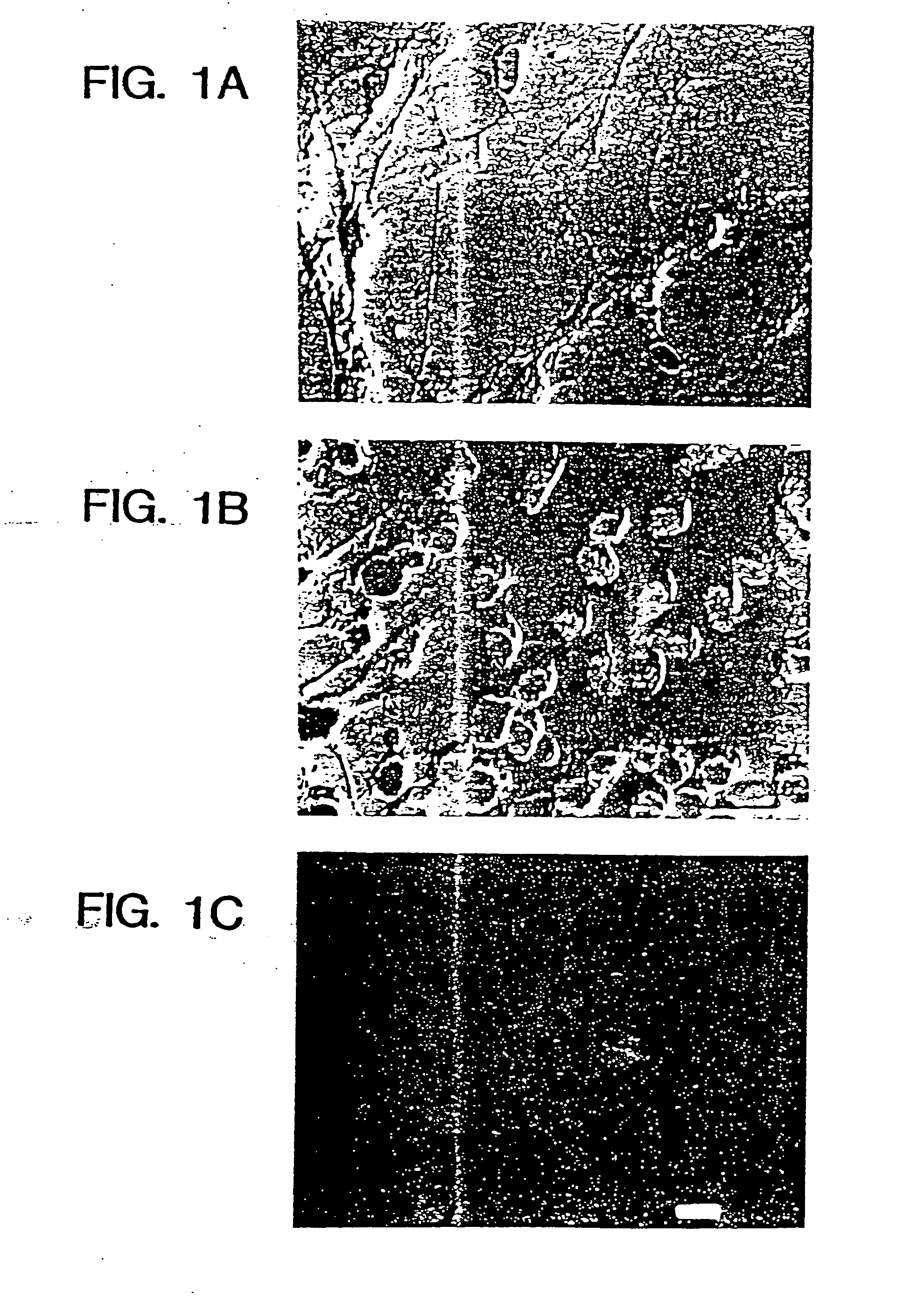
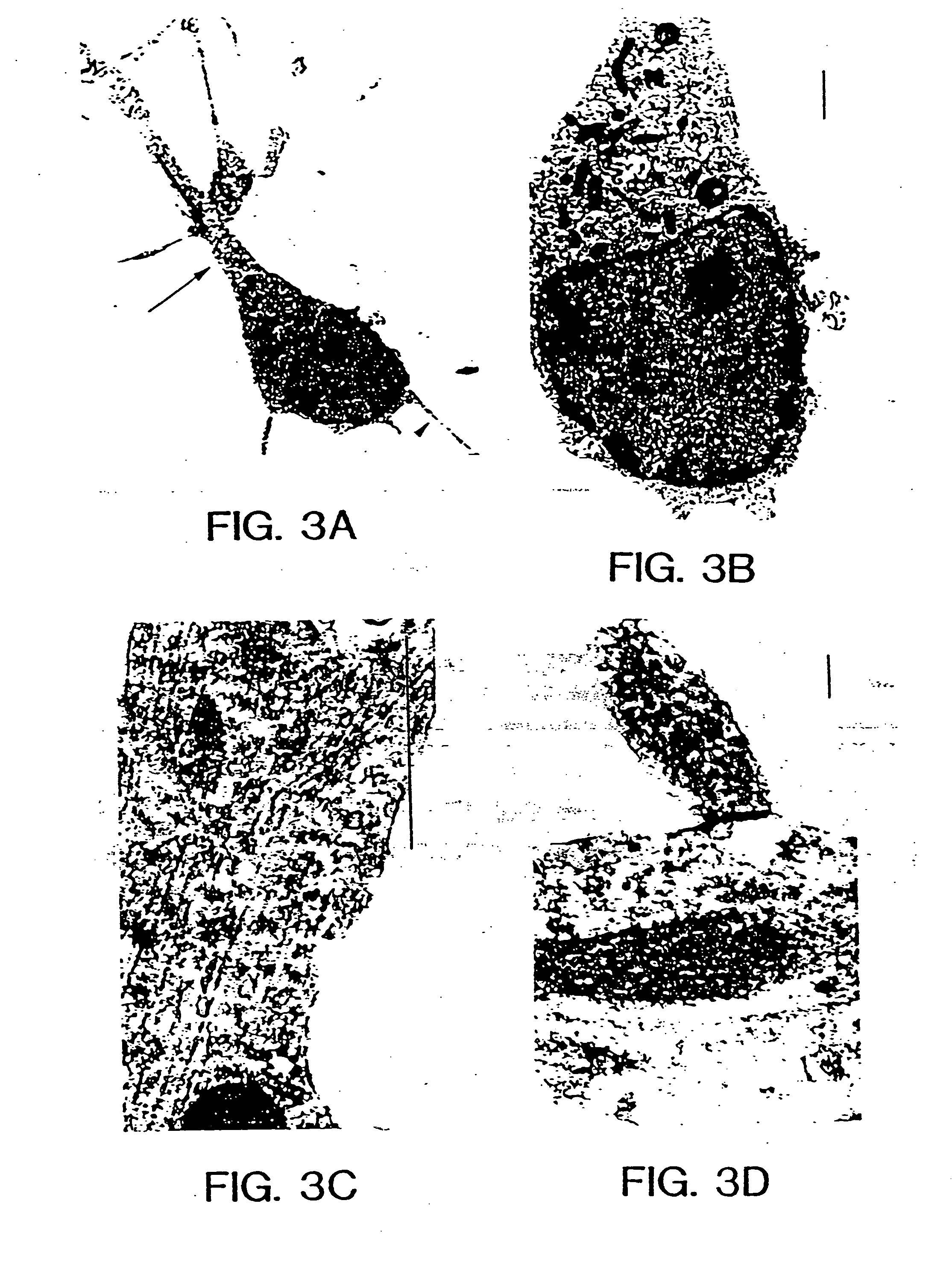
[0068] Several independent criteria were used to show that the cells in the cultures were indeed neurons. These included their morphological characteristics during growth, expression of neuronal markers and ultrastructural analysis by transmission and scanning electron microscopy.

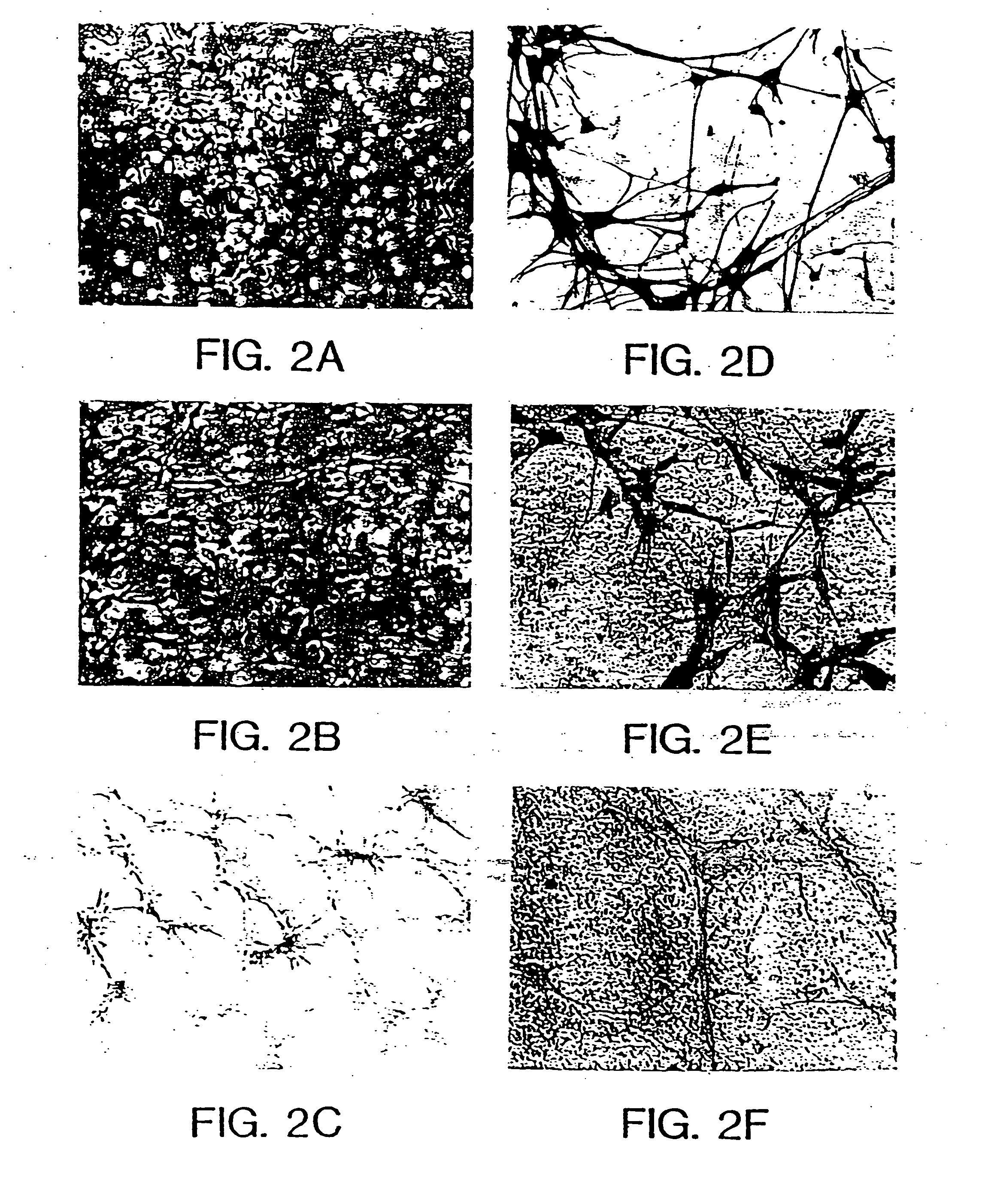
[0069] Cell morphology in culture was similar to that described for short-term cultures of neurons (Banker and Cowan, Brain Res., 126:397-425, 1977; Banker and Cowan, J. Comp. Neurol., 187:469-494, 1979) (FIGS. 2A, B, C). FIG. 2 illustrates photomicrographs showing the morphological changes that occur during the culture and passaging of primary neurons. A shows primary cell culture after 4 days of plating in N2+bFGF contained numerous proliferating and process-bearing cells. B shows primary cells 4 days in culture after passage (passage 3). Cells were larger and interconnected by processes that also increased in size. Small proliferating cells were visible in the culture. C shows ...


PUM

PropertyMeasurementUnit
doubling timeaaaaaaaaaa
concentrationaaaaaaaaaa

Abstract

A method for producing a neuroblast and a cellular composition comprising an enriched population of neuroblast cells is provided. Also disclosed are methods for identifying compositions which affect neuroblasts and for treating a subject with a neuronal disorder, and a culture system for the production and maintenance of neuroblasts.

Description

[0001] This application is a Continuation of U.S. application Ser. No. 10 / 622,206, filed Jul. 18, 2003, which is a Continuation of U.S. application Ser. No. 09 / 915,229, filed Jul. 24, 2001, now U.S. Pat. No. 6,599,695, which is a Continuation of U.S. application Ser. No. 08 / 884,427, filed Jun. 27, 1997, now U.S. Pat. No. 6,265,175, which is a Continuation of U.S. application Ser. No. 08 / 445,075, filed May 19, 1995, now abandoned, which is a Divisional of U.S. application Ser. No. 08 / 147,843, filed Nov. 3, 1993, now U.S. Pat. No. 5,766,948, which is a Continuation-in-part of U.S. application Ser. No. 08 / 001,543, filed Jan. 6, 1993, now abandoned. The contents of each of these prior applications are incorporated by reference herein in their entirety for any purpose.BACKGROUND OF THE INVENTION [0002] 1. Field of the Invention [0003] This invention relates generally to cell populations derived from neurons, which are denoted neuroblasts, methods for the production and long-term in vitro...

Claims


Application Information

Patent Timeline
Patent Type & Authority Applications(United States)
IPC IPC(8): A61K35/30C12N5/08A61K35/12A61K45/00C12N15/09A61K48/00A61P25/08A61P25/14A61P25/16A61P25/28C12N5/079C12N5/0793C12N5/0797C12N5/10C12P21/08C12Q1/02
CPCA61K35/12C12N2533/32C12N5/0618C12N5/0619C12N5/0623C12N2500/25C12N2500/99C12N2501/11C12N2501/115C12N2501/13C12N2501/235C12N2501/392C12N2502/08C12N2510/04A61K48/00C12N2500/90C12M23/20A61P25/08A61P25/14A61P25/16A61P25/28
Inventor GAGE, FREDRAY, JASODHARA
Owner RGT UNIV OF CALIFORNIA
Why Eureka
Try Eureka