
Temperature probe for insertion into the esophagus

a technology of temperature probes and esophagus, which is applied in the field of temperature probes, can solve the problems of irreparable damage to adjacent tissue areas, and achieve the effect of accurate acousti

Inactive Publication Date: 2007-03-22
SIEMENS HEALTHCARE GMBH
View PDF16 Cites 79 Cited by
  • Summary
  • Abstract
  • Description
  • Claims
  • Application Information

AI Technical Summary

Benefits of technology

[0015] According to a second aspect the invention achieves the object in that an ultrasound sensor is disposed on the tip of the catheter. Ultrasound catheters for insertion into the esophagus are already known and are referred to inter alia as TEE (TransEsophageal Echocardiogram) catheters. Combination with a temperature probe has the advantage that the temperature in the esophagus can be measured at the same time, while the ultrasound sensor is used to determine position and distance from a heat applicator or ablation catheter in the ventricle. The ultrasound sensor can therefore give a first indication of when the distance drops below a specified distance and it is therefore possible that a critical temperature threshold will be exceeded in the esophagus.
[0016] The inventive temperature probe also has an unfoldable balloon, on the outer skin of which one or more temperature sensors are disposed. This allows the temperature probe, after it has been inserted for example by way of the catheter into the body, to unfold or inflate in the manner of a balloon, such that the outer skin with the temperature sensors is as close as possible to the wall of the esophagus. The temperature sensors therefore do not measure the temperature changes in the lumen but the temperature changes in the esophageal wall, which are of actual relevance. This allows accurate acoustic, optical or tactile feedback to the electrophysiologist, when a temperature change is determined in the esophageal wall.
[0021] The catheter and / or balloon is / are preferably equipped with x-ray visible markers for precise positioning. This makes it possible to position the balloon in the esophagus under x-ray control, such that it is located in the most optimum manner possible on the adjacent epicardial tissue of the left atrium of the heart. During an ablation procedure the patient can therefore convey the catheter into the stomach initially by means of swallowing movements. The electrophysiologist then withdraws the catheter, until the x-ray visible markers are in the direct vicinity of the left atrium of the heart. To this end a contrast agent can optionally be briefly supplied to the left atrium.

Problems solved by technology

There is however the risk with ablation that adjacent tissue areas may be irreparably damaged.
However there are as yet no temperature probes, which are ideally suited for such an application.

Method used


Image

  • Temperature probe for insertion into the esophagus
  • Temperature probe for insertion into the esophagus
  • Temperature probe for insertion into the esophagus

Examples


Embodiment Construction

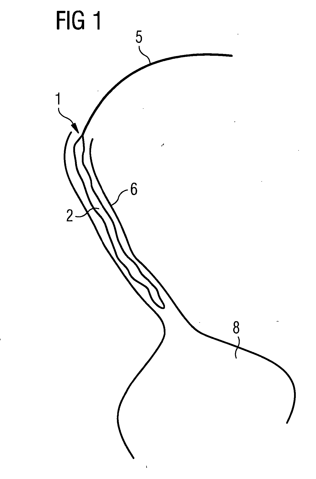
[0038]FIG. 1 shows a schematic diagram of an esophagus 6 opening into a stomach 8. Inserted into this is a catheter 5 with a temperature probe 1 with an unfoldable balloon 2. The drawing shows the balloon 2 in the folded state, in which it is inserted by way of the catheter 5 into the esophagus 6. This is preferably achieved by means of swallowing movements by the patient.

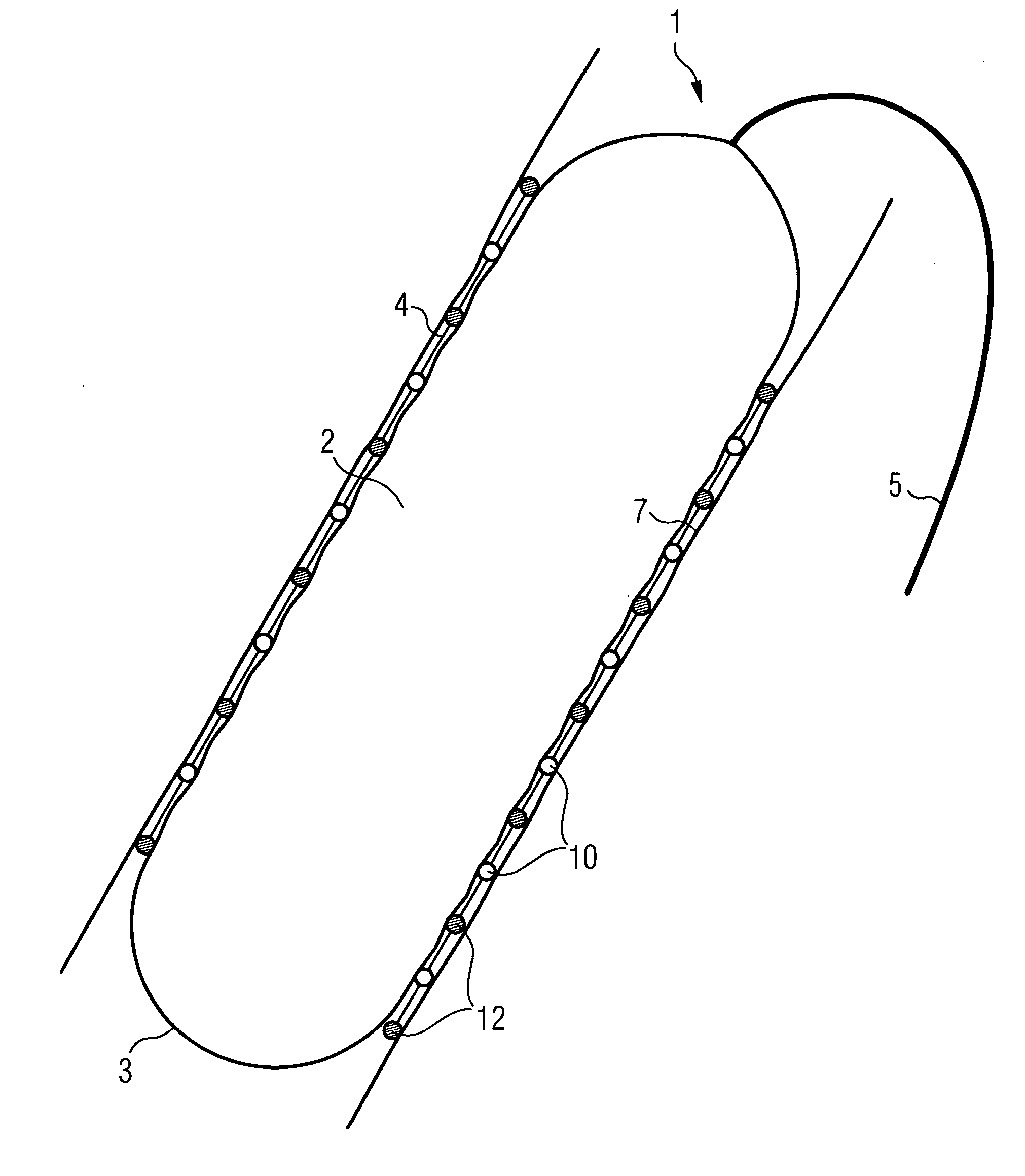
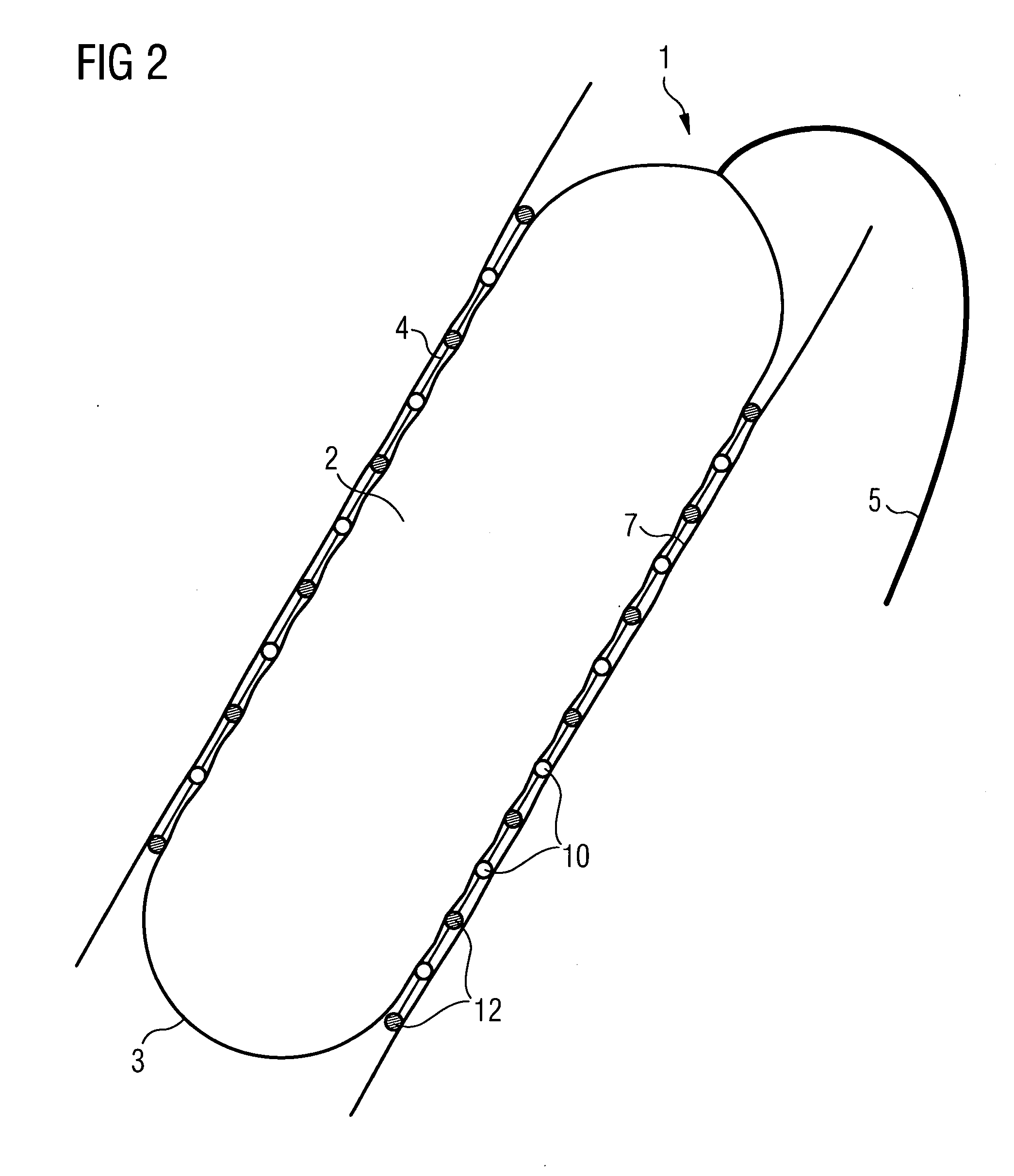
[0039]FIG. 2 shows the temperature probe 1 enlarged and in cross-section and in the unfolded state. 5 again shows the catheter, which for example contains a flexible tube, by way of which air can be supplied to inflate the balloon 2. The gas-filled balloon 2 is enclosed by an outer skin 3, made of an elastic plastic for example. A number of temperature sensors 10 and position sensors 12 are disposed on the outer skin 3, being pushed by the balloon 2 onto the wall 7 of the esophagus 6. The sensors 10, 12 can be attached in any manner to the outer skin; they can also be inside the balloon 2, as long as there is adeq...


PUM


Abstract

The invention relates to a temperature probe (1) for insertion into the human or animal body, having an unfoldable balloon (2) on a catheter (5), on the outer skin (3) of which balloon (2) one or more temperature sensors (10) is / are disposed. The catheter (5) can be moved by way of a cable (9, 11) and according to one aspect of the invention an ultrasound sensor is disposed on its tip. Position sensors (12) can also be provided on the outer skin of the balloon (2). The temperature probe (1) is used to monitor the temperature distribution in the esophagus (6) during a catheter ablation.

Description

CROSS REFERENCE TO RELATED APPLICATIONS [0001] This application claims priority of German application No. 10 2005 045 069.5 filed Sep. 21, 2005, which is incorporated by reference herein in its entirety. FIELD OF THE INVENTION [0002] The invention relates to a temperature probe for insertion into the esophagus of the human or animal body. The temperature probe has a catheter with an unfoldable balloon, on the outer skin of which one or more temperature sensors are disposed. The invention also focuses on a catheter system with a computation module for processing the temperature data transmitted by the temperature probe and a hyperthermia device with a heat applicator to heat a tissue section in the heart and a temperature probe for insertion into the esophagus. BACKGROUND OF THE INVENTION [0003] To treat cardiac arrhythmia for example diseased areas of the heart are eradicated by means of catheter ablation in an electrophysiological intervention. In this process one or more catheters...

Claims


Application Information

Patent Timeline
Patent Type & Authority Applications(United States)
IPC IPC(8): A61B18/04A61B18/18A61B5/00A61F2/958
CPCA61B1/273A61B5/01A61B2019/5454A61B18/1492A61B2017/00243A61B5/4233A61B2090/3954
Inventor RAHN, NORBERT
Owner SIEMENS HEALTHCARE GMBH
Learn More