
Method and system for processing an image of body tissues

a body tissue and image processing technology, applied in image analysis, image enhancement, instruments, etc., can solve the problems of difficult separation of liver from heart muscle, complicated removal of cages from cardiac ct scans, and complicated automatic detection of boundary between adjacent organs or tissues in scans, so as to achieve the effect of minimal cost and minimal cos

Inactive Publication Date: 2007-04-12
PHILIPS HEALTHCARE INFORMATICS INC
View PDF9 Cites 19 Cited by
  • Summary
  • Abstract
  • Description
  • Claims
  • Application Information

AI Technical Summary

Benefits of technology

[0011] In its second aspect, the invention provides a method for detecting a boundary between two body tissues or organs in a 3D image, such as a CT scan. In accordance with this aspect of the invention, a boundary between the body tissues or organs is detected in each of a plurality of two-dimensional slices of the image, where, for at least one of the slices, the boundary is found by the method of the first aspect of the invention. A boundary detected in one slice may be used to define the detection region for a neighboring slice in the plurality of slices, in order to decrease the size of the detection region and improve computability. The set of boundaries detected in the slices is interpolated in the 3D image to determine a surface in the 3D image that is the boundary between the two tissues.
[0049] (a) Obtaining a path of voxels in a detection region joining an initial point and an end point of minimal cost from among two or more paths joining an initial point and an end point, the cost of a path being obtained in a calculation based upon costs assigned to the voxels of the path; and

Problems solved by technology

Automatic detection of a boundary between adjacent organs or tissues in a scan is complicated by several factors.
For example, removing the cage from a cardiac CT scan is complicated, first of all, by the fact that the range of gray values of cardiac muscle differs from one scan to another, mainly due to variability among subjects.
Furthermore, separation of the liver from the heart muscle is difficult because these two tissues have very similar ranges of gray values.
This is especially difficult when removing the liver.

Method used


Image

  • Method and system for processing an image of body tissues
  • Method and system for processing an image of body tissues
  • Method and system for processing an image of body tissues

Examples


Embodiment Construction

[0062] The following is a detailed description of the preferred embodiments of the invention, reference being made to the drawings in which the same reference numerals identify the same elements of structure in each of the several figures.

[0063] In order to understand the invention and to see how it may be carried out in practice, a preferred embodiment will now be described, by way of non-limiting example only, with reference to the accompanying figures.

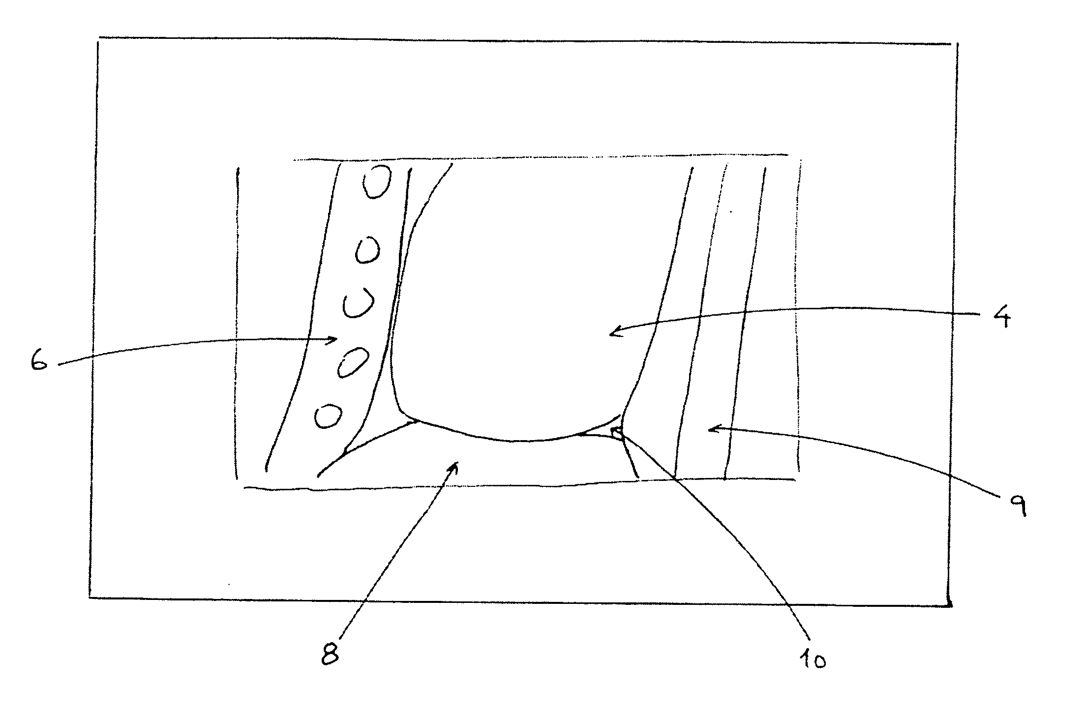
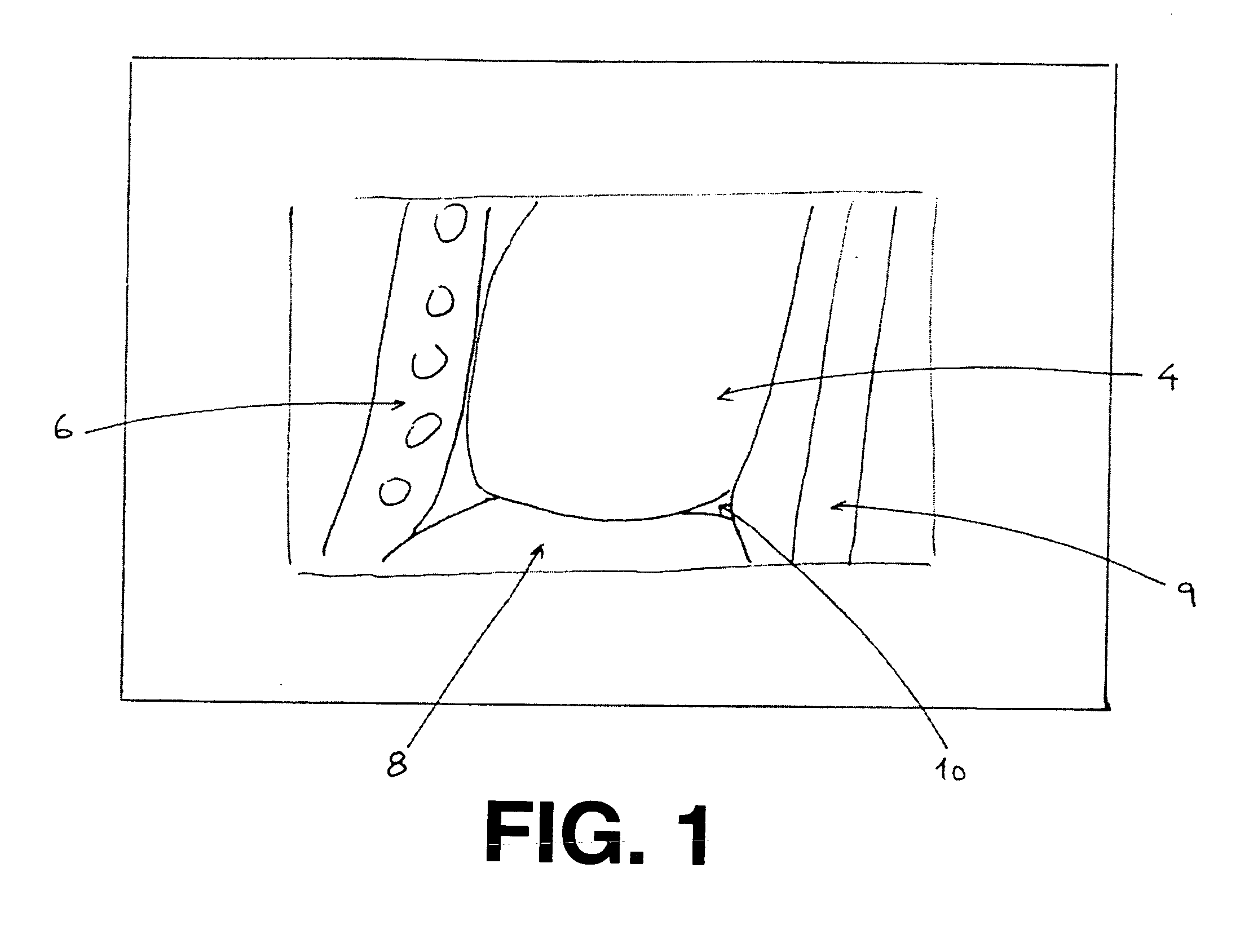
[0064] A method of the invention was used to detect the boundary between heart muscle tissue and liver tissue and between heart muscle tissue and the sternum in a 3D cardiac CT scan. FIG. 1 shows a schematic representation 2 of a saggital section of the 3D cardiac CT scan that was analyzed by the method of the invention. The scan included, in addition to cardiac tissue 4, sternum and ribs 6, liver 8, aorta 9 and epicardial fat 10. Lung tissue was also present in the 3D scan, although it is not present in the section of FIG. 1. The...


PUM


Abstract

A method and system for detecting a boundary between a first tissue and a second tissue in a two-dimensional image. A cost is assigned to each pixel in a detection region indicative of the likelihood that the pixel is not located on the boundary between the first tissue and the second tissue. The boundary between the first and second tissues or organs is a path of voxels of minimal cost in the detection region from among two or more paths, the cost of a path being obtained in a calculation based upon the costs assigned to the voxels of the path. The method may be used to detect a boundary between a first tissue or organ and a second tissue or organ in a three-dimensional scan by applying the method to each of one or more two-dimensional sections and joining the two dimensional boundaries into a three dimensional boundary in the three dimensional scan. The method may also be used in segmenting a three-dimensional body scan.

Description

FIELD OF THE INVENTION [0001] The present invention relates to methods for analyzing images of body tissues, such as an image obtained by radiographic techniques including computer tomography (CT). BACKGROUND OF THE INVENTION [0002] A computer tomography (CT) scan is three-dimensional (3D) image of at least a portion of a subject's body. The scan is a 3D array of integers each of which is the gray level of a body volume element called a voxel. Each voxel in the scan is addressed by means of a triplet of integral coordinates (x,y,z). A voxel in a 2D array or in an image is referred to as a “pixel”. The distance between adjacent voxels along the coordinate axes are referred to as “the x-mm-per-pixel”, the “y-mm-per-pixel” and the “z-mm-per-pixel”. The x- y- and z-mm-per-pixel are usually constant for a specific scan. The x- and y-mm-per-pixel are usually equal in which case they are both referred to simply as the “mm-perpixel”. The x- and y-dimensions of a scan are a property of the s...

Claims


Application Information

Patent Timeline
Patent Type & Authority Applications(United States)
IPC IPC(8): G06K9/00
CPCG06T7/0083G06T2207/10081G06T2207/30004G06T7/12
Inventor PORAT, HADARMILSTEIN, IDO I.AKERMAN, SHMUELMILLER, GAD G.
Owner PHILIPS HEALTHCARE INFORMATICS INC
Learn More