
Method for visually supporting an invasive examination or therapy of the heart with the aid of an invasive instrument

a technology of invasive examination and invasive therapy, which is applied in the field of visual support of invasive examination or heart therapy with the aid of an invasive instrument, can solve the problems of difficult ablation, time-consuming, and complex procedure for carrying out annular ablation

Active Publication Date: 2007-04-12
SIEMENS HEALTHCARE GMBH
View PDF13 Cites 6 Cited by
  • Summary
  • Abstract
  • Description
  • Claims
  • Application Information

AI Technical Summary

Benefits of technology

[0022] Steps d) and e) can preferably be repeated during the examination or therapy, that is, the visualization is subsequently transmitted to the invasive instrument (the ablation catheter). As a result thereof, the physician can make maximum use of the visualization when carrying out the therapy.
[0028] The pre-operative image data set can also be acquired using ECG gating, the X-ray image data set and the pre-operative image data set preferably being aligned with the same phase in the cardiac cycle. This makes it easier to align the images with one another.

Problems solved by technology

The procedure for carrying out annular ablation is very complex.
Here, too, ablation is very difficult to carry out because of the requirement for accurate targeting.
The problem lies in aligning three-dimensional image data with the position of the ablation catheter.
Aligning 3D mapping data with one another in the correct position and dimension is very time-consuming, however.

Method used


Image

  • Method for visually supporting an invasive examination or therapy of the heart with the aid of an invasive instrument
  • Method for visually supporting an invasive examination or therapy of the heart with the aid of an invasive instrument

Examples


Embodiment Construction

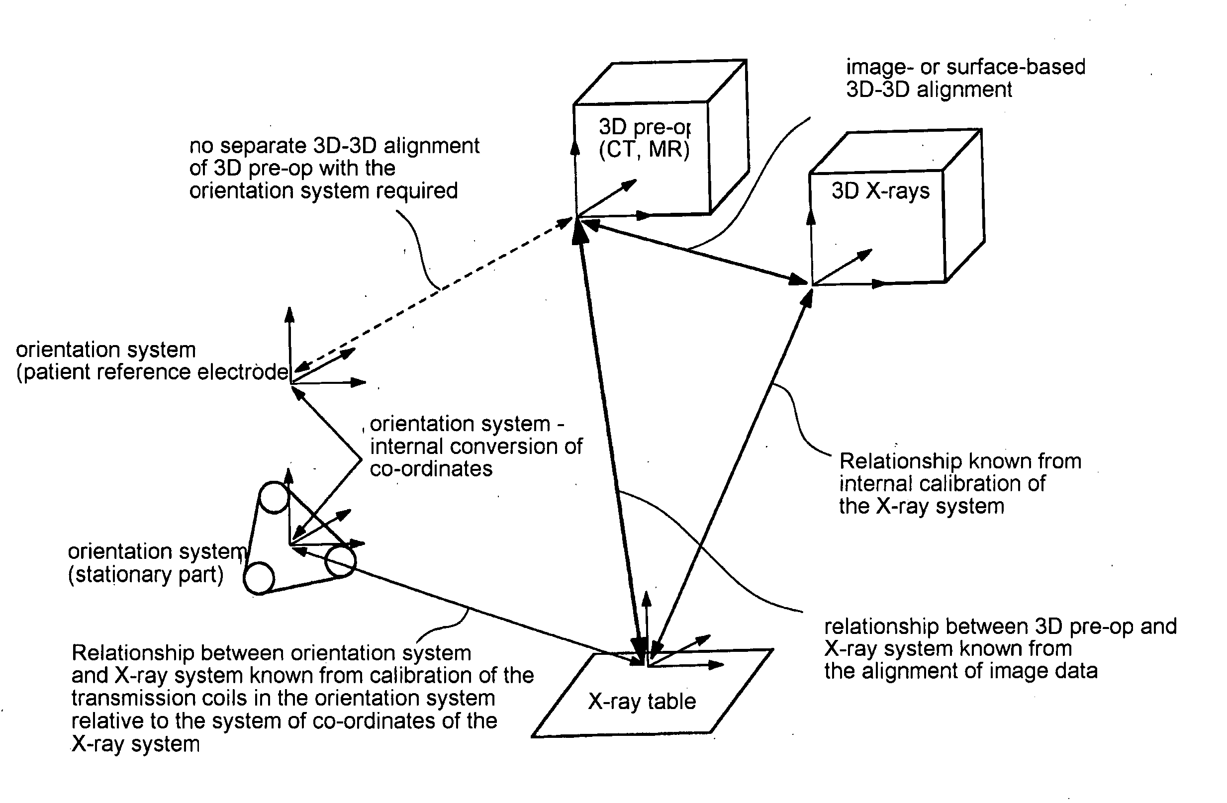
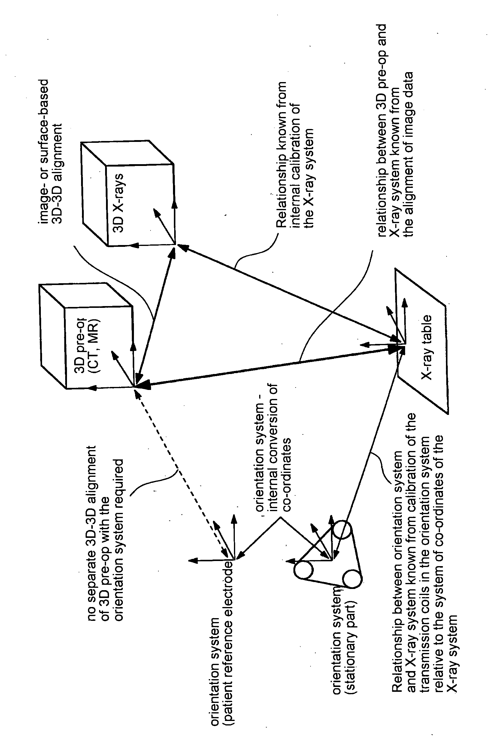
[0032] The figure shows three different systems for obtaining data to support a therapy of the heart, such as an ablation. Prior to the actual therapy, a three-dimensional image data set “3D pre-op” is acquired by means of computer tomography (CT) or magnetic resonance (MR). The system of co-ordinates is not at first correlated with other systems of co-ordinates. Furthermore, prior to or during therapy, a three-dimensional X-ray image data set of the heart is acquired using a technique as described in German patent application 10 2004 048209.8. The relationship between the system of co-ordinates of the three-dimensional X-ray image data set “3D X-rays” and an X-ray table on which the patient is lying, is known from the internal calibration within the X-ray system. The therapy of the heart ensues in particular with the aid of a catheter, for example an ablation catheter. An orientation system is available to pinpoint the position of the catheter. The orientation system comprises a st...


PUM


Abstract

To visually support a catheter ablation in the heart, three-dimensional image data have been used prior to the intervention. During ablation, the position of the catheter is pinpointed by an orientation system. The orientation system acquires electroanatomical 3D mapping data. the two-dimensional image data is assigned to the 3D mapping data in the correct position and dimensions which is a time-consuming step. The invention makes provision for the orientation system being in a fixed location relative to the X-ray system so that a positionally and dimensionally correct alignment of the X-ray image data set with the 3D mapping data is no longer required. An image or surface based 3D-3D alignment of the three-dimensional data acquired prior to the intervention with the three-dimensional X-ray image data is considerably less time-consuming than alignment thereof with the 3D mapping data and is more reliable because more structures is recognized in the three-dimensional X-ray image data.

Description

CROSS REFERENCE TO RELATED APPLICATIONS [0001] This application claims priority of German application No. 10 2005 045 073.3 filed Sep. 21, 2005, which is incorporated by reference herein in its entirety. FIELD OF THE INVENTION [0002] The invention relates to a method for visually supporting an invasive examination or therapy of the heart with the aid of an invasive instrument. BACKGROUND OF THE INVENTION [0003] The therapies involved are mainly those of the ablation kind with the aid of an ablation catheter. For the therapy of atrial fibrillation in the heart, for example, the pulmonary veins of the atrium are electrically isolated by annular ablations around the pulmonary veins. The procedure for carrying out annular ablation is very complex. [0004] Arrythmias in ventricular movement (ventricular fibrillation) can result from scar tissue in the myocardium. This scar tissue forms as a result of an infarction, for example. An accurately targeted ablation on the scar tissue may suppre...

Claims


Application Information

Patent Timeline
IPC IPC(8): G01N23/04A61B5/05
CPCA61B5/7285A61B6/12A61B6/541A61B8/543A61B19/52A61B19/5244A61B2017/00243A61B2019/5238A61B90/36A61B34/20A61B2090/376
Inventor BOESE, JANRAHN, NORBERT
Owner SIEMENS HEALTHCARE GMBH
Learn More