
Method and System For Estimating Cardiac Ejection Volume Using Ultrasound Spectral Doppler Image Data

a technology of ultrasound spectral doppler and cardiac ejection volume, which is applied in the field of method and system for estimating cardiac ejection volume using ultrasound spectral doppler image data, can solve the problems of limited measurement accuracy, limited range and effectiveness of possible measurements, and inapplicability of limitations on frequency of interrogation and angle of view, so as to achieve quantitative evaluation of the effect of a given therapy

Inactive Publication Date: 2007-04-12
ST JUDE MEDICAL ATRIAL FIBRILLATION DIV
View PDF99 Cites 11 Cited by
  • Summary
  • Abstract
  • Description
  • Claims
  • Application Information

AI Technical Summary

Benefits of technology

[0009] With the ability to measure cardiac ejection volumes and compare cycle-to-cycle variation, the effectiveness of a given therapy may be evaluated quantitatively. In particular, a clinician can evaluate the effectiveness of implanting a pacemaker electrode at a given cardiac location with the pacemaker programmed according to specific configuration parameters. By repeating the evaluation for more electrode locations and / or other specific pacemaker configuration parameters, the clinician can find an optimal combination of electrode location and pacemaker configuration. For the situation of comparing the relative effectiveness of two pacemaker configurations or electrode locations, the ejection volume area may be ignored when it does not change. Then the values of velocity-time integral may suffice when comparing the relative ejection effectiveness. The velocity-time integral may similarly suffice when comparing an image of the heart without a pacemaker with an image with one. The same technique may be used if the area does differ between two spectral images.

Problems solved by technology

However, until recent advances in miniaturized ultrasonic transducers, physicians were limited to only certain angles of view, thus limiting the range and effectiveness of possible measurements.
Further, given the depth of imaging required by such classical approaches, associated interrogation frequency limitations due to attenuation restricted the accuracy of measurement.
With the recent introduction of catheter based transducers for imaging the heart from either the vena-cava or even from within the heart, such limitations on frequency of interrogation and angle of view are not applicable.
One problem in the current therapy is the optimization of the placement of the left ventricular electrode so as to provide maximum therapy.

Method used


Image

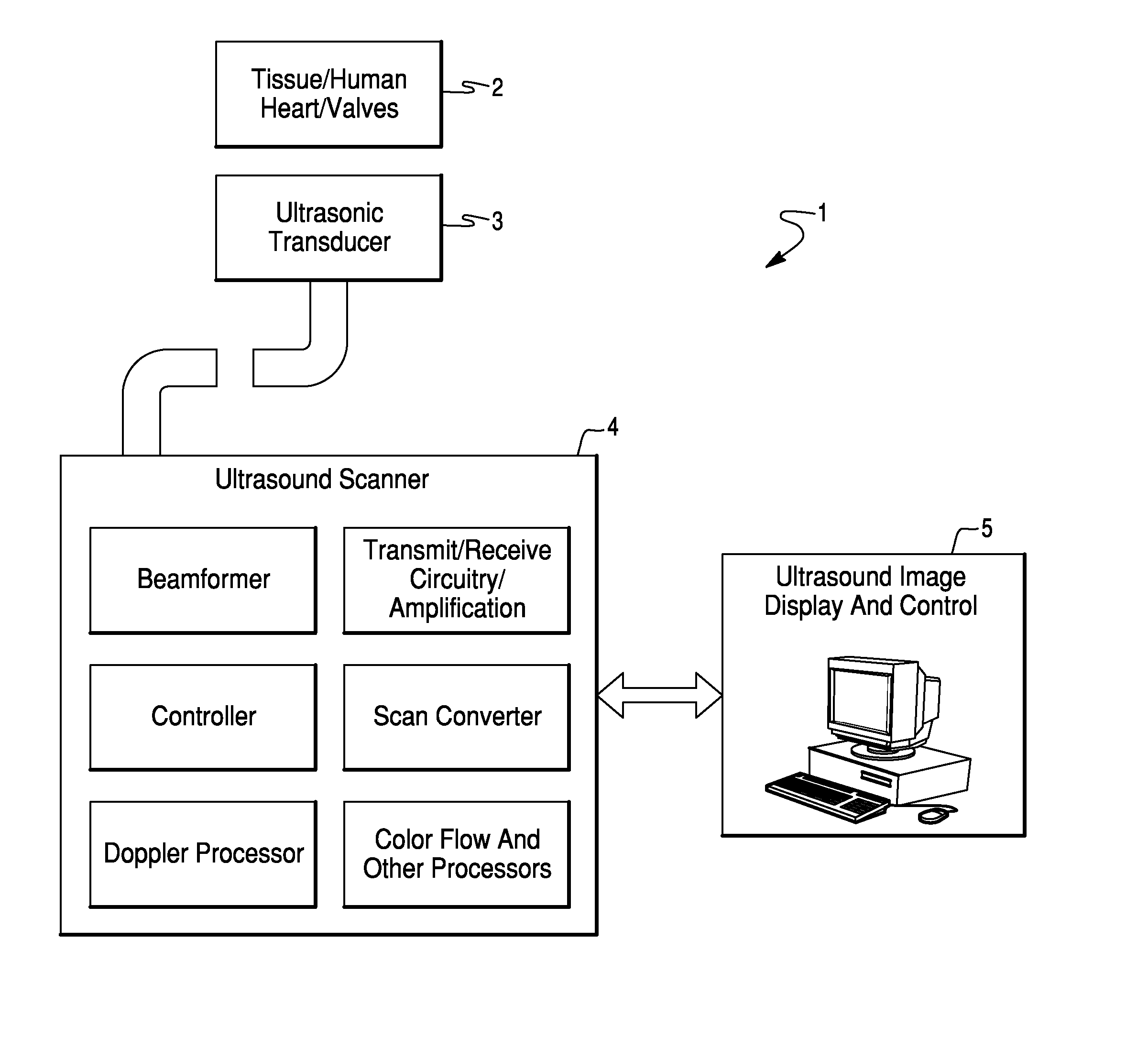
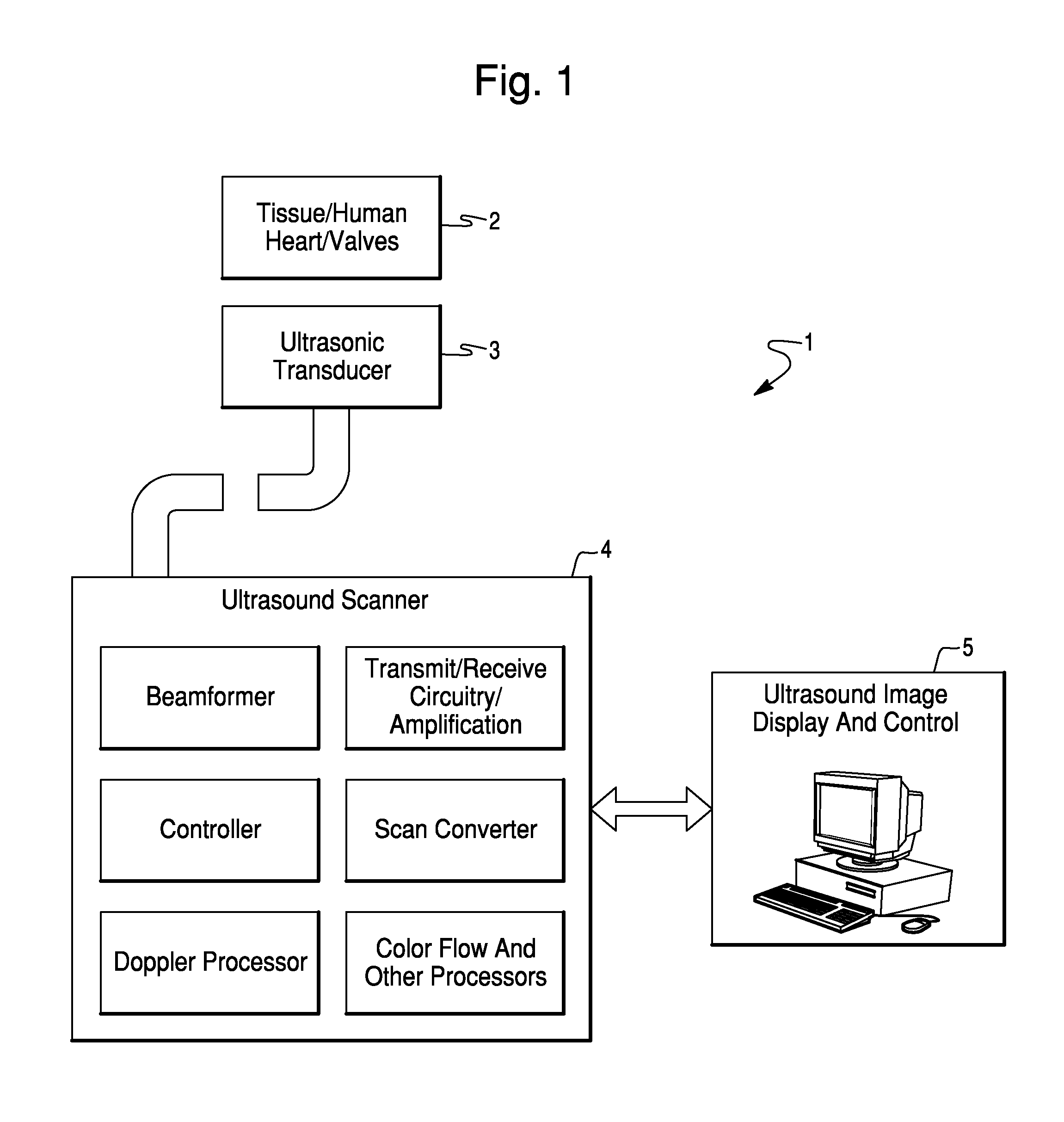
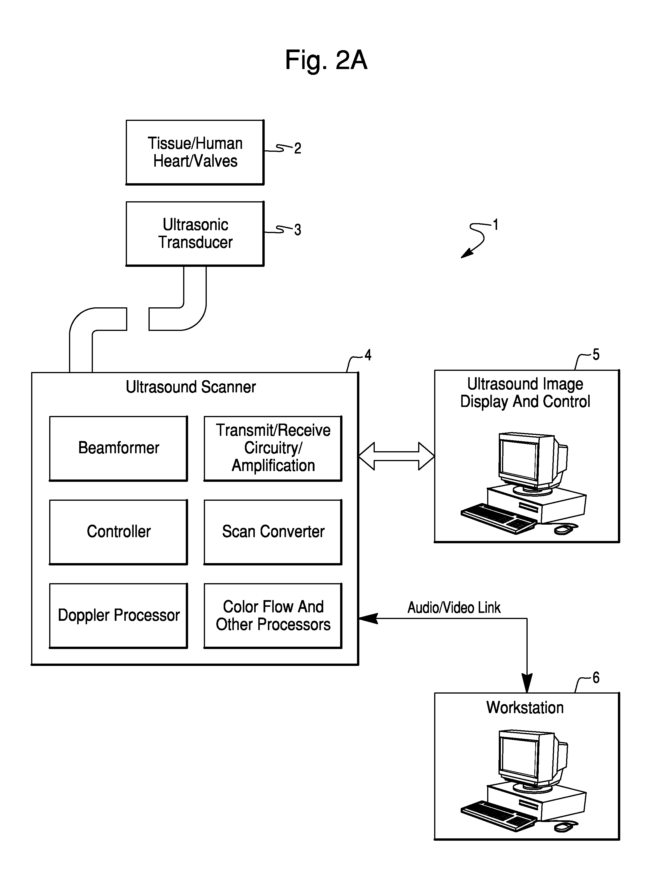
  • Method and System For Estimating Cardiac Ejection Volume Using Ultrasound Spectral Doppler Image Data
  • Method and System For Estimating Cardiac Ejection Volume Using Ultrasound Spectral Doppler Image Data
  • Method and System For Estimating Cardiac Ejection Volume Using Ultrasound Spectral Doppler Image Data

Examples


Embodiment Construction

[0022] Heart failure is a disease where the heart's main function, a pump for blood, is wearing down. The heart tissue can absorb fluid, the left ventricle does not allow quick electrical conduction, becomes enlarged, does not contract well, and becomes less efficient at pumping blood. A measurement for the cardiac output (volume of ejected blood) is called the “ejection volume” or “stroke volume”. The efficiency of the heart as a pump is called the “ejection fraction” or “EF”. EF is measured as the percentage of the blood volume contained in the ventricles which is ejected with each beat of the heart. A healthy, young heart will have an EF greater than 90 (i.e., 90 percent of the ventricular blood is pumped with each heart beat); an older, sick heart in heart failure can have an EF less than 30. Heart failure leads to an extremely diminished lifestyle, and, left untreated, can be a major cause of mortality.

[0023] A new therapy to treat heart failure is bi-ventricular pacing, or “r...


PUM


Abstract

A method and system for estimating the volume of blood ejected from a cardiac ventricle or atrium uses spectral Doppler ultrasound while imaging a portion of the heart. The method computes the mean ejection velocity Vavg(t) at time t from the spectral Doppler data. The process may further utilize a discrete graphical technique to compute a measure of cardiac output. Further, the measure of cardiac output can yield an approximation to the mean-ejection-velocity integral, which is the area under the curve Vavg(t) for all moments t within a cardiac cycle.

Description

RELATED APPLICATIONS [0001] This application is a continuation-in-part of U.S. patent application Ser. No. 11 / 428,517 filed Jul. 3, 2006, which is a continuation of U.S. patent application Ser. No. 10 / 620,517 filed Jul. 16, 2003, now U.S. Pat. No. ______, which claims the benefit of priority to U.S. Provisional Application Ser. No. 60 / 397,653, filed on Jul. 22, 2002, the entire contents of all of which previous applications are hereby incorporated by reference.FIELD OF THE INVENTION [0002] The specification herein relates to a technique for measuring cardiac output (i.e., total volume of blood ejected by the left ventricle in one cardiac cycle) using Doppler ultrasonic imaging, particularly for use in evaluating placement of cardiac pacing electrodes. BACKGROUND OF THE INVENTION [0003] Volumetric output of blood from the heart and / or circulatory system is of interest in various diagnostic and therapeutic procedures. Such measurements are of significant interest during electrophysiol...

Claims


Application Information

Patent Timeline
Patent Type & Authority Applications(United States)
IPC IPC(8): A61B8/00A61B8/06
CPCA61B8/065A61B8/0883A61B8/543
Inventor DALA-KRISHNA, PRAVEEN
Owner ST JUDE MEDICAL ATRIAL FIBRILLATION DIV
Learn More