

Method of off-resonance detection using complementary MR contrast agents
a contrast agent and off-resonance detection technology, applied in the field of magnetic resonance imaging (mri), can solve the problems of significant signal dephasing, agent cannot be distinguished from a void in the image, and negative contrast agents suffer from partial volume effects
- Summary
- Abstract
- Description
- Claims
- Application Information
AI Technical Summary
Benefits of technology
Problems solved by technology
Method used
Image

Examples
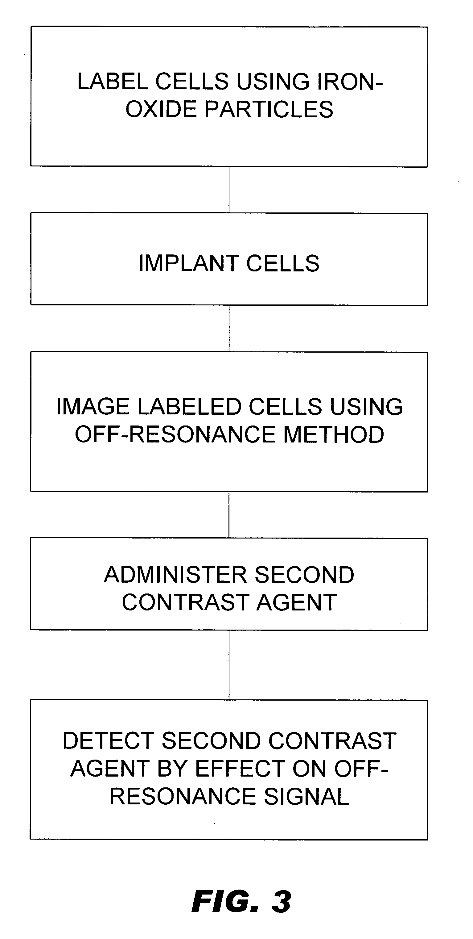
Embodiment Construction
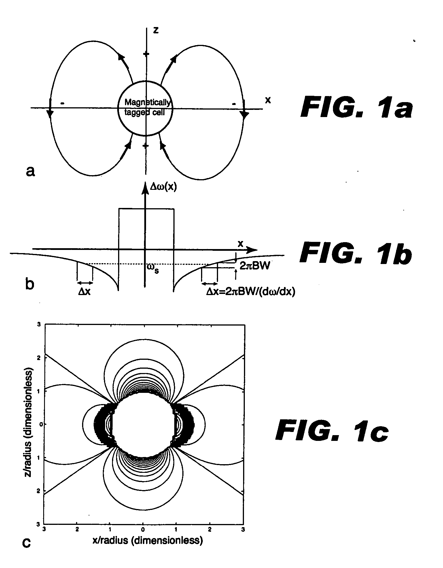
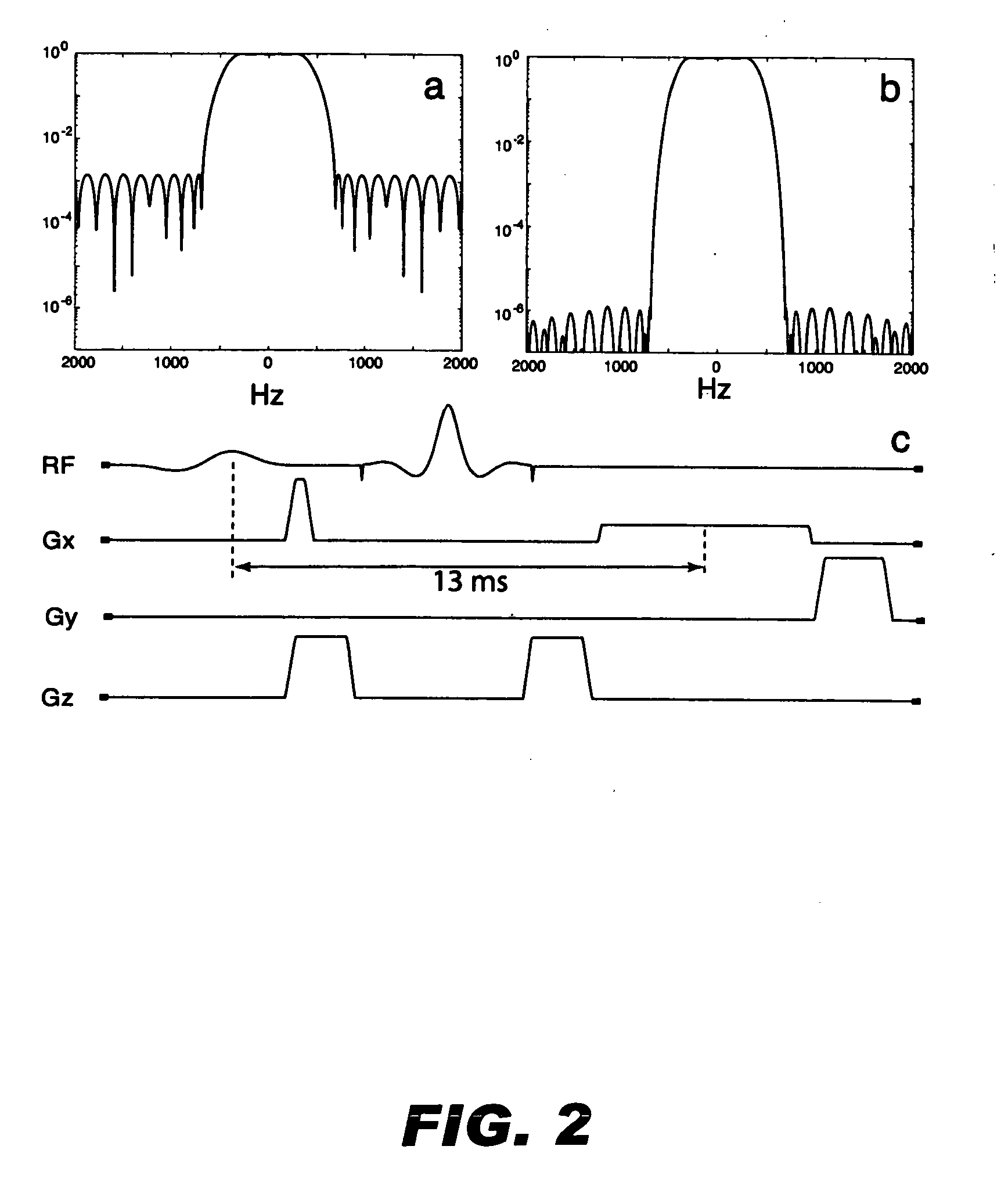
[0015] As described in application Ser. No. 10 / 849,068, supra, a collection of labeled cells will cast a field pattern in the water molecules immediately surrounding the cells. The field pattern can be approximated by a dipole field from a magnetized sphere. The dipole pattern demonstrates a classic field cross pattern, in which the local B field is enhanced in the north and south poles and suppressed along the equator. The polarity of the field perturbation would be reversed for a diamagnetic agent. The dipole field pattern intensity falls off quickly. The field perturbation varies as Δ Bz(r,θ)=Δ χ BO3(ar)3(3cos2θ-1)(1)
[0016] where Δχ is the difference in bulk magnetic susceptibility between the sphere and surroundings, a is the radius of the sphere, r is the distance from the sphere center, and θ is the angle relative to the main field, B o. Hence, the field pattern from a smaller collection of cells will fall off more steeply than that from a larger collection. In pra...
PUM
 Abstract
Description
Claims
Application Information