
Compositions for nanoconfinement induced contrast enhancement and methods of making and using thereof

a technology of contrast enhancement and nanoconfinement, which is applied in the direction of in-vivo testing preparations, powder delivery, pharmaceutical delivery mechanisms, etc., can solve the problems of short imaging lifetime, long-term risk of secondary cancer, and intrinsically poor ct contrast discrimination of soft tissues with similar effective electron densities, etc., to achieve the effect of enhancing ct contras

Active Publication Date: 2017-09-21
YALE UNIV
View PDF1 Cites 0 Cited by
  • Summary
  • Abstract
  • Description
  • Claims
  • Application Information

AI Technical Summary

Benefits of technology

This patent text is about a new way to make contrast agents for CT scans. These agents are made by combining certain molecules with iodine, which helps to make the scans clearer. Using lower concentrations of these agents reduces the risk of toxic effects.

Problems solved by technology

Although X-ray-based computed tomography (CT) is one of the most commonly used diagnostic tools in clinical practice, CT is intrinsically poor in discriminating soft tissues with similar effective electron densities, e.g. an early-stage tumor from surrounding tissue, or normal tissue from tissue following ischemic injury.
In addition, the relatively high dose of ionizing radiation imparted by the X-rays during CT is carcinogenic, which carries a long-term risk of developing secondary cancers, especially for patients in need of multiple CT scans or with existing health conditions that are sensitive to ionizing radiation.
However, despite the increased radiographic contrast obtained with conventional iodinated CT contrast agents, rapid clearance and vascular permeation of these agents leads to short imaging lifetimes, necessitating higher concentrations (˜300 mg I / mL) in clinical applications.
This level of contrast agent often results in severe adverse side effects in patients, particularly those with impaired renal function, such as nephrotoxicity.

Method used


Image

  • Compositions for nanoconfinement induced contrast enhancement and methods of making and using thereof
  • Compositions for nanoconfinement induced contrast enhancement and methods of making and using thereof
  • Compositions for nanoconfinement induced contrast enhancement and methods of making and using thereof

Examples


example 1

of Iodinated Dendrimers (ID)

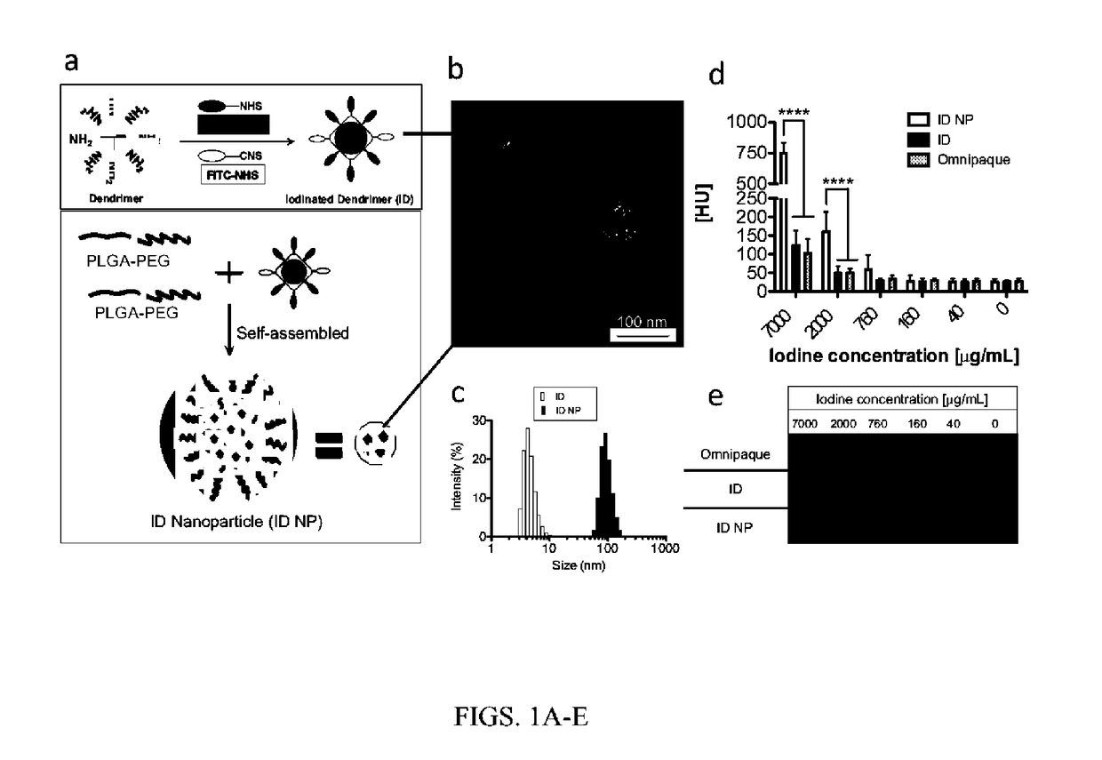
[0083]Triiodobenzoic acid (TIBA) was conjugated to the amino termini of a generation 4 dendrimer (Diameter32˜10 nm, number of branches=64) (FIG. 1a).

[0084]The dendrimer primary surface amines were first partially acetylated with sulfo-N-hydroxysuccinimide (NHS)-acetate. Five hundred milligrams of ethylenediamine-core PAMAM generation 4 dendrimers (G4) were desiccated in a 100 mL round-bottom flask and subsequently dissolved in 100 mM sodium bicarbonate buffer (pH 9.0) to a final concentration of 10 mg / mL with magnetic stirring. To this dendrimer solution, 36 mol equiv of sulfosuccinimidyl acetate were added as a solid and allowed to dissolve, The pH of the reaction mixture was immediately adjusted to 8.5 with 1 N NaOH and the reaction was allowed to proceed for 2 h at 25° C. The partially acetylated G4 (G4-Ac) product was purified by ultrafiltration with deionized water using 10K MWCO Amicon Ultra-15 filters and lyophilized to obtain a white crystalline s...

example 2

on of Iodinated Dendrimer-Encapsulated Particles

[0087]The iodinated dendrimer prepared in Example 1 were encapsulated in a biocompatible, biodegradable polymer to form polymeric nanoparticles (ID-NP). Specifically, the iodinated dendrimers were encapsulated in PLGA-PEG block copolymers using self-assembly (FIG. 1a). The PLGA-PEG block copolymers were prepared using literature procedures.

[0088]Acid-terminated PLGA (500 mg) together with 10-fold excess of NHS and DCC were dissolved in 10 mL anhydrous DCM. After being stirred at room temperature for four hours, the reaction solution was filtered through a PTFE filter to remove the precipitate. The NHS-activated PLGA was obtained through precipitation in cold ethyl ether. After dried under vacuum, NHS-activated PLGA was dissolved in anhydrous DCM with equivalent mole of NH12-PEG-COOH and the solution was stirred at room temperature. The conjugate was precipitated in cold ethyl ether and dried under vacuum. To encapsulate ID into nanopar...

example 3

ization of Iodinated Dendrimer-Encapsulated Particles

[0089]Particle Size

[0090]The diameter of the iodinated dendrimers and the iodinated dendrimer-encapsulated nanoparticles were determined by scanning transmission electron microscopy (STEM) imaging and Dynamic light scattering (DLS). DLS measurements and STEM images showed the particle size of the ID and ID encapsulated nanoparticle (ID NP), ranging from 5-13 nm and 70-135 nm, respectively (FIGS. 1b and 1c).

[0091]CT Contrast

[0092]The CT contrast of the ID NP with different iodine concentrations was compared with a commercial CT contrast agent, Omnipaque, using a micro CT (eXplore CT 120, GE Healthcare) (FIGS. 1d and 1e). The CT number of the ID NP at the higher iodine concentration (7 mg / mL) was approximately 750 HU, an increase of more than five-fold over that of ID (˜130 HU) or Omnipaque (˜100 HU). At a lower iodine concentration of 2 mg / mL, the CT number of the ID NP was approximately 170 HU, whereas the CT numbers of ID or Omni...


PUM

PropertyMeasurementUnit
pHaaaaaaaaaa
diameteraaaaaaaaaa
diameteraaaaaaaaaa

Abstract

Multivalent CT or MR contrast agents and methods of making and using thereof are described herein. The agents contain a moiety, such as a polymer, that provides multivalent attachment of CT or MR contrast agents. Examples include, but are not limited to, multivalent linear polymers, branched polymers, or hyperbranched polymers, such as dendrimers, and combinations thereof. The dendrimer is functionalized with one or more high Z-elements, such as iodine. The high Z-elements can be covalently or non-covalently bound to the dendrimer. The dendrimers are confined in order to enhance CT contrast. In some embodiments, the moiety is confined by encapsulating the dendrimers in a material to form particles, such as nanoparticles. In other embodiments, the dendrimer is confined by conjugating the moiety to a material, such as a polymer, which forms a gel upon contact with bodily fluids.

Description

CROSS-REFERENCE TO RELATED APPLICATION[0001]This application claims priority to and the benefit of U.S. Ser. 61 / 992,552 filed May 13, 2014, the disclosure of which is expressly incorporated hereby by reference.STATEMENT REGARDING FEDERALLY SPONSORED RESEARCH[0002]This invention was made with government support under Grant No. U19 203-0025 awarded by the National Institutes of Health. The government has certain rights in the invention.FIELD OF THE INVENTION[0003]This invention is in the field of compositions for nanoconfinement induced contrast enhancement.BACKGROUND OF THE INVENTION[0004]Although X-ray-based computed tomography (CT) is one of the most commonly used diagnostic tools in clinical practice, CT is intrinsically poor in discriminating soft tissues with similar effective electron densities, e.g. an early-stage tumor from surrounding tissue, or normal tissue from tissue following ischemic injury. In addition, the relatively high dose of ionizing radiation imparted by the X-...

Claims


Application Information

Patent Timeline
Patent Type & Authority Applications(United States)
IPC IPC(8): A61K49/04C08K3/16C08K9/10
CPCA61K49/0485A61K49/0423C08K3/16A61K49/0438C08K9/10A61K49/0442C08J3/124A61K49/1887
Inventor FAHMY, TAREKSINUSAS, ALBERTKIM, DONGIN
Owner YALE UNIV
Learn More