Nitric Oxide Device and Method for Wound Healing, Treatment of Dermatological Disorders and Microbial Infections

a technology of nitric oxide and wound healing, applied in the direction of immobilised enzymes, peptide/protein ingredients, capsule delivery, etc., can solve the problems of affecting disrupting normal wound healing, and chronic wounds, etc., to improve the shelf life of red meat products, preservation or physical appearance

Inactive Publication Date: 2011-05-05
MICROPHARMA
View PDF31 Cites 133 Cited by
  • Summary
  • Abstract
  • Description
  • Claims
  • Application Information

AI Technical Summary

Benefits of technology

[0044]first exposing the wound to a device of the application to produce a high concentration of nitric oxide gas that contacts the wound for a first treatment period without inducing toxicity to the subject or healthy tissue; and
[0045]second exposing the wound to a second device of the application to produce a low concentration of nitric oxide gas that contacts the wound for a second treatment period.
[0046]In a further aspect, the disclosure provides a method of improving red meat product shelf life, preservation, or physical appearance comprising exposing the red meat product to a device of the application, wherein NO contacts the red meat product.

Problems solved by technology

Wound healing is a complicated process relying heavily on the integration of a multitude of control mechanisms, events, and factors.
Several pathological conditions, including diabetes and venous stasis, are associated with a number of changes at the molecular level which ultimately disrupt normal wound healing and can lead to the formation of chronic wounds (Blackytny et al.
If a wound fails to heal, however, or becomes infected, the body maintains the circulating NO at a high level and the wound is then caught in a vicious cycle preventing it from healing (Stenzler et al.
Infected wounds pose a specific and significant problem to wound care specialists treating a chronic wound, non-healing ulcer, or healthy post surgical wound for that matter.
Systemic and topical antibiotics, as well as other topical anti-microbial agents such as colloidial silver polymyxins or dye compounds, however, have become increasingly less effective against common pathogens.
P. aeruginosa infection is particularly problematic, as patients are often immune suppressed or are severely disabled and artificially ventilated.
Thus, as the common antimicrobial agents begin to fail, alternative treatments which do not rely on conventional antibiotics are needed.
Another problem in treating infected chronic wounds with systemic antibiotics is that such wounds often accompany reduced local and regional circulation.
Patients with venous stasis ulcers have venous thrombosis, reduced circulation and poor regional blood flow; and patients with diabetic foot ulcers suffer from poor microcirculation due to deposition of glucose and reduced circulation.
Systemic antibiotics can exacerbate this problem, due to constriction of the capillaries and small blood vessels, causing a further reduction in blood flow to the wound and reduced delivery of the antimicrobial agent.
Topical agents are often more effective at concentrating the antimicrobial agent at the wound site; however, they are often less effective at eliminating infection for other reasons which include reduced circulation once again.
Thus, traditional therapies often leave an infected wound untreated and a patient's limb or life in danger.
In addition to poor circulation and resistant infection, many chronic wounds simply fail to heal in the face of daily wound care or treatment with advanced wound care therapy.
Diabetic foot ulcers and venous stasis ulcers pose a great difficulty to patients and clinicians alike.
Most often this condition results from inactivity and poor eating habits.
These patients become bed ridden, immobilized, and emaciated while trying to stay off the wounds on their lower extremities, only worsening their problem of sedentary living.
Clinicians frequently appeal to surgeons to bypass arteries or provide surgical coverage of wounds; however, the patients frequently have multiple co-morbidities, are not well nourished, and are poor surgical candidates.
This leaves the patient and clinician with the only remaining option of treating the chronic wound with daily dressing changes, a time consuming, costly, and relatively ineffective practice.
However, with a lack of compliance, poor circulation, poor nutrition, non-sterile conditions, and simply the time it takes to heal wounds in this way they often stay open for years and even decades.
The NO delivery device, however, utilized many bulky and costly components including air pump systems, gNO source cylinders, internal pressure sensors, mechanical pressure regulators, and plastic foot boot with inflatable cuff to cover the patient's lower extremity (Stenzler et al.
Another drawback with the delivery of gNO is that NO rapidly oxidizes in the presence of oxygen (O2) to form NO2, which is highly toxic, even at low levels.
Third, in-vitro studies demonstrated that inhibition of NO synthases resulted in impaired cytokine-mediated activation of phagocytic cells and reduction of bactericidal and bacteriostatic activity (Adams et al 1990).
And fourth, direct administration of NO-donor compounds in-vitro, induced microbial stasis and death.
Moreover, nitrosylation of free thiol groups may result in inactivation of metabolic enzymes (Fang 1997).
Dysregulation of NOS2 expression is often correlated with impairment of barrier function in dermatitis.
Unfortunately, humans have lost the ability to make vitamin C, the predominant antioxidant in skin, due to a specific gene mutation.
This, however, is a costly solution employing bulky “gas-diluting delivery systems” and “single use plastic boots”.
Other devices, utilizing a chemical reaction to produce the gas, may have solved the difficulties of cost and convenience; however, are unable to provide a constant concentration over time.

Method used


Image

  • Nitric Oxide Device and Method for Wound Healing, Treatment of Dermatological Disorders and Microbial Infections
  • Nitric Oxide Device and Method for Wound Healing, Treatment of Dermatological Disorders and Microbial Infections
  • Nitric Oxide Device and Method for Wound Healing, Treatment of Dermatological Disorders and Microbial Infections

Examples


example 1

Results

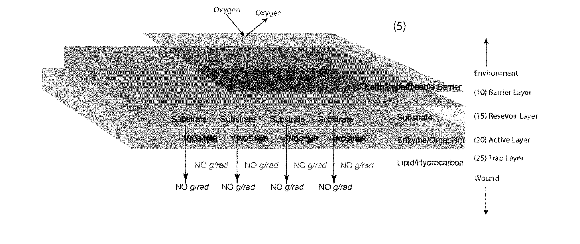
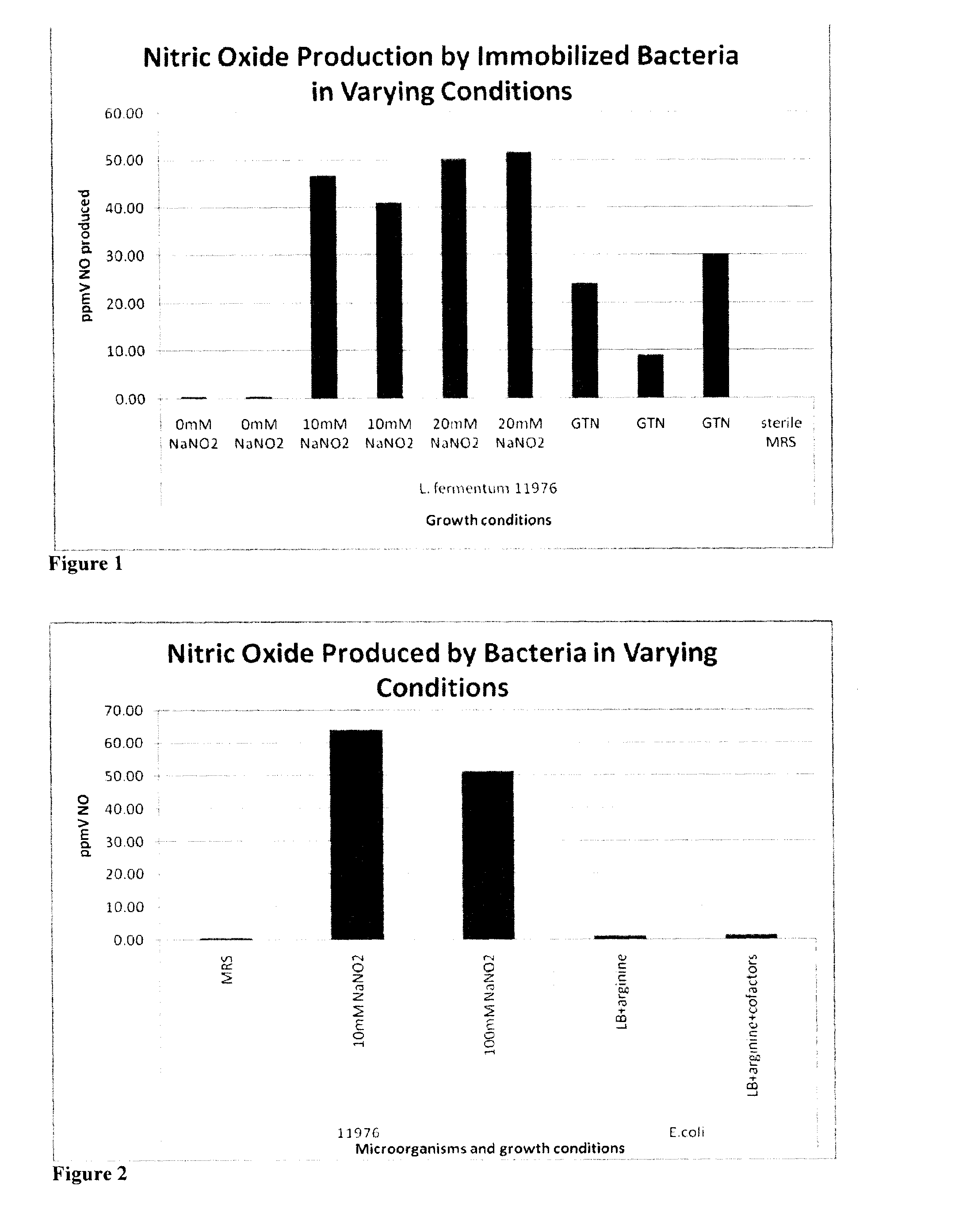
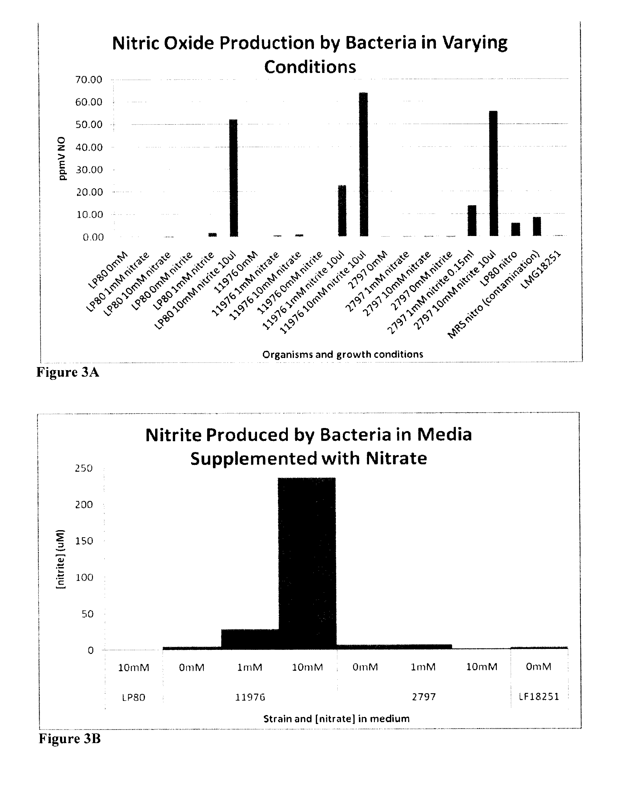
[0166]Tables 2-4 show the reaction that produces nitric oxide from a precursor. The results also show that live bacteria are able to produce nitric oxide gas (gNO) when immobilized in a slab-like piece of agarose supplemented with MRS growth media and either nitrite or a nitroglycerine patch (FIG. 1). The results in FIG. 2 show that live bacteria are able to produce nitric oxide gas when grown in media with the indicated cofactors. Without wishing to be bound by theory, the most probable mechanism for nitric oxide production from nitrite is the reduction of the salt to gNO by lactic acid produced by the metabolically active bacteria. The most probable mechanism of gNO production from nitroglycerine is that the organisms produce lactic acid which reduces nitroglycerine to nitrite and the resulting nitrite is reduced to nitric oxide again by lactic acid. In this way, the immobilized bacteria are capable of releasing gNO from a medical device or composition and onto affected tis...

example 2

Results

[0185]The gNO-producing patches showed a bactericidal effect on E. coli (FIG. 15), S. aureus (FIG. 16), P. aeruginosa (FIG. 17), A. baumannii (FIG. 18), and MRSA (FIG. 21). The gNO-producing patches showed a fungicidal effect on T. rubrum (FIG. 19) and T. mentagrophytes (FIG. 20). The gNO-producing patches also showed bacteriostatic effects on E. coli (FIG. 22 (left)), S. aureus (FIG. 22 (middle)), and P. aeruginosa (FIG. 22 (right)).

Materials and Methods

[0186]Patch Preparation: A one-sided gas permeable pocket was created by heat sealing 3 sides of a rectangular gas permeable membrane (Tegaderm) with a heat sealable plastic film. The resulting pocket was filled up with an alginate-immobilized L. Fermentum wafer and a glucose / NaNO2 solution and the fourth side of the pocket was heat sealed. A layer of aluminized tape was applied to the plastic film to avoid loss of gas. Control patches are made with a glucose solution that does not contain the NO donor NaNO2.

[0187]Bactericida...

example 3

Pilot Pre-Clinical Study

[0190]A pilot study was performed to provide information on the ability of nitric oxide to improve wound healing. The model uses the ischemic ear model in the rabbit, a well-validated model of ischemic wounds. Establishing ischemia involves a minor surgical procedure on the ear and the healing characteristics are similar to human healing in that it requires the generation of granulation tissue and reepithelization.

Results

[0191]This pilot study provided very promising data on the efficacy and safety of the nitric oxide producing dressing. It was found that treated ischemic wounds healed faster than controls and that improvements could also be seen in the histological evaluation of the wounds.

[0192]It was found that non-ischemic wounds closed between 10 and 15 days post-surgery, whether infected or not. The treatment of non-ischemic wounds with gNO marginally accelerated healing, as compared to the vehicle control (see FIG. 23, lower panels). Furthermore, the t...


PUM

PropertyMeasurementUnit
concentrationsaaaaaaaaaa
transparentaaaaaaaaaa

Abstract

The present disclosure provides a device having a casing with a barrier surface and a contact surface and a composition in the casing having a nitric oxide precursor and an isolated enzyme or live cell expressing an endogenous enzyme, for converting the nitric oxide gas precursor to nitric oxide gas or having activity on a substrate that produces a catalyst that causes the conversion of the nitric oxide gas precursor to nitric oxide gas. The present disclosure also provides methods and uses for treating wounds, microbial infections and dermatological disorders and for preserving meat products.

Description

FIELD OF THE DISCLOSURE[0001]The present disclosure relates to methods, devices and compositions for the treatment of wounds, dermatological disorders and microbial infections with nitric oxide. In particular, the disclosure relates to methods, devices and compositions for topical administration of nitric oxide.BACKGROUND OF THE DISCLOSURE[0002]Wound healing is a complicated process relying heavily on the integration of a multitude of control mechanisms, events, and factors. Inflammatory cells, keratinocytes, fibroblasts, and endothelial cells, as well as many enzymes and growth factors, must interact seamlessly for the normal healing process to occur (Blackytny et al. 2006). These factors will act together during the processes of clot formation, inflammation, re-epithelialisation, angiogenesis, granulation, contraction, scar formation, and tissue remodelling to ensure adequate wound healing. Several pathological conditions, including diabetes and venous stasis, are associated with ...

Claims


Application Information

Patent Timeline
Patent Type & Authority Applications(United States)
IPC IPC(8): A61K9/70A61K9/127A61K9/14A61K9/16A61K35/00A61K38/43A61K38/44A61K38/45A61K38/46A61K38/48A61K35/74A61P31/00A61P17/00A61P17/02A61M37/00A23L5/41A23L13/00A61K35/747
CPCA23B4/16A23B4/22A61K9/7007A61K9/703A61K33/00A61M35/00A61K45/06C12N11/00C12P3/00A61K38/465A61K35/747A61K38/44A61P3/10A61P17/00A61P17/02A61P17/04A61P17/06A61P17/08A61P17/10A61P17/12A61P19/02A61P19/10A61P29/00A61P31/00A61P31/02A61P31/04A61P31/06A61P31/08A61P31/10A61P31/12A61P31/22A61P33/00A61P33/02A61P33/04A61P33/06A61P33/14A61P35/00A61P37/08A61P41/00A61P43/00Y02A50/30
Inventor JONES, MITCHELL LAWRENCEPRAKASH, SATYA
Owner MICROPHARMA
Why Eureka
Try Eureka