Devices For Fixing A Sensor In A Lumen

a sensor and lumen technology, applied in the field of implantable medical devices, can solve the problems of blood clots, perforations or aneurysms, and the sensing device may not be adequately implanted in the vessel segment, so as to reduce the chance of embolism, increase the stability of the sensing device, and reduce the risk of embolism.

Active Publication Date: 2007-06-07
REMON MEDICAL TECH
View PDF94 Cites 80 Cited by
  • Summary
  • Abstract
  • Description
  • Claims
  • Application Information

AI Technical Summary

Benefits of technology

[0015] While the present inventions should not be so limited in their broadest aspects, in accordance with the afore-described methods, the only portion of the sensing device within the bifurcation between the main anatomical vessel and the anatomical branch is the connecting element, which may have a relatively low profile. As a result, very little force will be applied to the sensing device by fluid flowing between the anatomical branch and the main anatomical vessel, thereby increasing the stability of the sensing device within the main anatomical vessel, and in the case where the fluid is blood, decreasing the chance of embolism tha

Problems solved by technology

However, high local or radial force on the relative weak pulmonary artery vessel wall may cause perforation or aneurysm.
If, however, the length of the vessel segment is limited, the sensing device may not be adequately implanted within the vessel segment without crossing the branch.
In this case, the implanted sensing device may block future access to the vessel branch, e.g., during catheterization, may be unstable due to the transverse blood flow through the branch, and worse yet, may cause blood clots that may potentially result in an embolism.
In ad

Method used


Image

  • Devices For Fixing A Sensor In A Lumen
  • Devices For Fixing A Sensor In A Lumen
  • Devices For Fixing A Sensor In A Lumen

Examples


Embodiment Construction

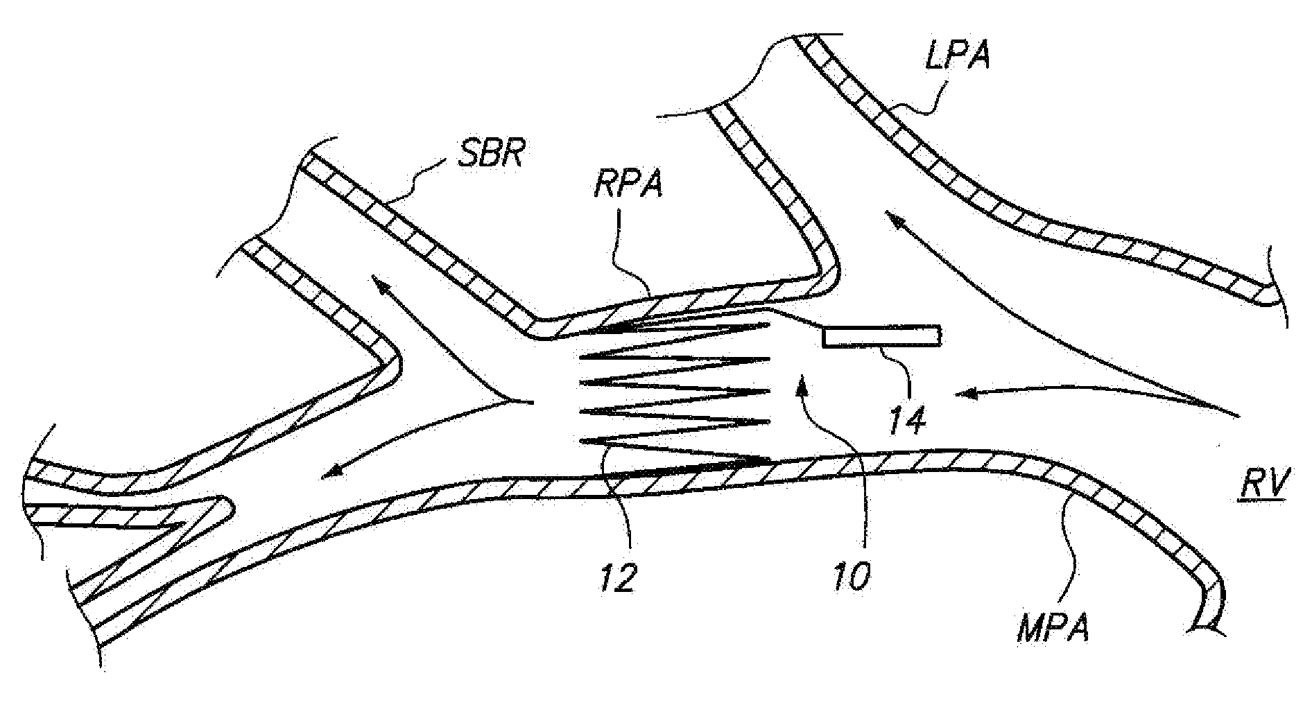
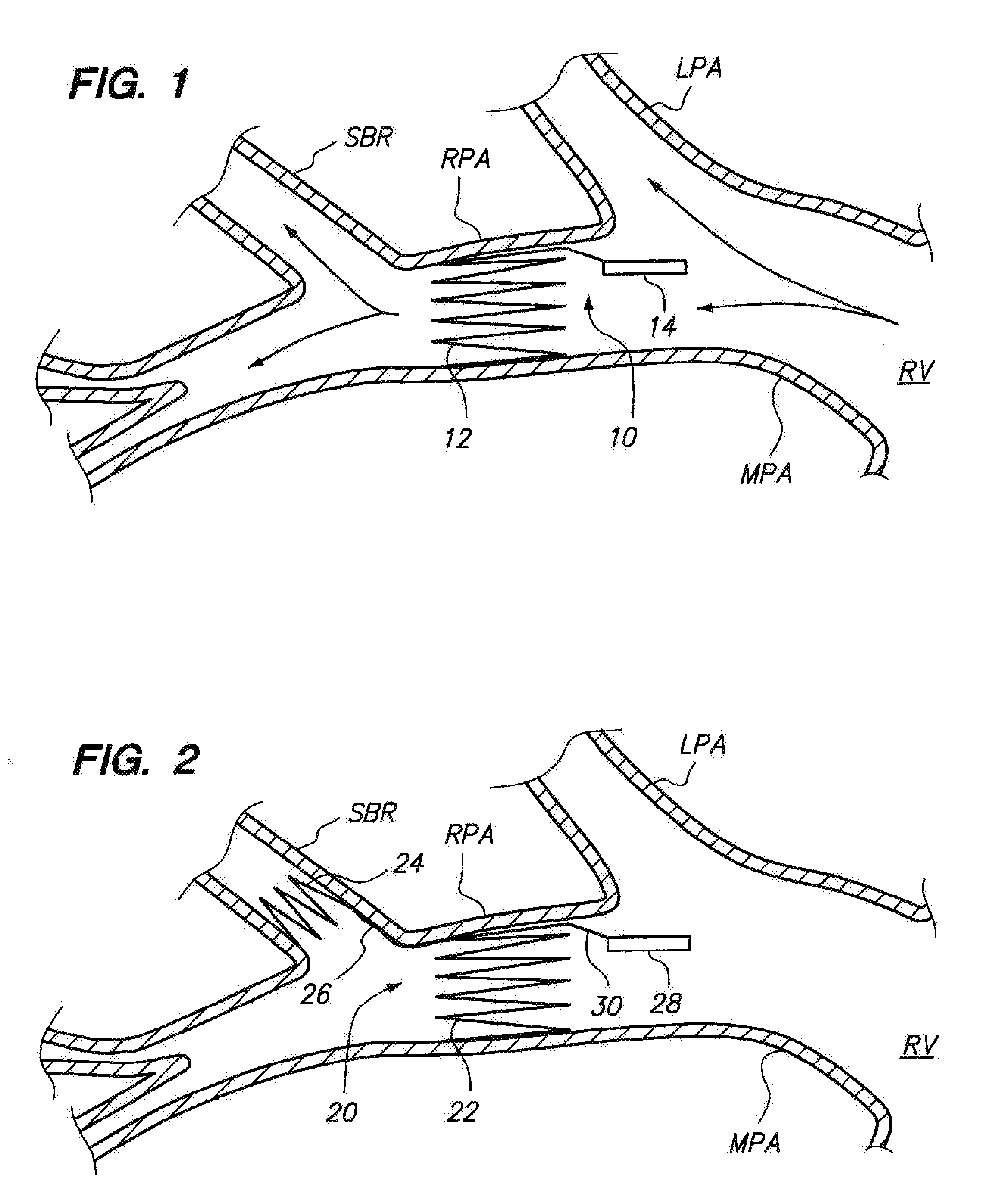
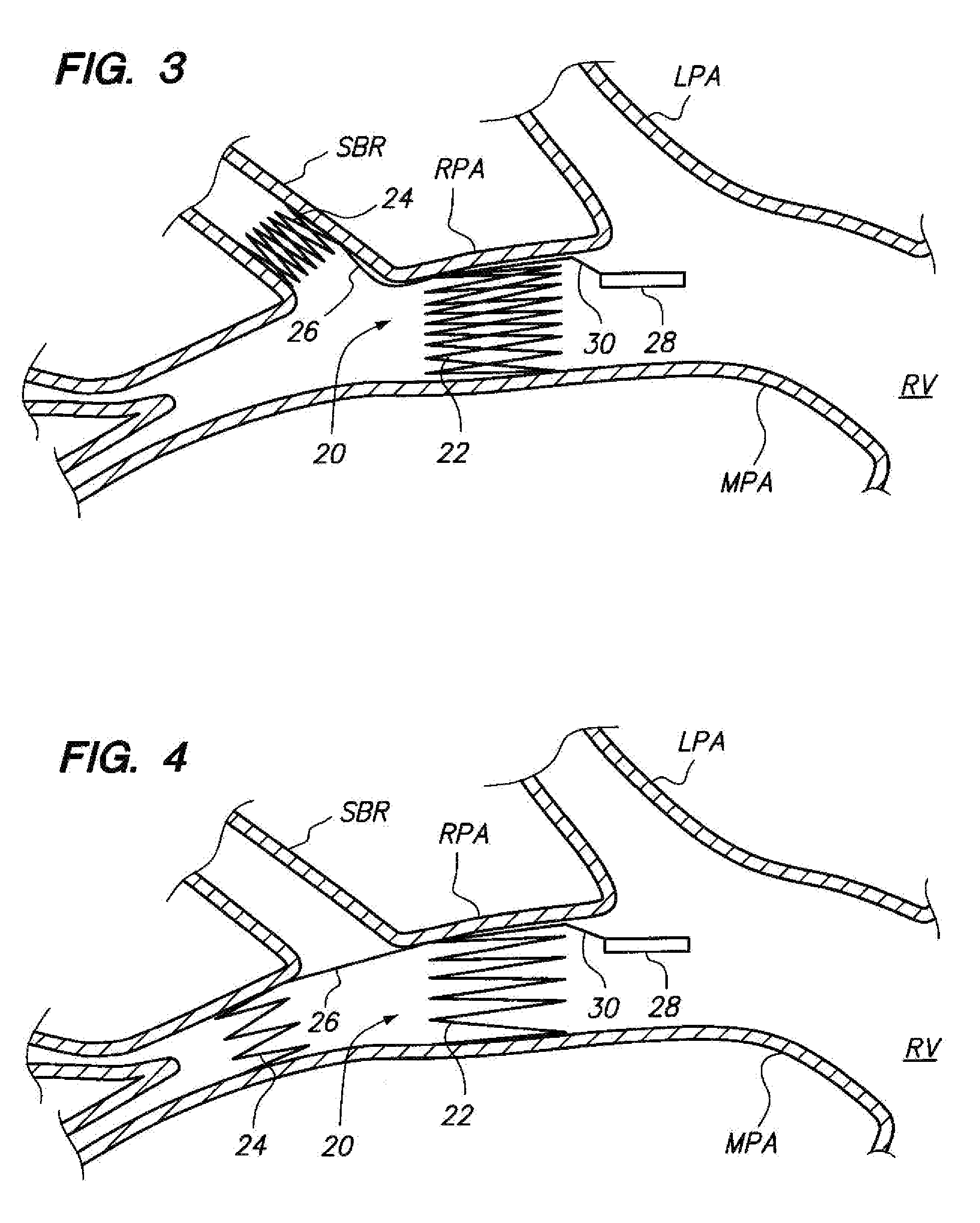
[0037] Referring to FIG. 2, an implantable sensing device 20 constructed in accordance with the present inventions will now be described. The sensing device 20 is shown implanted within an anatomical vessel network, and in particular, the pulmonary arterial network of a patient. As previously described, blood flows from the right ventricle RV of the heart H, out through the main pulmonary artery MPA, which branches into the right pulmonary artery RPA and a left pulmonary artery LPA. The blood from the right pulmonary artery RPA further flows into various pulmonary artery branches PBR of the right pulmonary artery RPA.

[0038] The sensing device 20 generally comprises a proximal fixation element 22, a distal fixation element 24, a connecting element 26 mechanically coupling the proximal fixation element 22 to the distal fixation element 24, and a sensing element 28 mechanically coupled to the proximal fixation element 22 via another connecting element 30 opposite the connecting elemen...


PUM


Abstract

Sensing devices and methods of implanting sensing devices within an anatomical vessel network of a patient are provided. In one method, a fixation element (e.g., a stent or coil) of the sensing device is expanded into firm contact with a wall of a main anatomical vessel (e.g., a right pulmonary artery), and a stabilization element of the sensing device is placed into contact with a wall of an anatomical vessel branch of the main anatomical vessel. In another method, a first fixation element of the sensing device is expanded into firm contact with the wall of the main anatomical vessel (e.g., a right pulmonary artery) at a longitudinal location proximal to the anatomical vessel branch, and a second fixation element of the sensing device is expanded into firm contact with the wall of the main anatomical vessel at a longitudinal location distal to the anatomical vessel branch.

Description

RELATED APPLICATIONS [0001] The application claims priority from U.S. Provisional Patent Application Ser. No. 60 / 737,131, filed Nov. 15, 2005, and is a continuation-in-part of U.S. patent application Ser. No. 11 / 034,502, filed Jan. 12, 2005, both of which are expressly incorporated herein by reference.FIELD OF THE INVENTION [0002] The invention relates to the field of implantable medical devices, and in particular, to devices and methods for positioning a sensor within the lumen of a blood vessel. BACKGROUND OF THE INVENTION [0003] The use of sensing devices in anatomical lumens is well known. For example, U.S. Pat. No. 4,485,813 describes a cardiac pacemaker sensor that can be permanently implanted in a specific location within a patient's body. U.S. Pat. Nos. 6,645,143, 6,053,873, and 6,442,413 and U.S. Patent Publication No. 2002 / 0188207 describe medical monitoring sensors designed to be permanently implanted in blood vessels and capable of sensing and transmitting via a telemetr...

Claims


Application Information

Patent Timeline
IPC IPC(8): A61B5/02A61B5/00
CPCA61B5/02152A61B5/6882A61F2/82
Inventor WOLINSKY, LONEBEN-YOSEPH, ALONPENNER, ABRAHAM
Owner REMON MEDICAL TECH
Why Eureka
Try Eureka