Targeting gli proteins in human cancer by small molecules
a technology of gli protein and human cancer, which is applied in the field of inhibiting tumor growth and tumor survival, can solve the problems of advanced disease, limited understanding of the molecular mechanisms and tumorigenic programs that are activated in response to hh-signaling and gli-activity in human cancer, and disadvantages in approaches, so as to inhibit the activity of a gli protein, inhibit the expression of a gene, and inhibit the signaling of a cell by a gli protein
- Summary
- Abstract
- Description
- Claims
- Application Information
AI Technical Summary
Benefits of technology
Problems solved by technology
Method used
Image

Examples
example 2
Mimicking the Peptide FDAII of the Gli3 Transcription Activation Domain
[0296] In order to mimic the FDAII motif of the transcriptional activation domain of GLI3 by small molecules the 3D structure of the FDAII was modeled. As suggested by Yoon et al. (1998, J Biol Chem 273:3496-3501), the FVAIL motif within the GLI1 polypeptide resulted in an α-helix structure. It was plausible to also model an a-helical structure for the FDAII motif of the GLI3 polypeptide. Thus, an α-helical structure of the FDAII motif of the GLI3 polypeptide was generated and rational chemical design was employed to design small molecule compounds mimicking the structure of this motif. Small molecule compounds comprising a pyrazoline structure were designed. FIG. 3 shows a RMS overlay between the small molecule compound FN1-5 (described herein) and the FDAII motif of the GLI3 polypeptide modeled as an α-helix.
example 3
Preparation of Small Molecule Compounds
a) Preparation of ethyl 1,3,5-triphenyl-4,5-dihydropyrazole-4-carboxylate (FN1-1)
[0297]
[0298] A mixture of ethyl cinnamate (7, 2.55 g), benzaldehyde phenylhydrazone (2, 4.21 g), chloramine-T trihydrate (6.45 g), and methanol (45 mL) were heated under refluxing for 22 hours. The reaction mixture was diluted with 50% ethyl acetate in n-hexanes (400 mL), filtered, and evaporated. The residue was purified by column chromatography (silica gel 200 g, eluent: 0 to 10% ethyl acetate gradient in hexanes) and evaporated to give FN1-1 (8, 1.25 g) as colorless crystal. 1H NMR (CDCl3, 400 MHz) δ 7.66-7.64 (m, 1H), 7.46-7.22 (m, 13H), 6.88 (t, J=7.2 Hz, 1H), 4.81 (d, J=4.0 Hz, 1H), 4.70 (d, J=4.0 Hz, 1H), 4.07 (quartet, J=7.2 Hz, 2H), 0.97 (t, J=7.2 Hz, 3H). MS: 371.9 (M+H), 325.5, 297.5.
b) Preparation of 1,3,5-Triphenyl-4,5-dihydropyrazole-4-carboxylic acid (FN1-2)
[0299] A mixture of FN1-1 (53 mg), methanol (0.5 mL), 1,4-dioxane (0.5 mL), and 10% (w / v)...
example 4
Most Cancer Cell Lines Express Gli3
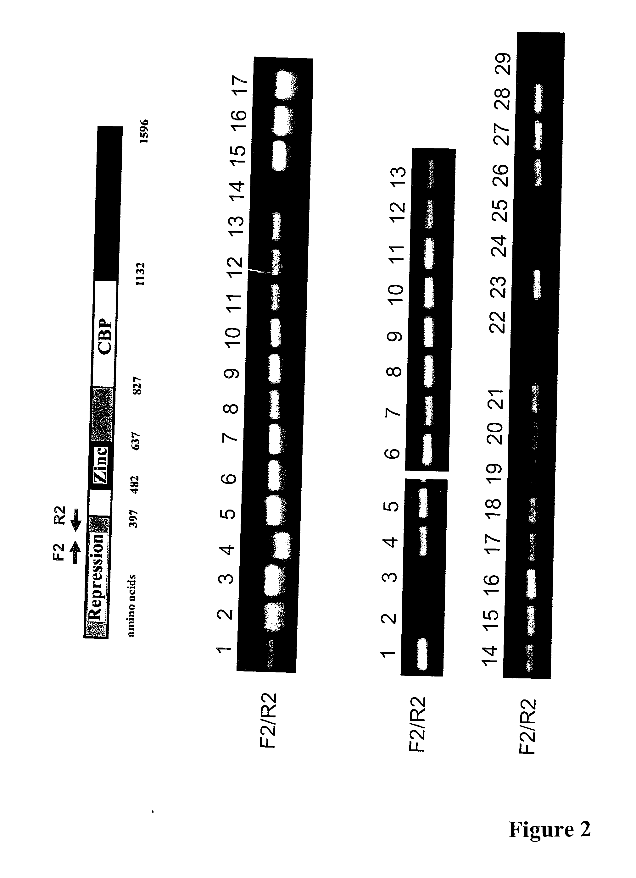
[0319] Several cancer cell lines and primary tissue samples were analyzed by semi-quantitative RT-PCR to investigate expression of Gli3. An exemplary analysis of an RT-PCR detecting Gli3 expression is shown in FIG. 2. The cell lines analyzed and found to express detectable levels of Gli3 MRNA included: normal liver (FIG. 2, middle panel, lane 1); NSCLC: H1703, H460 and A549 (FIG. 2, middle panel, lanes 2, 3, 4); breast cancer: HuL100, BT474 and MCF-7 (FIG. 2, middle panel, lanes 5, 6, 7); mesothelioma: Met5A, H290, REN, H513, H28, and 211H (FIG. 2, middle panel, lanes 8-13); colon cancer: SW480 (FIG. 2, middle panel, lane 15); a pair of primary NSCLC tissue samples (FIG. 2, middle panel, lane 16 and 17 are normal and cancer tissues, respectively). LARK1A did not show detectable expression of Gli3 mRNA (FIG. 2, middle panel, lane 14).
[0320] In another set of similar RT-PCR experiments Gli3 expression was analyzed and detected in colon cancer cell...
PUM
| Property | Measurement | Unit |
|---|---|---|
| subject weight | aaaaa | aaaaa |
| diameter | aaaaa | aaaaa |
| temperature | aaaaa | aaaaa |
Abstract
Description
Claims
Application Information