Computer-aided detection system utilizing temporal analysis as a precursor to spatial analysis

a technology of temporal analysis and computer-aided detection, applied in the field of computer-aided detection (cad) in medical imagery, can solve the problems of no current method of providing feedback to a radiologist, abnormal tissue may or may not appear different from normal tissue, etc., and achieve the effect of improving the interpretability of results and speeding up analysis

Active Publication Date: 2007-07-19
GENERAL ELECTRIC CO
View PDF8 Cites 60 Cited by
  • Summary
  • Abstract
  • Description
  • Claims
  • Application Information

AI Technical Summary

Benefits of technology

[0013] In yet another aspect of the invention, a preliminary temporal analysis on a four-dimensional series of medical images can be performed and used in two ways: one, to select voxels for which detailed spatial and temporal analysis needs to be performed, thereby greatly speeding up the analysis; and two, by limiting the display of the results of the spatial and temporal analysis of voxels to those that meet certain criteria, thus improving the interpretability of the results.

Problems solved by technology

Thus, prior to contrast agent administration, abnormal tissue may or may not appear different from normal tissue.
Since CAD systems typically base display decisions on computed numerical scores from a plurality of measurements, there is no current method for providing feedback to a radiologist regarding why a particular image region was marked or not.

Method used


Image

  • Computer-aided detection system utilizing temporal analysis as a precursor to spatial analysis
  • Computer-aided detection system utilizing temporal analysis as a precursor to spatial analysis
  • Computer-aided detection system utilizing temporal analysis as a precursor to spatial analysis

Examples


first embodiment

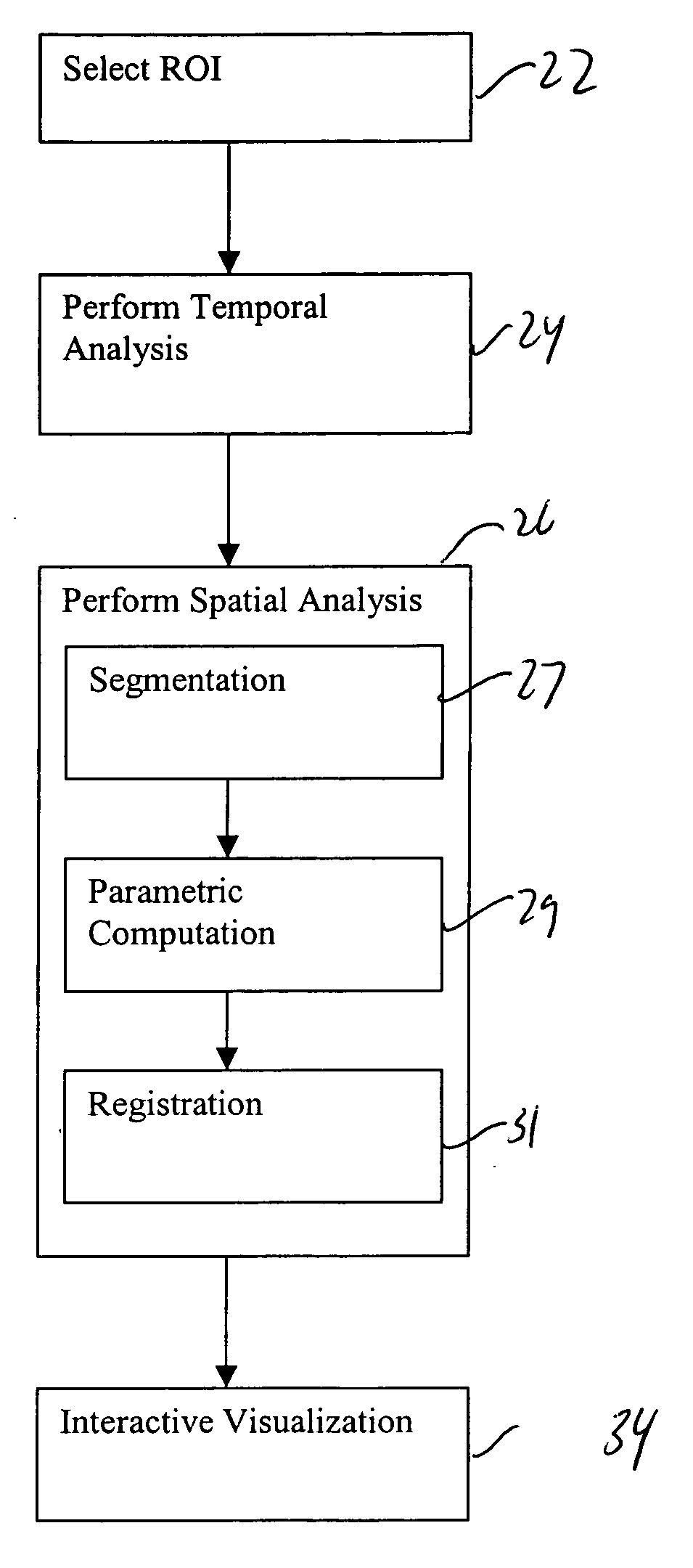
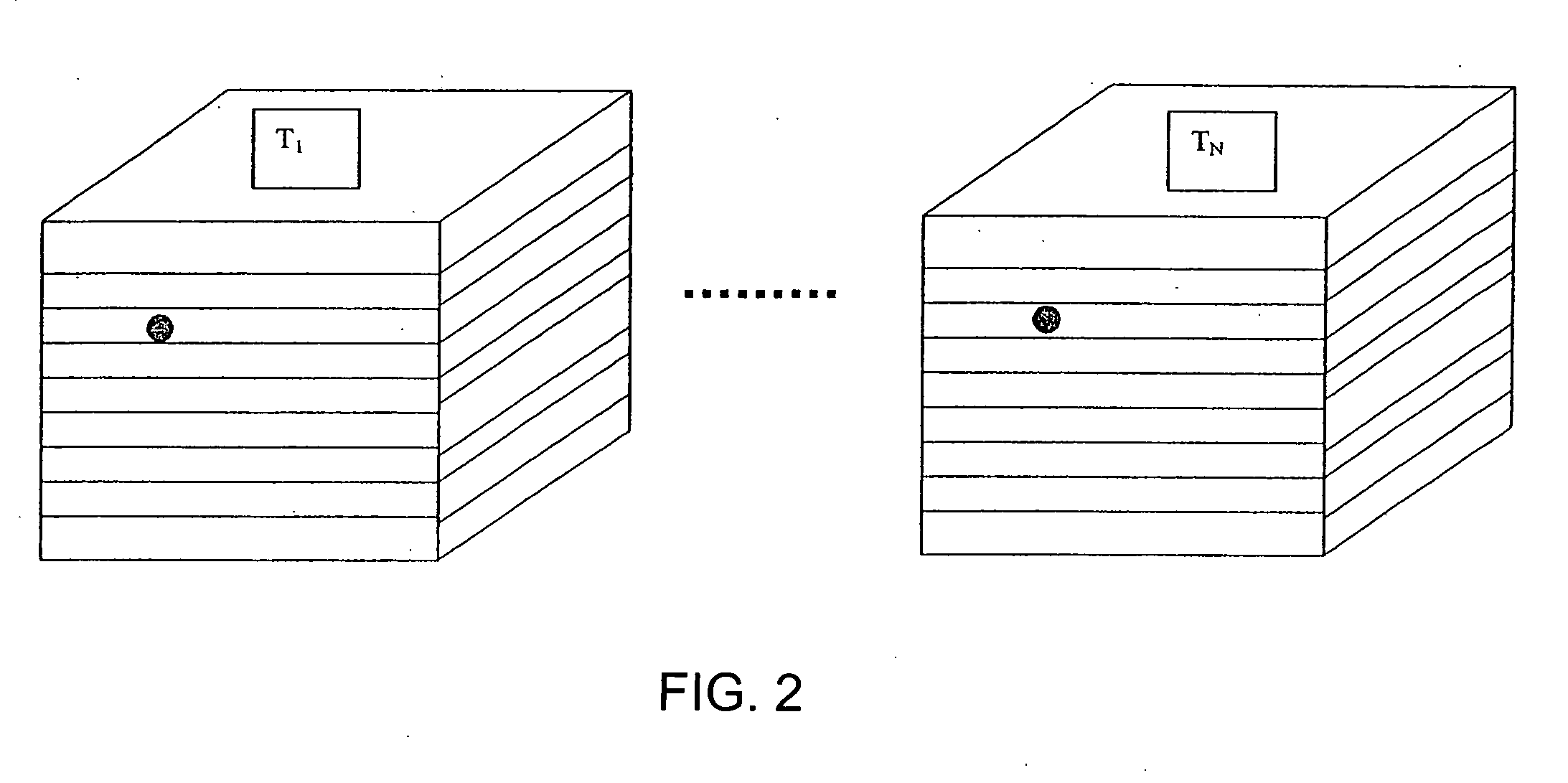
[0034] Referring now FIG. 5, an algorithm useful in a computer-aided detection system of the present invention is shown. After the data of FIG. 2 is acquired, it can be transmitted or otherwise transferred to the memory component 14 of the computer-aided detection system 10, or can be stored in a remote location accessible to the computer-aided detection system 10. After the four dimensional series of imaging data is provided to the computer system 10 in step 22, a region of interest is selected on the original acquired images. The specific region of interest (ROI) can be selected either spatially or temporally. To select the ROI spatially, a user can use the user input device 16, which can be, for example, a keyboard, mouse, joystick, or other device, to select boundaries on the original image either by drawing the boundary or by selecting the boundary using numerical coordinates. This selection can also be automated to separate, for example, body from background. To select the ROI...

second embodiment

[0040] Referring now to FIG. 6, in a second embodiment, the computer-aided detection system 10 processes the data through a series of iterations in which the scale space, e.g. the number of voxels evaluated in a given image, is varied between a minimum coarse level (as low as a 1×1) and a maximum fine level (such as 256×256). Iterations can be made at predetermined steps in scaled space. Here, an initial scale space and iteration level are selected prior to processing. After these parameters are selected, a specific region of interest (ROI) is selected either spatially or temporally, as described above with reference to step 22, and the image in the selected region of interest is then evaluated.

[0041] After a region of interest is selected, the temporal analysis 24 and spatial analysis 26 are performed as described above for the selected scale space. Then, in step 32, a determination is made as to whether the analysis has been performed at all the predetermined scale space steps. If...


PUM


Abstract

A computer-aided detection system for evaluating tissue based on a series of timed images acquired both before and after a contrast agent is administered performs a temporal analysis of the tissue and then a spatial analysis in which the tissue is categorized. After the temporal and spatial analysis is performed, the results can be displayed. The displayed results can include both tissue characterization results, underlying curves used to determine the characterization, and confidence data regarding how good the characterization is expected to be. The confidence data can be provided, for example, by using variations in color schemes, displaying numerical confidence levels, or providing graphical features such as piecewise linear models.

Description

TECHNICAL FIELD [0001] The present invention generally relates to computer-aided detection (CAD) in medical imagery and, in particular, relates to an improved method for providing computer-aided detection of lesions and tumors by analyzing dynamic properties in contrast enhanced medical images. BACKGROUND OF THE INVENTION [0002] Medical imaging technologies, such as Computed Tomography (CT), Positron Emission Tomography (PET), Single Photon Emission Computed Tomography (SPECT), and Magnetic Resonance Imaging (MRI) detect and record tissue responses in the presence of applied signals and / or injected or ingested substances, and generate visual representations indicative of such responses. This data can be analyzed to differentiate or monitor various types of tissue, and can be used to diagnose malignancies in the tissue. [0003] In typical medical imaging techniques a volume of tissue is scanned within a region of anatomical interest. The acquired scan data can then be transformed into...

Claims


Application Information

Patent Timeline
Patent Type & Authority Applications(United States)
IPC IPC(8): G06K9/00G06V10/25
CPCA61B5/055A61B19/52A61B2019/5289G06K9/3233G06K2209/05G06T7/0012A61B5/7264G06T7/0087G06T2207/10076G06T2207/20104G06T2207/30068G06T2207/30096G06T7/0081A61B90/36A61B2090/364G06T7/11G06T7/143G06V10/25G06V2201/03
Inventor GERING, DAVID THOMASAVINASH, GOPAL B.GUPTA, SANDEEP N.
Owner GENERAL ELECTRIC CO
Why Eureka
Try Eureka