
Systems, methods and apparatus for an endo-rectal receive-only probe

Active Publication Date: 2007-07-19
RGT UNIV OF CALIFORNIA +1
View PDF28 Cites 13 Cited by
  • Summary
  • Abstract
  • Description
  • Claims
  • Application Information

AI Technical Summary

Benefits of technology

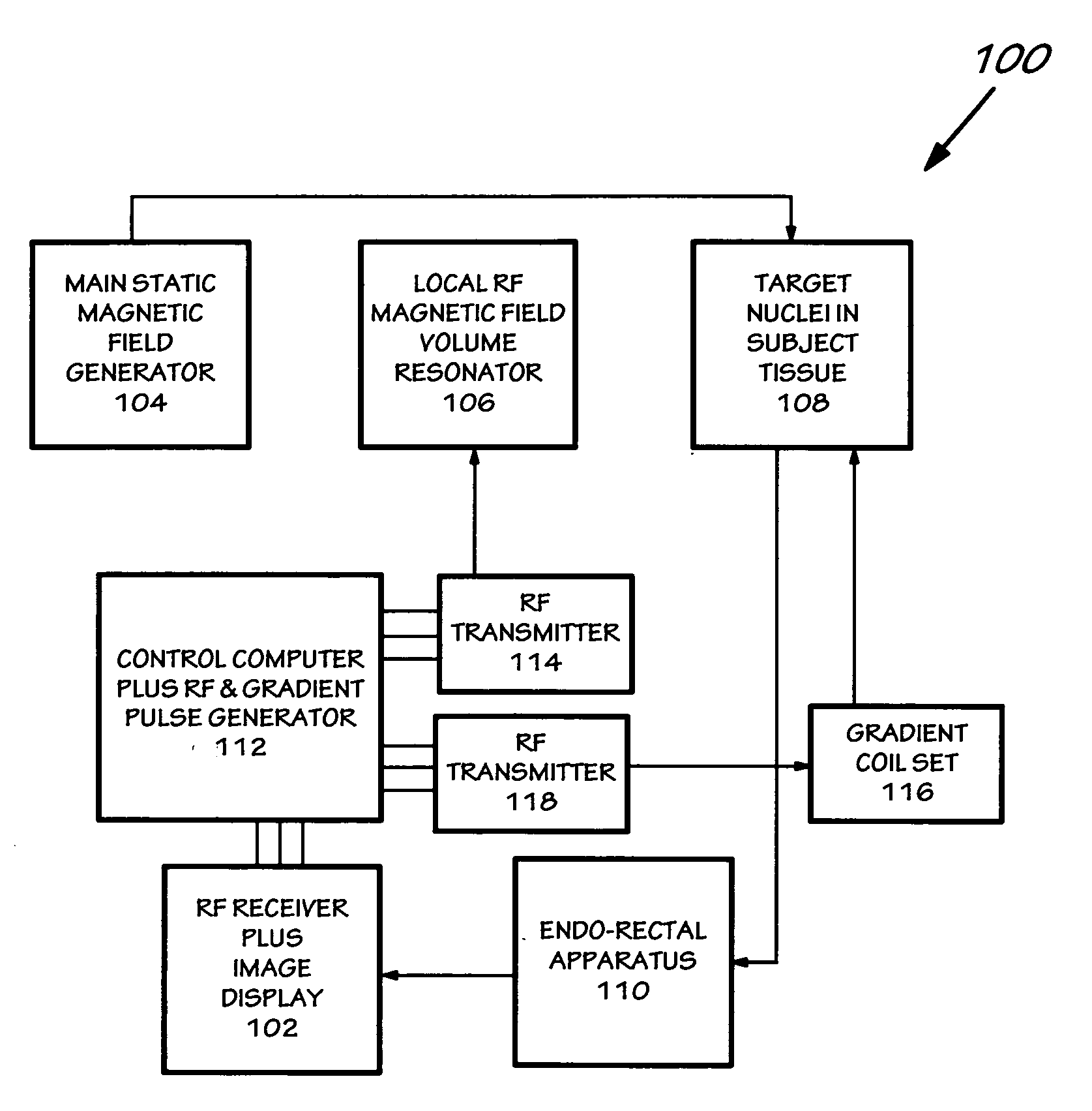
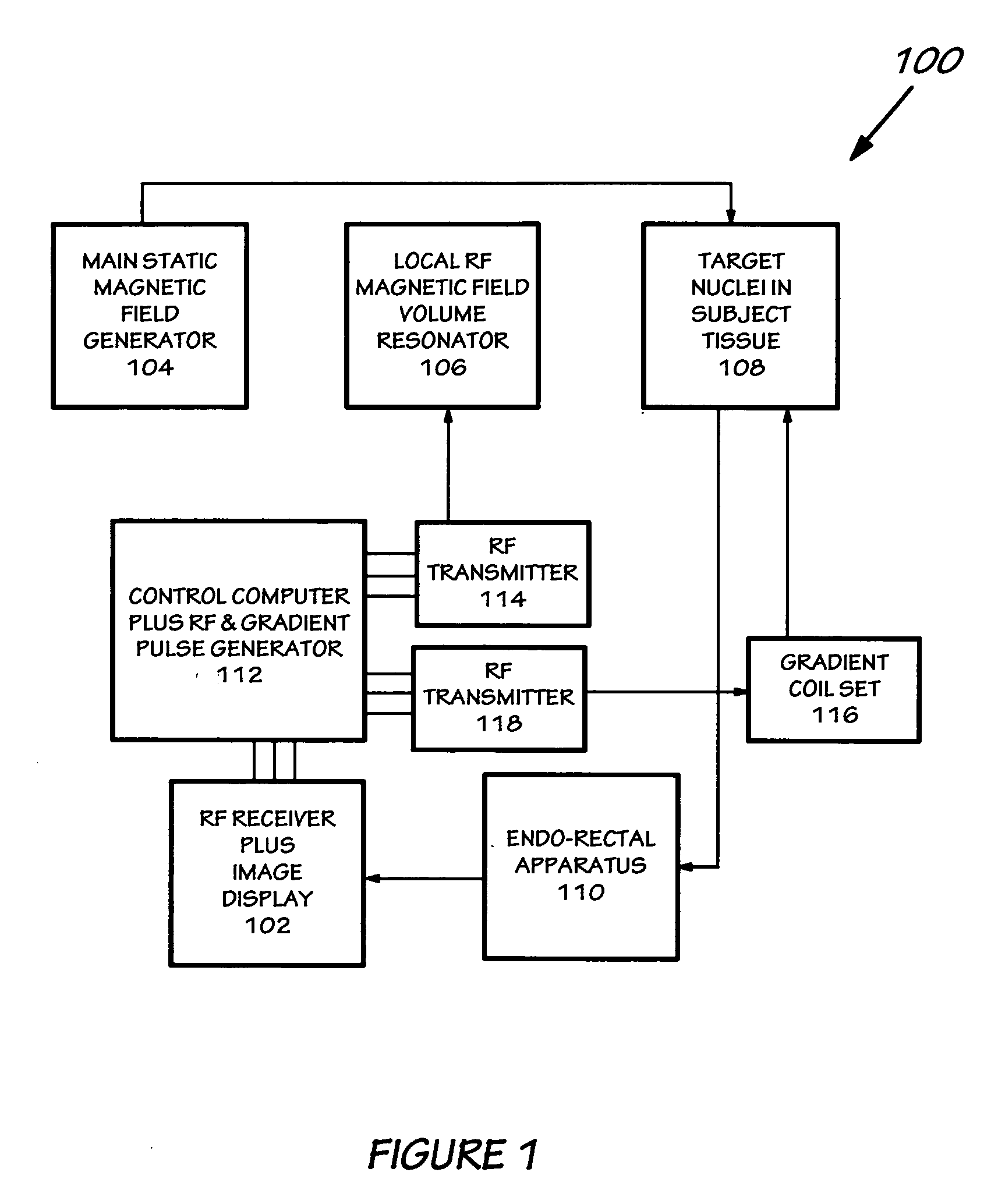
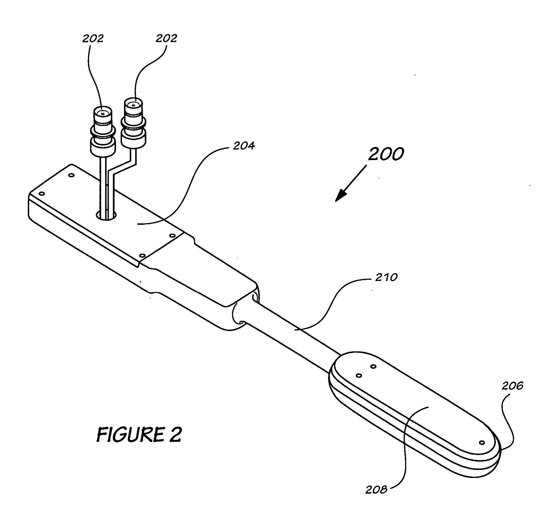
[0014] The endo-rectal receive-only probe includes the insertable pod and an attached handle pod to facilitate insertion and positioning and to carry cables with radio frequency signal data to an imaging device. The housing of the endo-rectal receive-only probe is made of a sturdy, benign material, such as solid delrin rendering the endo-rectal receive-only probe both re-usable and safe. The solid construction of the housing of the insertable pod portion of the endo-rectal receive-only probe is also an improvement over the currently used inflatable endo-rectal probe, which causes static magnetic susceptibility at the interface between antenna and tissue, perturbing the homogeneity of the main static magnetic field, and causing degradation of image quality and spectral quality.

Problems solved by technology

The distance from a large radio-frequency near-field antenna (or ‘coil’ as it is known in the art) to the tissue in question can be so great as to render said coil unable to provide the necessary strength of signal required for the level of detail and accuracy needed for proper evaluation of the target tissue.
But present endo-rectal technology can not reliably make use of two such signals—proton and carbon-13—during the same test.
One issue is the signal quality limitations resulting from the circuitry design of today's endo-rectal devices, which cannot adequately block the wrong RF signals while gathering the right ones.
The design of the body of the apparatus is also a source of error.
This creates a large discontinuity in static magnetic susceptibility at the interface between antenna body and tissue, resulting in degradation of image quality and spectral quality.
The solid construction of the housing of the insertable pod portion of the endo-rectal receive-only probe is also an improvement over the currently used inflatable endo-rectal probe, which causes static magnetic susceptibility at the interface between antenna and tissue, perturbing the homogeneity of the main static magnetic field, and causing degradation of image quality and spectral quality.

Method used


Image

  • Systems, methods and apparatus for an endo-rectal receive-only probe
  • Systems, methods and apparatus for an endo-rectal receive-only probe
  • Systems, methods and apparatus for an endo-rectal receive-only probe

Examples


Embodiment Construction

[0023] In the following detailed description, reference is made to the accompanying drawings that form a part hereof, and in which is shown by way of illustration specific embodiments which may be practiced. These embodiments are described in sufficient detail to enable those skilled in the art to practice the embodiments, and it is to be understood that other embodiments may be utilized and that logical, mechanical, electrical and other changes may be made without departing from the scope of the embodiments. The following detailed description is, therefore, not to be taken in a limiting sense.

[0024] The detailed description is divided into several sections. In the first section, a system level overview is described. In the second section, apparatus of embodiments and methods are described. Finally, in the third section, a conclusion of the detailed description is provided.

[0025] The object of a clinical MRS / MRI procedure is to enable an examination which combines a preliminary st...


PUM


Abstract

Systems, methods and apparatus are provided through which a compact pod insertable into the rectum for Magnetic Resonance Imaging / Magnetic Resonance Spectroscopy (MRI / MRS) examination of the prostate and containing two receive coils, each connected to transmit blocking and pre-tuned trap circuitry which can be superimposed within the pod in close proximity, can, without either circuit interfering with the other, efficiently gather, for imaging and tissue analysis, radio frequency signals emanating from magnetically disturbed nuclei in prostate tissue.

Description

FIELD OF THE INVENTION [0001] This invention relates generally to Magnetic Resonance Imaging and Magnetic Resonance Spectroscopy medical diagnostic devices, and more particularly to Magnetic Resonance Imaging and Magnetic Resonance Spectroscopy medical devices used to diagnose and assess pelvic pathologies, including prostate cancer. BACKGROUND OF THE INVENTION [0002] The value of Magnetic Resonance Imaging (MRI) devices for medical use was recognized almost immediately after they first appeared in the 1970s. Because they appear both to do no harm to the human body, and to create better images of the body's interior than the best X-ray technology, they have gained widespread use for diagnosis, pre-operative examination and even for assistance during surgical procedures. While MRI provides information on size and location of pathological abnormalities such as tumors, a variation of magnetic resonance technology called Magnetic Resonance Spectroscopy (MRS)—which identifies various bio...

Claims


Application Information

Patent Timeline
IPC IPC(8): A61B5/05
CPCA61B1/31A61B5/055A61B5/4381G01R33/3657A61B2562/02G01R33/287G01R33/3635A61B5/68
Inventor TROPP, JAMES S.CALDERON, PAUL D.VIGNERON, DANIEL BLACKBURNKARPODINIS, KONSTANTINOS GEORGECARVAJAL, LUCAS GUILLERMO
Owner RGT UNIV OF CALIFORNIA
Learn More