Method and apparatus for ECG-synchronized optically-based image acquisition and transformation

a technology optical coherence tomography, which is applied in the field of optical coherence tomography (optical coherence tomography) methods and apparatuses, can solve the problems of coronary artery narrowing, stenoses, lipid-filled plaque deposits, and inability to provide information regarding the morphology (appearance and extent) of the lesion

Inactive Publication Date: 2007-07-19
SIEMENS HEALTHCARE GMBH
View PDF6 Cites 55 Cited by
  • Summary
  • Abstract
  • Description
  • Claims
  • Application Information

AI Technical Summary

Benefits of technology

[0016] It is an object of the present invention to provide an optically-based slice imaging apparatus and method (such as an OCT or OFDI method and apparatus) that allow a meaningful image of a pathological vessel segment to be obtained without a positioning system and without additional radiation exposure to the patient, wherein the pathological vessel segment is seen in an intuitively recognizable manner, with a resolution that is sufficient for a therapy selection.

Problems solved by technology

Arteriosclerotic pathologies of the coronary vessels are the primary cause of death in industrialized nations.
Narrowing of the coronary vessels (stenoses) or lipid-filled plaque deposits are the most frequent cause of heart attacks.
Angiography equipment available today provided information only about the vessel itself, but does not supply information regarding the morphology (appearance and extent) of the lesion.
The lack of information regarding the vessel structure in the region of the lesion, and the two-dimensional nature of angiography exposures are limiting factors in assessing a therapy using this technique.
Errors primarily occur in the length measurement, due to the lesion not lying precisely in the plane of the angiography image, and thus appearing foreshortened in the angiography image.
Despite the aforementioned advantages of IVUS, the evaluation of IVUS pullback sequences is subject to limitations.
Automatic quantifications of IVUS images would require an automatic segmentation of the IVUS images, which currently can be achieved only to a limited extent due to the limited resolution, the fluctuating intensities and contrasts, and by artifacts in the IVUS images.
Such a biplanar x-ray system, however, is available only in a limited number of catheter laboratories, and also requires an additional radiation exposure for the patient.

Method used


Image

  • Method and apparatus for ECG-synchronized optically-based image acquisition and transformation
  • Method and apparatus for ECG-synchronized optically-based image acquisition and transformation
  • Method and apparatus for ECG-synchronized optically-based image acquisition and transformation

Examples


Embodiment Construction

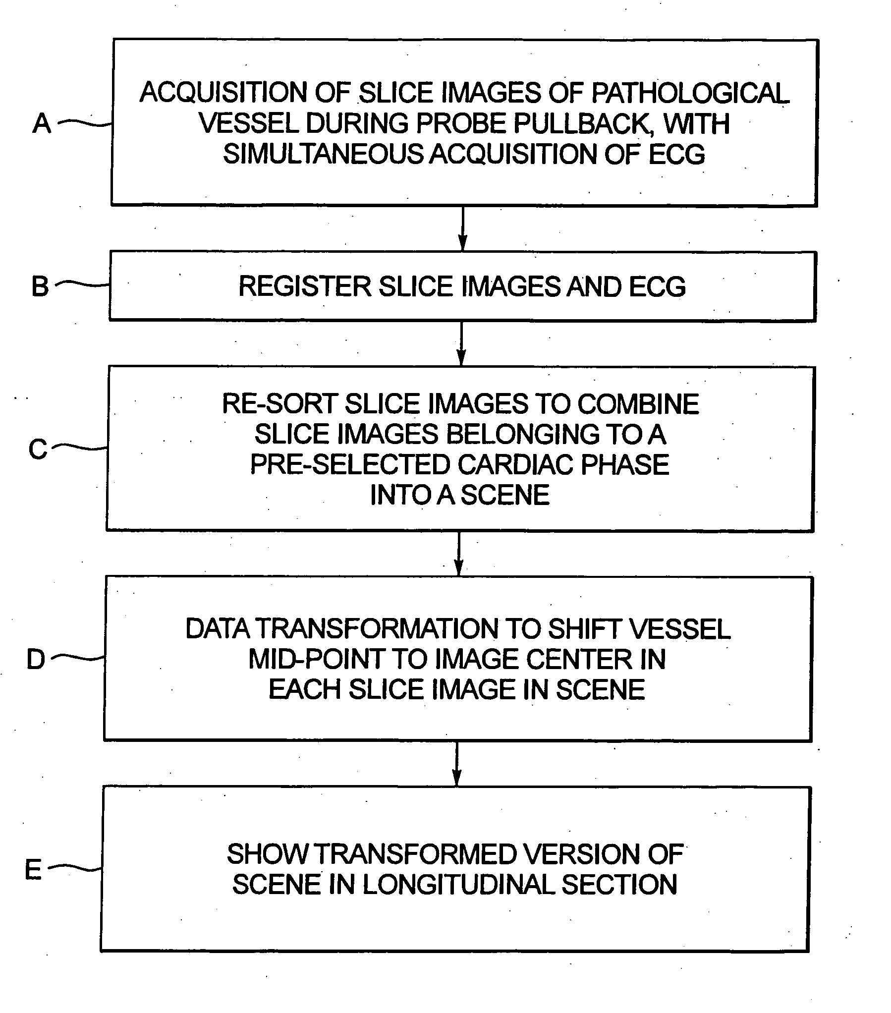
[0026] The basic steps of an optically-based slice imaging method, for acquisition of the slice images as well as combining the slice images into a diagnostic image, according to the invention are shown in FIG. 1.

[0027] In step A, a series of slice images of a pathology-containing vessel are acquired during pullback of an optical probe, such as an OCT probe or an OFDI probe. The optical probe can be positioned in the vessel at a location downstream from the suspected location of a lesion (pathology) so that during pullback a succession (series) of slice images are acquired not only from the lesion-containing portion of the vessel, but also from portions downstream and upstream therefrom. As also indicated in step A, an ECG signal is acquired from the subject during pullback of the optical probe. Preferably a centering catheter is used sot that the slice images will all be oriented perpendicularly to the center axis of the vessel. Alternatively, known external position and orientati...


PUM


Abstract

In a method and apparatus for optically-based acquisition of slice images from a vessel of a subject, and for combining the slice images to provide an intuitively recognizable visualization of a pathology in the vessel, slice images of the vessel are acquired during pullback of an optical probe of the optically-based slice imaging system, while simultaneously acquiring an ECG signal from the subject. The slice images and the ECG signal are registered, and slice images acquired at a selected cardiac phase are combined into a scene. The slice images in the scene are subjected to a first data transformation to shift the vessel midpoint in each slice image to the image center of each slice image. After the first data transformation, the slice images in the scene are subjected to a second data transformation to produce the visualization. The second transformation, for example, can be a curved planar reformation to allow the vessel to be shown in longitudinal section.

Description

BACKGROUND OF THE INVENTION [0001] 1. Field of the Invention [0002] The present invention is directed to an OCT (optical coherence tomography) method and apparatus, and in particular to such a method and apparatus for obtaining a sequence of OCT images and for subsequently combining those images into a diagnostic image. [0003] 2. Description of the Prior Art [0004] Optical coherence tomography (OCT) is an invasive imaging modality that is currently used for visual assessment of lesions or pathologies in blood vessels, such as stenoses and problematic plaque deposits. To improve such assessment, it is desirable to produce a diagnostic image wherein the portion of the vessel in question is displayed so that the pathology can be intuitively recognized without additional technical outlay, and without additional radiation exposure to the patient. [0005] Arteriosclerotic pathologies of the coronary vessels are the primary cause of death in industrialized nations. Narrowing of the coronary...

Claims


Application Information

Patent Timeline
Patent Type & Authority Applications(United States)
IPC IPC(8): A61B6/00
CPCA61B5/0066A61B5/7289A61B5/6852A61B5/0402A61B5/318
Inventor REDEL, THOMASCAMUS, ESTELLEMEISSNER, OLIVERKLINGENBECK-REGN, KLAUS
Owner SIEMENS HEALTHCARE GMBH
Why Eureka
Try Eureka