Apparatus and methods for endometrial biopsies

a biopsies and endometrial technology, applied in the field of endometrial biopsies, can solve the problems of high patient discomfort, abnormal uterine bleeding in the procedure, and cramps in the uterine cavity, so as to reduce patient discomfort and increase the operating flexibility of the clinician

Inactive Publication Date: 2007-07-26
FEMSUITE
View PDF32 Cites 17 Cited by
  • Summary
  • Abstract
  • Description
  • Claims
  • Application Information

AI Technical Summary

Benefits of technology

[0013]In view of the foregoing, it is an object of the present invention to provide apparatus and methods for endometrial biopsies that overcome the drawbacks of the prior art and that increase the operating flexibility of the clinician while reducing patient discomfort.
[0014]It is also an object of the present invention to provide apparatus and methods for endometrial biopsies that enable a secure retention of the plastic acorn in the cervical os during application of the anesthetic.
[0015]It is another object of the present invention to provide apparatus and methods for endometrial biopsies that enable a clinician to retain the cervical os in a dilated position without the use of supplemental devices.
[0016]It is a further object of the present invention to provide apparatus and methods for endometrial biopsies that reduce the number of instruments that must be separately inserted and removed from the uterine cavity.
[0017]These and other objects of the present invention are accomplished by providing apparatus and methods for endometrial biopsies having an applicator disposed on the distal end of an outer tube and an injection device connected to the proximal end of the same tube. The applicator is configured to be securely positioned in the cervical os and to maintain the os in a dilated position during the injection of an anesthetic medication and the later insertion of a tissue-removing catheter. The apparatus and methods of the present invention is configured to minimize discomfort while facilitating the operation of the clinician.
[0018]In the first embodiment, the outer tube has a length of 20-30 cm and a diameter of 3-5 mm, and is preferably manufactured from plastic material. A frustoconical applicator, preferably of a silicone material, surrounds the distal end of the outer tube and has one or more grooves disposed on the conical wall in a helical pattern, giving the applicator a screw-like pattern that provides for a tight fit with the cervical os and prevents the undesired release of the applicator. The cervical os is then retained in the desired dilated condition by a secure anchoring of the applicator grooves into the os walls, facilitating the activity of the clinician.

Problems solved by technology

Most women undergoing this procedure suffer from abnormal uterine bleedings or from amenorrhea, a prolonged absence of the menstrual period.
Unfortunately, endometrial biopsies can be highly uncomfortable to the patient, in spite of the use of anesthetics.
The placement of the catheter through the cervix and inside the uterine cavity can produce cramps, in spite of the use of painkillers prior to the procedure and of the spraying with a numbing medication during the procedure.
The activity of the clinician can be complicated by the effort to maintain a proper positioning of the catheter inside the uterus during the procedure, in spite of constrictive actions of the cervical os and of movements of the patient's body.
One disadvantage of endometrial biopsies in the prior art is that some leakage of anesthetic may occur due to an imperfect contact of the acorn with the cervical os, reducing the duration and extent of the relief from pain that is available during the extraction of tissue samples.
Another disadvantage of endometrial biopsies in the prior art is that multiple devices must be inserted serially into the uterus to obtain the desired sample, increasing the complexity of the procedure and the discomfort to the patient.
A further disadvantage of endometrial biopsies in the prior art is the level of difficulty in maintaining the cervical os in a dilated position during the procedure.

Method used


Image

  • Apparatus and methods for endometrial biopsies
  • Apparatus and methods for endometrial biopsies
  • Apparatus and methods for endometrial biopsies

Examples


Embodiment Construction

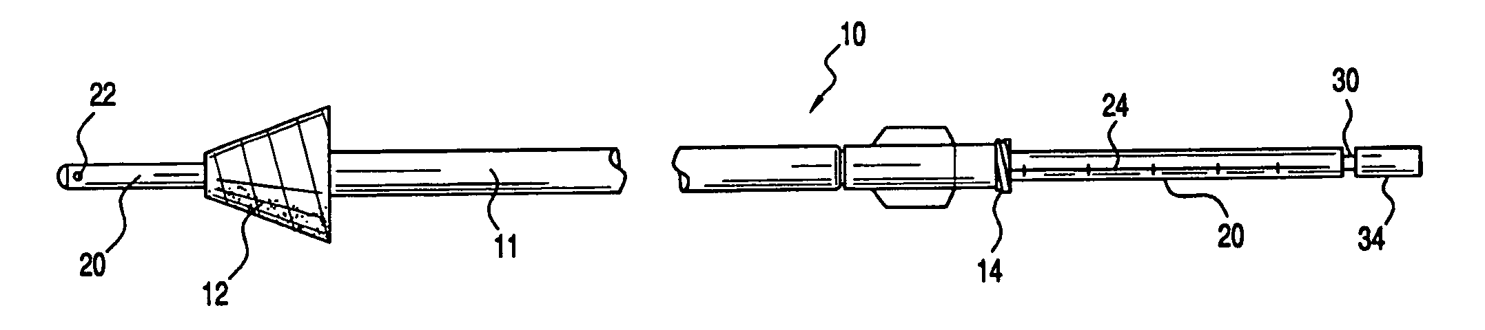
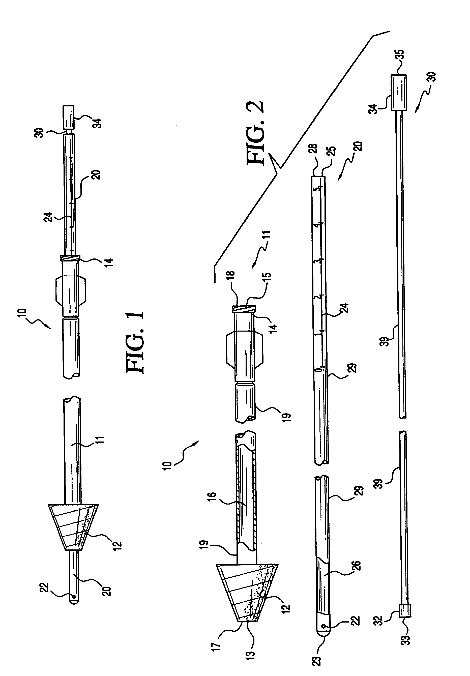
[0028]The present invention is directed to apparatus and methods for endometrial biopsies that increase the flexibility of operation of a clinician while reducing patient discomfort. In a preferred embodiment, an apparatus is provided that includes an applicator, through which a catheter is inserted and an anesthetic is injected. Grooves on the outer surface of the applicator enable the clinician to maintain the applicator in the cervical os in a secure position and also to maintain the cervical os in a dilated position during the entire procedure.

[0029]Referring to FIGS. 1 and 2, an exemplary embodiment of an apparatus for endometrial biopsies constructed in accordance with the principles of the present invention is described. Apparatus 10 includes four basic members, outer tube 11, applicator 12, inner tube 20, and injection device30. Applicator 12 is positioned at distal end 13 of outer tube 11, and Luer-Lok fitting 14 disposed at proximal end 15 of outer tube 11. Lumen 16 extend...


PUM


Abstract

Apparatus and methods for endometrial biopsies, wherein an applicator is disposed on the distal end of an outer tube and an injection device is connected to the proximal end of the same tube. The applicator comprises one or more ridges on its outer surface that anchor the applicator in the cervical os, and that cause the applicator to remain securely positioned and maintain the os in a dilated position during the entire procedure. The ridges may be shaped like protrusions, scalloped edges or grooves, and may be disposed in a helical or circular pattern. A method for performing endometrial biopsies is also provided. The apparatus and methods according to the present invention are configured to minimize discomfort to the patient while facilitating the operation of the clinician.

Description

RELATED APPLICATIONS[0001]This application is a continuation-in-part of U.S. patent application Ser. No. 11 / 245,498, filed on Oct. 7, 2005.FIELD OF THE INVENTION[0002]The present invention relates to apparatus and methods for performing endometrial biopsies. More particularly, the present invention relates to apparatus and methods for applying an anesthetic and removing uterine cells that provide the clinician with improved flexibility while at the same time reducing discomfort to the patient.BACKGROUND OF THE INVENTION[0003]Endometrial biopsies are procedures employed for evaluating uterine tissue for the presence of cancerous or precancerous cells. Most women undergoing this procedure suffer from abnormal uterine bleedings or from amenorrhea, a prolonged absence of the menstrual period. Other women undergoing this procedure include women with a high risk of endometrial cancer such as hereditary colorectal cancer, or with infertility problems.[0004]Endometrial biopsies essentially ...

Claims


Application Information

Patent Timeline
Patent Type & Authority Applications(United States)
IPC IPC(8): A61B10/00
CPCA61B10/0283A61B10/0291A61B17/3415A61B17/42A61B2010/0074A61B2017/00862A61M2210/1433A61B2017/349A61B2017/4216A61B2017/4225A61B2019/462A61M1/008A61B2017/3488A61B2090/062A61M1/84
Inventor FEUER, GERALDSANDERS, GERALD J.
Owner FEMSUITE
Why Eureka
Try Eureka