

Markers for detection of gastric cancer
a gastric cancer and marker technology, applied in the field of cancer detection, can solve the problems of insufficient sensitivity and specificity of available markers, and achieve the effect of reducing the frequency of false positives and false negative test results
- Summary
- Abstract
- Description
- Claims
- Application Information
AI Technical Summary
Benefits of technology
Problems solved by technology
Method used
Image

Examples
example 1
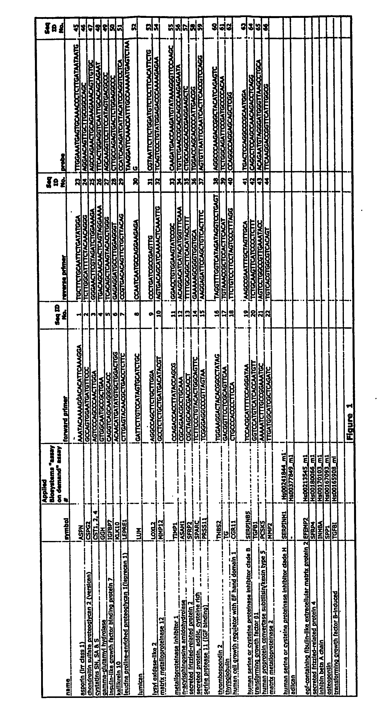
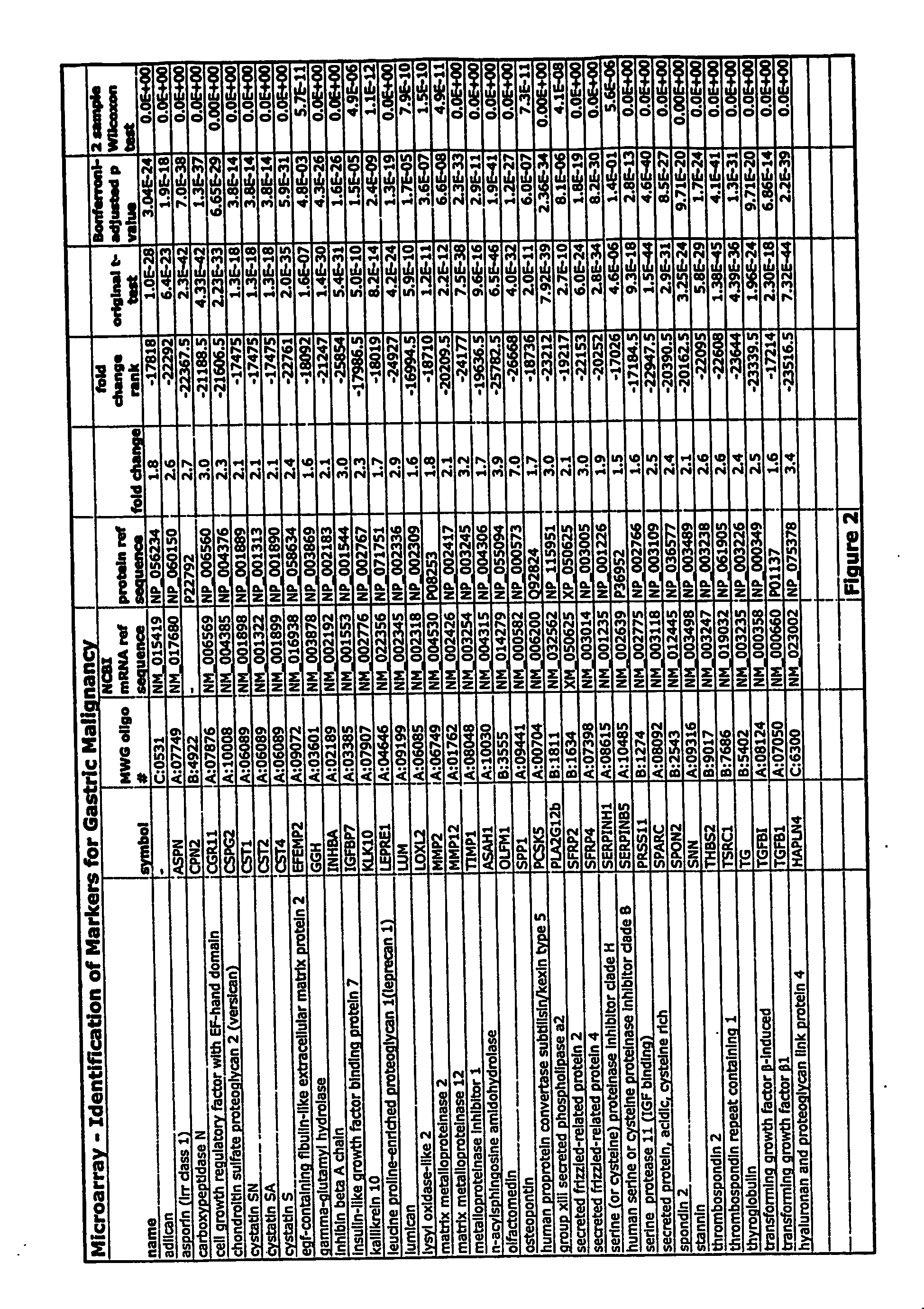
Identification of Markers for Gastric Malignancy
[0253]FIG. 2 depicts a table that shows results of studies using 38 markers for gastric malignancy selected using the above criteria. The FIG. 2 includes the symbol for the gene (“symbol”), the MWG oligo number, the NCBI mRNA reference sequence number, the protein reference sequence number, the fold change between tumor and non-tumor gene expression, the fold change rank relative to other genes in the microarray analysis, the results of an original, unadjusted Student's t-test, the results of the Bonferroni-adjusted p value and the results of the 2-sample Wilcoxon test.
[0254] The median fold change (tumor: non malignant tissue) for these 34 genes ranged from 1.6 to 7 and the median change in fold change rank ranged from −16,995 to −25,783. The maximum possible change in fold change rank was −29,718. For each of the markers shown, the statistical significance of their specificity as cancer markers was found to be extremely high. The B...
example 2
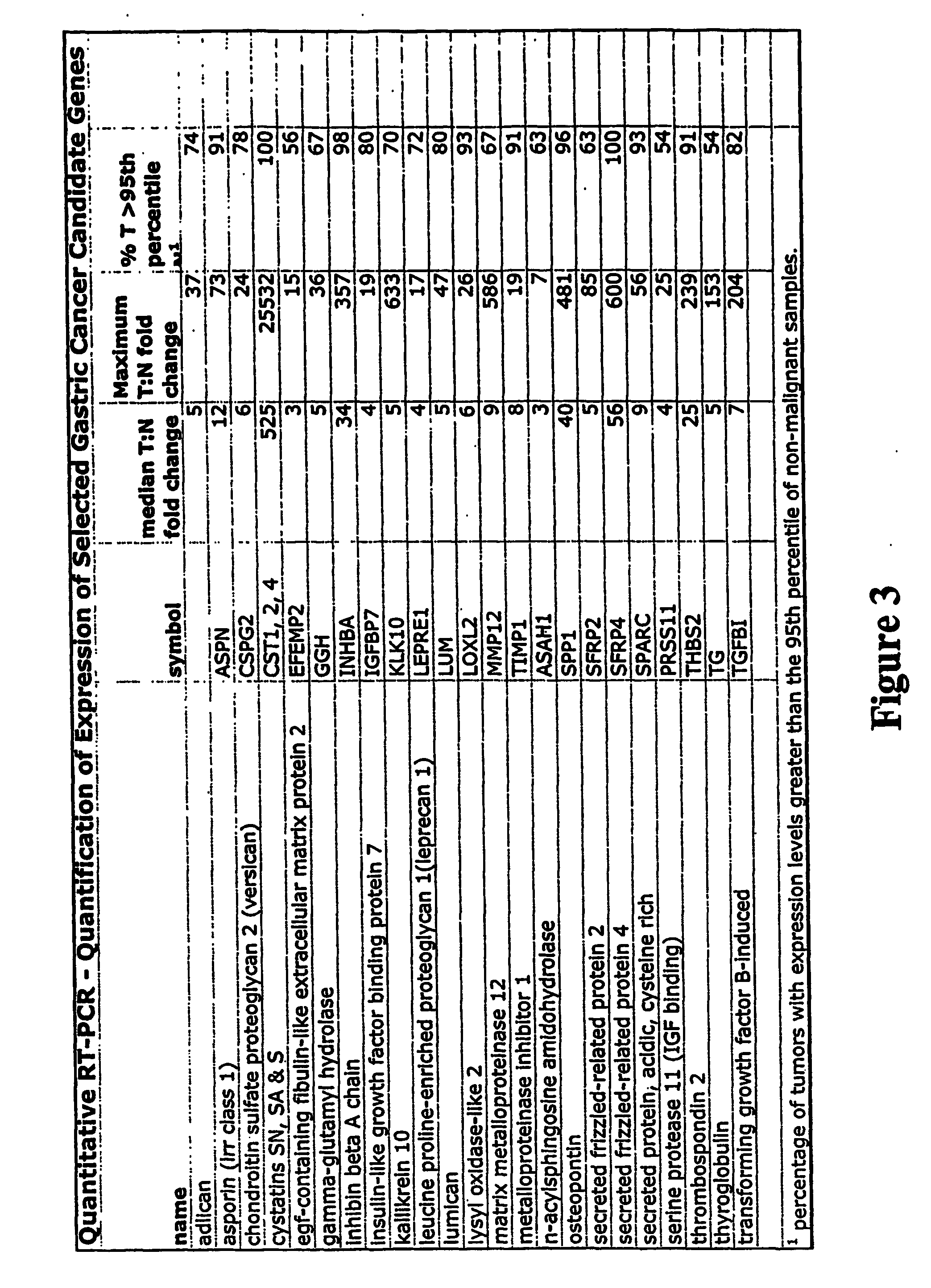
qPCR Analysis
[0258] More sensitive and accurate quantitation of gene expression was obtained for a subset of the genes shown in FIG. 3 using qPCR. RNA from 46 tumor and 49 non-malignant samples was analyzed for 23 genes identified by the microarray analysis (FIG. 2) and results are shown in FIG. 3. FIG. 3 includes the gene symbol, median fold change between cancer and normal tissue, and the % of tumor samples with expression levels greater than the 95th percentile of expression levels in non-malignant samples. 12 tumor samples and 9 normal samples were excluded from the analysis because of high (>75%) normal cell contamination, a high degree of necrosis (>40%), or poor hybridization signal on the microarrays. The median fold change (tumor tissues compared to the median non-malignant tissue expression) for these 23 genes ranged from 3 to 525 fold FIG. 3).
[0259] The level of expression of genes ASPN, CST1,2,4, LOXL2, TIMP1, SPP1, SFRP4, INHBA, THBS2 and SPARC was greater in tumors t...
example 3
Validation of Array Data Using qPCR
[0260] Array data was validated using quantitative, real-time PCR (qPCR) on the tumor and non-malignant samples with probes for 24 genes. Of all 24 genes studied, 20 showed a strong correlation between the two techniques. Four of these analyses are show in FIGS. 4a-4d, which depict graphs of the relative expression for the 4 selected cancer markers detected using array and qPCR methods. For each graph in FIG. 4, the horizontal axis represents the array log2 fold change in gene expression, and the vertical axis represents the qPCR log2 fold change in gene expression. We found that there was a strong correlation between the two methods, as indicated by the co-variant relationship between the methods. The strong correlation indicates that both microarray fold change analysis and qPCR are suitable methods for detecting changes in the expression of gastric cancer marker genes and therefore can be used as an accurate, sensitive screening method. It can ...
PUM
| Property | Measurement | Unit |
|---|---|---|
| molecular weights | aaaaa | aaaaa |
| molecular weights | aaaaa | aaaaa |
| molecular weights | aaaaa | aaaaa |
Abstract
Description
Claims
Application Information