Methods for modulating angiogenesis

a technology of angiogenesis and angiogenesis, applied in the field of modulating angiogenesis, can solve the problems of further harming patients, and achieve the effect of stimulating or inhibiting angiogenesis

Inactive Publication Date: 2007-08-16
TRUSTEES OF DARTMOUTH COLLEGE THE
View PDF3 Cites 3 Cited by
  • Summary
  • Abstract
  • Description
  • Claims
  • Application Information

AI Technical Summary

Benefits of technology

[0013] Angiogenesis is the process of blood vessel growth towards a tissue in need of oxygen or an injured tissue. Angiogenesis can be either harmful or beneficial, for example, in cases such as tumor growth, angiogenesis towards the tumor can supply the tumor with nutrients and support its growth, thus further harming the patient. However, in occl

Problems solved by technology

Angiogenesis can be either harmful or beneficial, for example, in cases such as tumor growth, angiogenesis to

Method used


Image

  • Methods for modulating angiogenesis
  • Methods for modulating angiogenesis

Examples


example 1

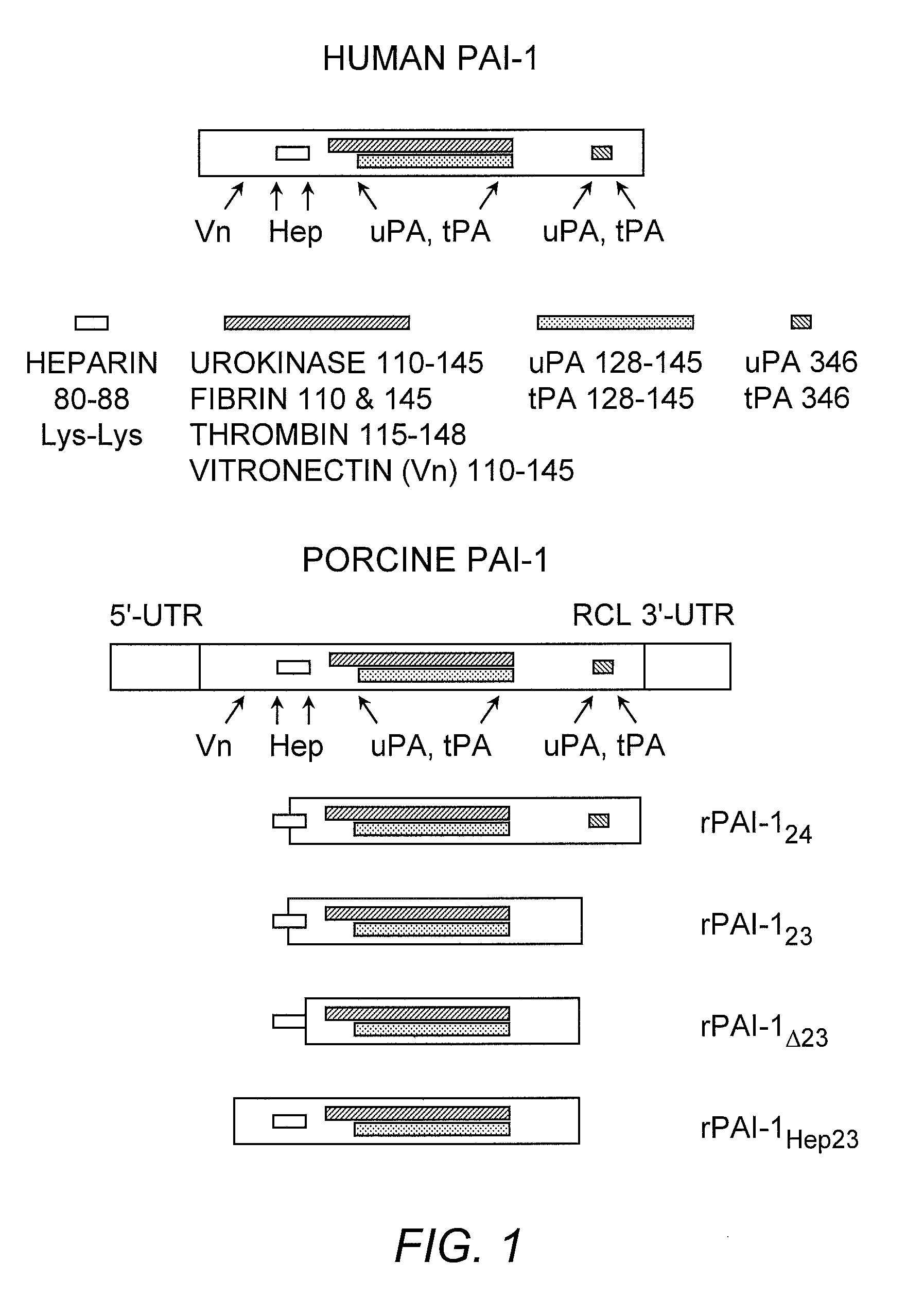
PAI-1 Gene Deletions

[0114] The DNA encoding the truncated PAI-1 proteins was obtained by deleting the porcine PAI-1 gene (poPAI-1) (Bijnens, et al. (1997) Thromb. Haemost. 77:350-356; Accession number Y11347; SEQ ID NO:11) . The selection of gene fragments was based on the poPAI-1 sequences that correspond to the human PAI-1 gene (huPAI-1) (Bosma, et al. (1988) J. Biol. Chem. 263:9129-9141; Accession number J03764; SEQ ID NO:12) sequence reported to code for functional domains in human PAI-1 (Reilly and Hutzelmann (1992) supra) . Each DNA fragment was isolated from porcine aortic endothelial cells by reverse transcribing RNA into cDNA. The cDNA was made double-stranded in a polymerase chain reaction (PCR) containing porcine PAI-1-specific primers. Primers for amplification of the cDNA encoding rPAI-123, were 5′-primer, 5′-GGAATTCAAGGAGCTATGG-3′ (SEQ ID NO:13) and 3′-primer, 5′-GCTCTAGATTTCCACTGGTGATG-3′ (SEQ ID NO:14). The resulting product corresponds to nucleotides 444-999 of poP...

example 2

Anti-Angiogenic Effects of rPAI-1 Proteins

Characterization of the rPAI-1 Protein Interactions with Heparin, uPA, and Plasminogen.

[0115] The functionality of the rPAI-1 proteins was first tested by incubating each truncated protein with uPA and plasminogen to assess the proteolytic activity of the products of the reaction, as known in the art, for example Mulligan-Kehoe, et al. ((2001) supra) . The functionality of each rPAI-1 isoform was then tested in a reaction with heparin bound to Sepharose beads. First, the rPAI-123, rPAI-1Δ23, rPAI-1Hep23, and rPAI-124 proteins (20 μg) were each incubated with 50 μl of heparin-bound Sepharose beads (Amersham Pharmacia Biotech, Piscataway, N.J.) for 2 hours at 37° C. The unbound protein was separated from the heparin-Sepharose-bound protein complex by microcentrifugation at 4° C. for 15 minutes. The proteins bound to the Sepharose beads were washed in TBS / Tween-20 followed by TBS washes. Bound samples were reacted with uPA (0.5 IU / 1 μg or rP...

example 3

VEGF-A Interactions

Biochemical Interactions of a VEGF-Heparin Complex with rPAI Proteins, uPA, and Plasmin.

[0126] VEGF was isolated from bovine aortic endothelial cells by first incubating the cells overnight in serum-free DMEM. The medium was changed before adding 100 μg / ml heparin (Sigma, St. Louis, Mo.) for 4 hours at 37° C. The serum-free DMEM containing the VEGF-heparin complex was precipitated in 80% ethanol. The serum-free medium from which the VEGF-heparin complex was isolated, was probed for VEGF-A121 (Santa Cruz Biotechnology, Inc., Santa Cruz, Calif.) to insure that his non-heparin-binding VEGF isoform did not precipitate with the VEGF-heparin complex. VEGF-A121 was not detected. The heparin-VEGF complex was incubated with rPAI-123, rPAI-1Δ23, rPAI-1Hep23, or rPAI-124 protein (3, 15, and 30 nM) for 2 hours at 37° C. Either uPA (0.25 IU) or plasmin (made from 1.0 IU plasminogen and 0.25 IU uPA) was added to the VEGF-heparin-rPAI-1 reaction. After an additional 2 hours i...


PUM

PropertyMeasurementUnit
Cell proliferation rateaaaaaaaaaa

Abstract

Recombinant plasminogen activator inhibitor-1 (PAI-1) isoforms which lack the reactive center loop and contain the complete heparin-binding domain or lack at least a portion of the heparin-binding domain are described. The rPAI-1 isoforms disclosed herein may be used to modulate angiogenesis through blocking release of VEGF from a VEGF-heparin complex. Furthermore, the rPAI-1 proteins may be used to inhibit cell proliferation and migration, induce apoptosis, and produce proteolytic fragments corresponding to angiostatin kringles 1-3 and kringles 1-4. A truncated proteolytic plasmin protein of 34 kDa is also provided.

Description

[0001] This application is a continuation-in-part application of U.S. patent application Ser. No. 10 / 506,225, filed Dec. 20, 2004, which claims the benefit of priority from PCT / US2003 / 009981, filed Apr. 1, 2003, and U.S. Provisional Application Ser. Nos. 60 / 369,392, filed Apr. 1, 2002 and 60 / 448,301, filed Feb. 14, 2003, the contents of which are incorporated herein by reference in their entireties.[0002] This invention was supported in part by funds from the U.S. government (NIH Grant Nos. RO1-HL59590 and R01 HL069948) . Therefore, the U.S. government may have certain rights in this invention.BACKGROUND OF THE INVENTION [0003] Angiogenesis is the formation of new capillary blood vessels as outgrowths of pre-existing vessels. The tightly regulated process plays a vital role in many physiological processes, such as embryogenesis, wound healing and menstruation. Angiogenesis is also important in certain pathological events. In addition to a role in solid tumor growth and metastasis, o...

Claims


Application Information

Patent Timeline
IPC IPC(8): A61K38/18
CPCA61K38/57
Inventor MULLIGAN-KEHOE, MARY JOPOWELL, RICHARD J.
Owner TRUSTEES OF DARTMOUTH COLLEGE THE
Why Eureka
Try Eureka