
Method for processing tissue samples in preparation for histological examination

a tissue sample and histological examination technology, applied in the field of devices and methods for processing tissue samples for histological examination, can solve the problems of specimen damage and cross contamination during processing steps

Active Publication Date: 2007-08-23
LEICA BIOSYST RICHMOND INC
View PDF26 Cites 2 Cited by
  • Summary
  • Abstract
  • Description
  • Claims
  • Application Information

AI Technical Summary

Benefits of technology

The device effectively retains and orients tissue samples, minimizing damage and cross-contamination, ensuring accurate anatomical orientation and facilitating precise histological examination.

Problems solved by technology

In addition, movement of the specimen during preparation, either during collection and transfer to the lab or during laboratory processing in a tissue processing cassette, may damage the specimen.
Lastly, cross contamination may occur during the processing steps if a standard tissue sample processing cassette is utilized because very small specimens may slip through the usual openings of the cassette.

Method used


Image

  • Method for processing tissue samples in preparation for histological examination
  • Method for processing tissue samples in preparation for histological examination

Examples


Embodiment Construction

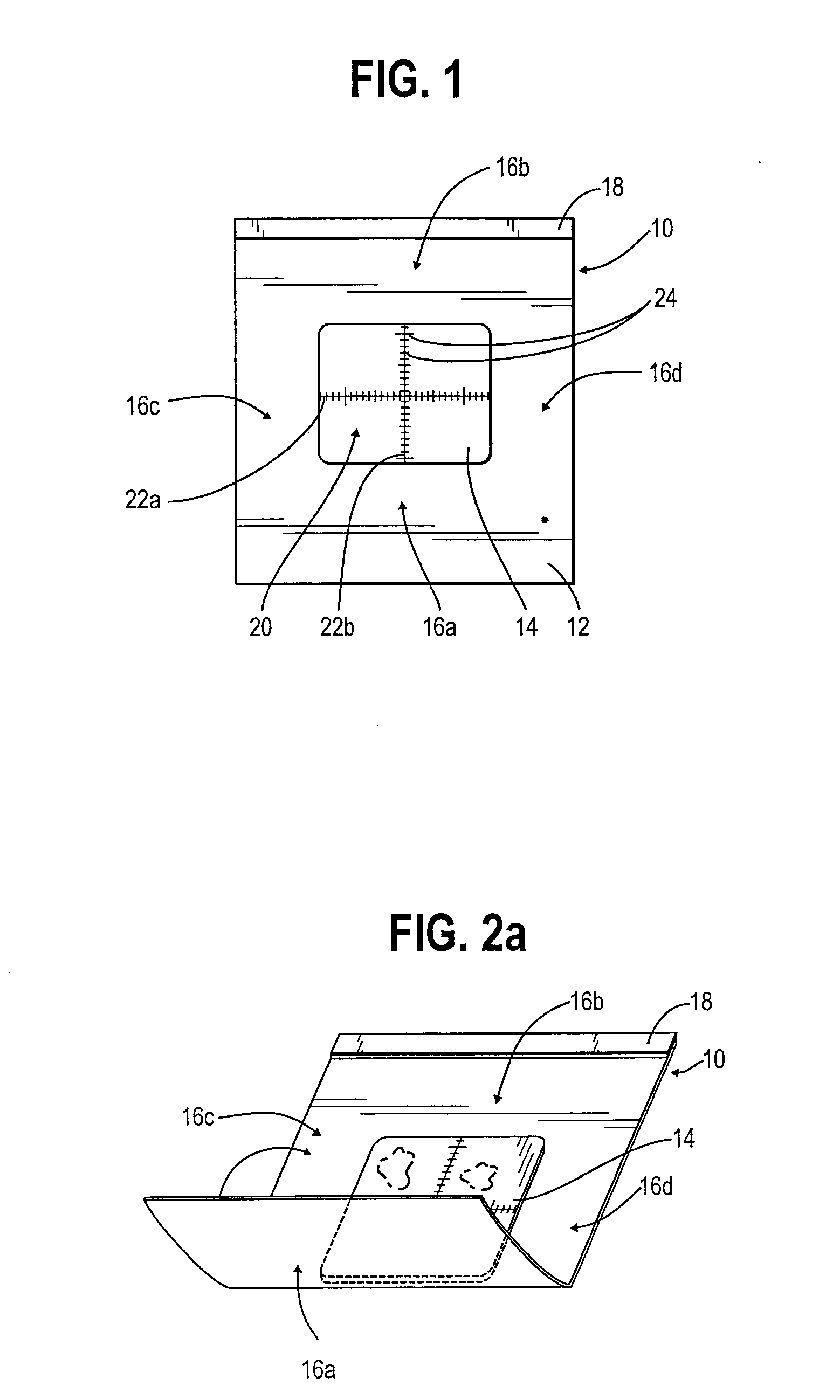
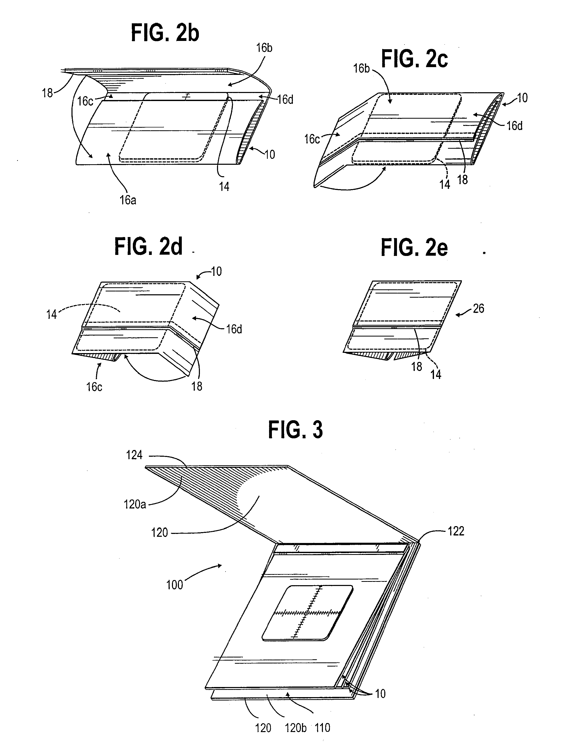
[0017] Referring to FIG. 1, there is illustrated an exemplary histological retaining device 10 embodying the features of this present invention. Device 10 generally consists of four parts: a permeable sheet 12, a permeable target 14, extended flap portions 16a-d, and a malleable securing strip 18.

[0018] More specifically on FIG. 1, permeable sheet 12 is a sheet of filter-type paper or similar porous paper that is permeable to processing fluids and / or molten embedding wax, but retains a tissue specimen during processing. Because permeable, filter-type papers come in a wide range of permeability, thickness, and strengths, many types of papers will work as permeable sheet 12 as long as the sheet is porous to the typical processing fluids used in histological preparation in an efficient manner according to routine histological procedures while retaining tissue specimens 1 mm or smaller in size.

[0019] Common lens paper is an example of a typical media suitable for permeable sheet 12; p...


PUM

PropertyMeasurementUnit
sizeaaaaaaaaaa
thickaaaaaaaaaa
thickaaaaaaaaaa

Abstract

A histological specimen retaining device for processing tissue. The histological specimen retaining device comprises a foldable permeable sheet having edges and a permeable target disposed on the foldable permeable sheet within the edges of said sheet thereby providing extended flap portions. The extended flap portions are foldable to overlap the target. The histological specimen retaining device also comprises a malleable securing strip attached to the permeable sheet of a length sufficient to secure said folded flap portions overlapping said target.

Description

FIELD OF THE INVENTION [0001] The present invention is generally directed to a device and a method for processing tissue samples for histological examination. More specifically, the invention relates to an improved device and method for retaining and processing tissue samples with various fluids prior to embedding the tissue samples in paraffin or the like in preparation for microscopic examination. BACKGROUND OF THE INVENTION [0002] Standard procedures for preparing tissue samples for microscopic examination involve contacting the tissue sample with various fluids, embedding the tissue samples in paraffin, and slicing paraffin-embedded tissue samples very thinly with a microtome. Detailed examples of the preparation of tissue samples for histological examination are fully described in U.S. Pat. Nos. 5,080,869; 5,665,398; and 5,928,934; which are incorporated herein by reference. [0003] In the practice of histopathology and the preparation of cellular tissue materials for examinatio...

Claims


Application Information

Patent Timeline
Patent Type & Authority Applications(United States)
IPC IPC(8): G01N33/48C12M3/00G01N1/31
CPCY10T436/25G01N1/31
Inventor MCCORMICK, JAMES B.
Owner LEICA BIOSYST RICHMOND INC
Learn More