Apparatus and methods for examining, visualizing, diagnosing, manipulating, treating and recording of abnormalities within interior regions of body cavities

Inactive Publication Date: 2007-09-13
GYNTEC MEDICAL
View PDF21 Cites 225 Cited by
  • Summary
  • Abstract
  • Description
  • Claims
  • Application Information

AI Technical Summary

Benefits of technology

[0014] In some embodiments of the present invention, a lens is mounted on the head, and a camera and a light emitter can be disposed in the head and/or the body. Especially preferred heads have a base with a substantially round outer boundary, wherein a tissue stabilizer (or “stabilizing ring”) includes extendable arms which are configured to retract to provide a substantially continuous outer boundary with the base. While not limiting to the inventive subject matter, the extendable arms can comprise a first, a second, and a third arm. Additionally or alternatively, the frame extends the arms outward from a retracted position about th

Problems solved by technology

While such configurations can have various advantages in stationary use, the light source and image analysis system required for such systems often prevent mobile use.
Unfortunately, Kirsner's device fails to provide any test results to a patient, and only advises the patient whether to see the physician or not.
Moreover, implementation of Kirsner's device to detect early signs

Method used


Image

  • Apparatus and methods for examining, visualizing, diagnosing, manipulating, treating and recording of abnormalities within interior regions of body cavities
  • Apparatus and methods for examining, visualizing, diagnosing, manipulating, treating and recording of abnormalities within interior regions of body cavities
  • Apparatus and methods for examining, visualizing, diagnosing, manipulating, treating and recording of abnormalities within interior regions of body cavities

Examples


Embodiment Construction

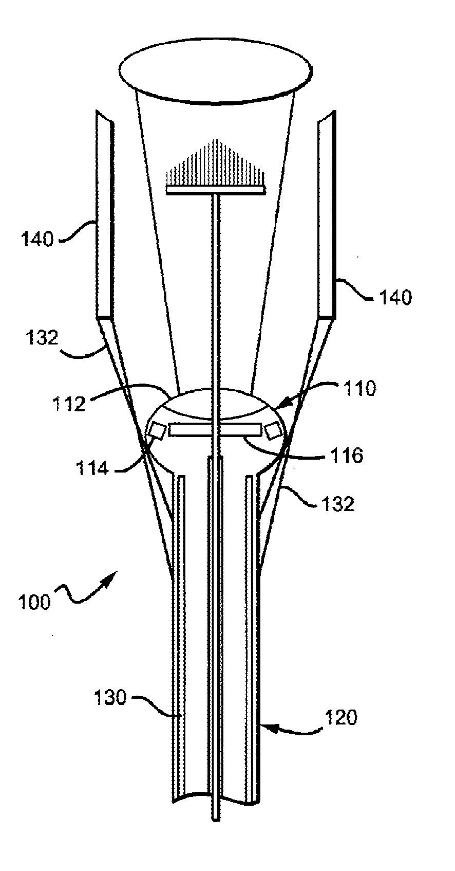
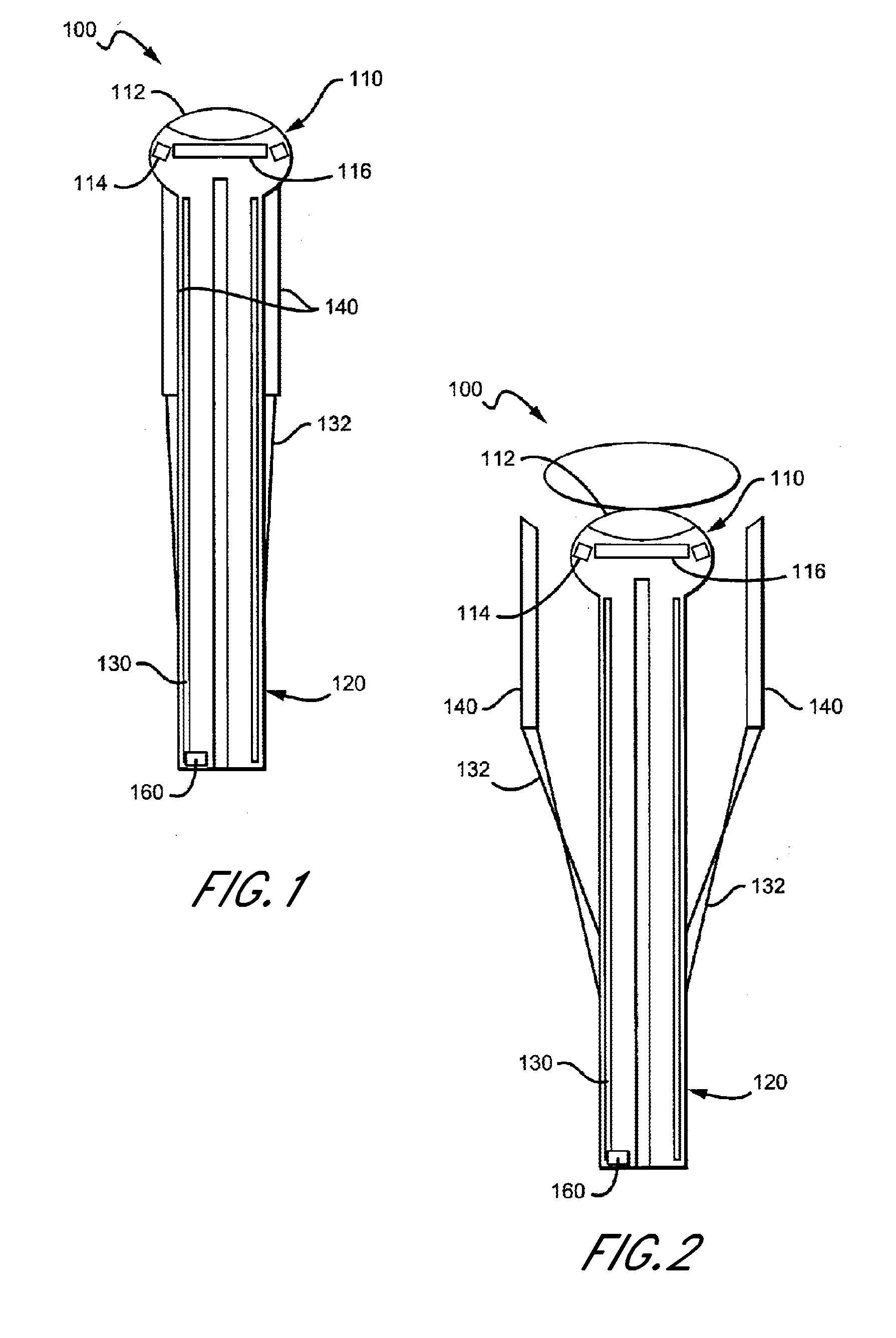
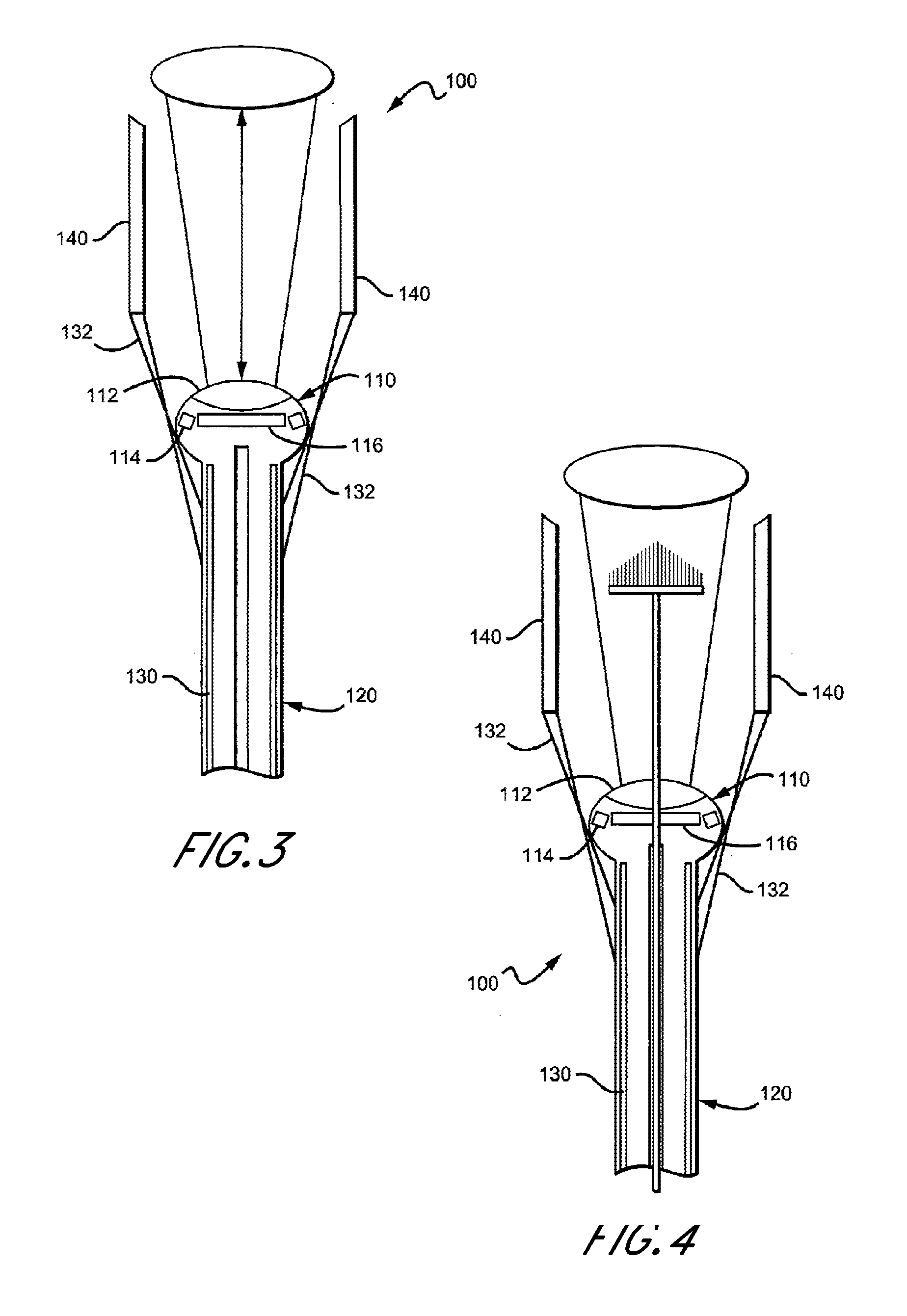
[0051] As should be understood in view of the following detailed description, this application is primarily directed to apparatuses, systems and methods for examining, visualizing, diagnosing, manipulating, treating and recording of abnormalities with interior regions of body cavities. As used herein, “distal end” may be understood to apply to a distal region, and is not necessarily limited to a distal surface or a distal tip, but can refer to the entire region near a distal end. Likewise, “proximal end” can be treated in a similar manner.

1. Multi-Functional Video Scope Embodiments

[0052] The inventor has discovered that an endoscopic device can be configured to allow mobile, office or home use of the device in situ in multiple configurations, and especially to allow examination of a tissue using a plurality of perspective positions of the optical head relative to the examined tissue while maintaining the device in substantially the same position. Contemplated devices further advan...


PUM


Abstract

A portable multi-functional endoscopic device and method for use in the examination of tissue within a corporeal orifice to permit diagnostic, therapeutic or anatomical assessment data to be transmitted, recorded, or analyzed. The device includes a base unit sized and configured to be held in a human hand to permit functional and directional control of the device, an interchangeable head assembly sized and configured to be inserted into the orifice being removably connectable to the base unit, and an inflatable tissue stabilizer disposed external to a distal end of the device. In preferred aspects, the endoscopic device has an image sensor, light source, lens, air pump, and working tools.

Description

CROSS-REFERENCE TO RELATED APPLICATIONS [0001] This application claims the benefit of priority as a Continuation-in-part of U.S. application Ser. No. 11 / 225,381, filed on Sep. 12, 2005, which claims priority from U.S. Provisional application No. 60 / 608,810, filed on Sep. 10, 2004, all of which are incorporated by reference in their entireties, herein. This application also claims the benefit of priority as a Continuation-in-part of U.S. application Ser. No. 10 / 938,688, filed on Sep. 10, 2004, which claims priority from U.S. Provisional application No. 60 / 510,706, filed on Sep. 9, 2003, all of which are incorporated by reference in their entireties, herein.BACKGROUND OF THE INVENTION [0002] 1. Field of the Invention [0003] The field of the invention is endoscopic devices. In particular, a small, portable, light-weight endoscopic device for a mammalian orifice or cavity is presented. In various embodiments, the cavities include, but are not limited to, a vagina, uterus, anus, urethra,...

Claims


Application Information

Patent Timeline
IPC IPC(8): A61B1/06
CPCA61B1/00087A61B1/00096A61B1/05A61B1/0684A61B1/00101A61B1/0676A61B1/0638
Inventor SQUICCIARINI, JOHN B.
Owner GYNTEC MEDICAL
Why Eureka
Try Eureka