That higher pressure causes the
muscle to exert more force, resulting in hypertrophy of the bladder
muscle.
Symptoms of bladder
muscle hypertrophy include increased wall thickness and increased
mass.
Hyperdistension refers to the case in which the bladder is allowed to fill to such an extreme that excessive
bladder pressure builds which can cause potential
renal damage, renal failure and even patient death from autonomic dysreflexia if the patient has
spinal cord damage.
At low bladder volumes, bladder
distension information is not as useful.
Thus, it is more difficult to establish a volume threshold at which over-
distension occurs or when incontinence occurs.
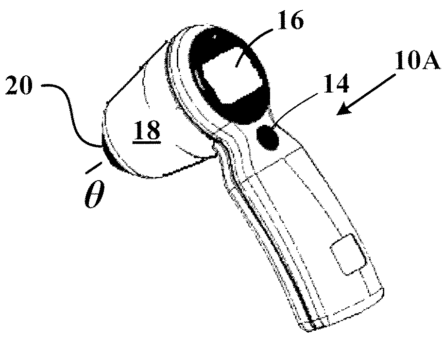
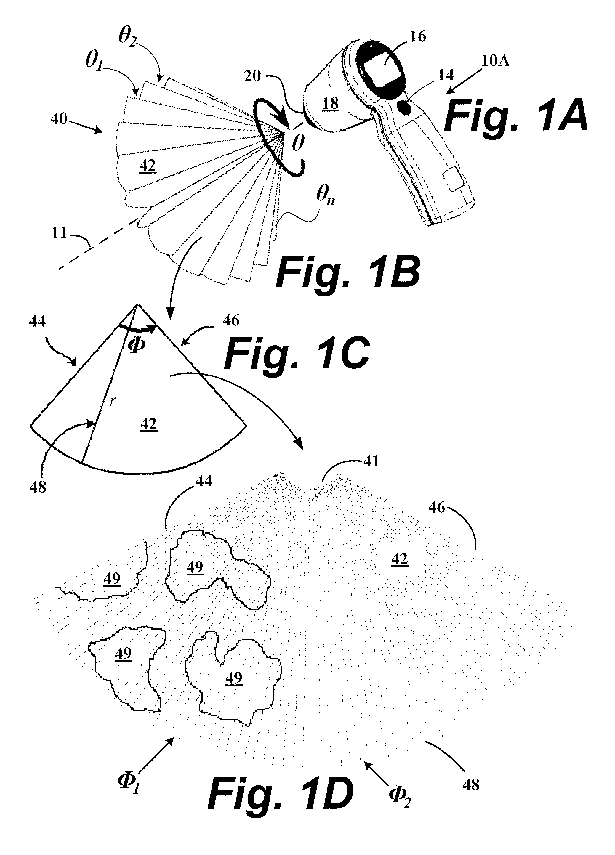
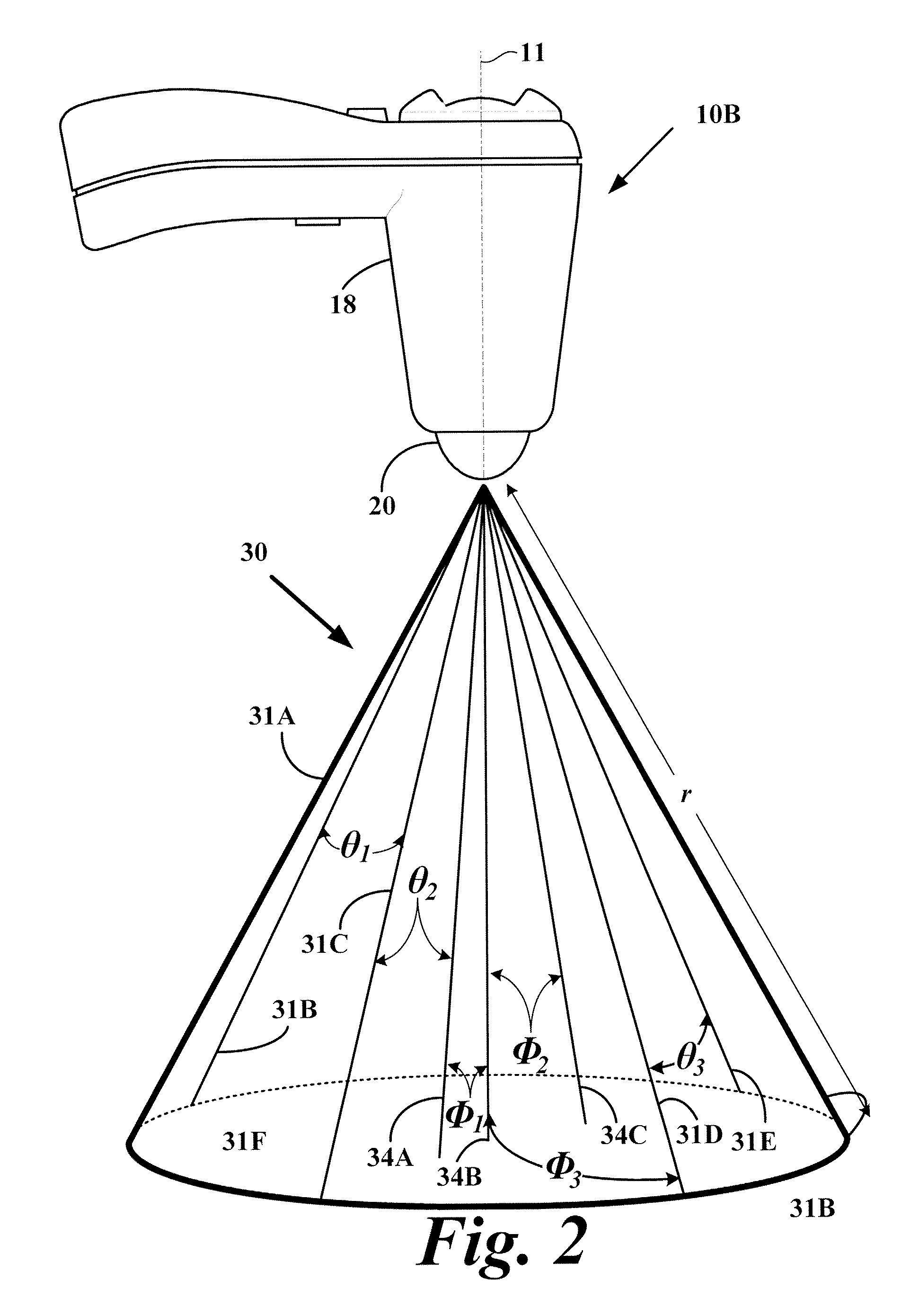
Current methods to measure bladder wall thickness rely on one-dimensional (A-mode) and two-dimensional (B-mode) ultrasound and are greatly susceptible to operator error,
time consuming, and inaccurate.
Furthermore, the limitations of one and two-dimensional ultrasound require inaccurate
spherical model assumptions for the bladder.
Presumably for these and other reasons, the industry has concluded that measuring bladder wall thickness is an unreliable or ineffective means to quantize bladder distension.
Accordingly, an undesired hypertrophy of the bladder muscle often results.
When hyperdistension occurs, the bladder fills with an excessive amount
urine and generates an internal
bladder pressure that may cause serious adverse effects, including
renal damage, renal failure, or even death of the patient from autonomic dysreflexia if the patient has
spinal cord damage.
At low bladder volumes, bladder distension information is not typically useful since normal humans have widely varying bladder capacities.
Thus, it is more difficult to establish a volume threshold at which over-distension occurs or when incontinence occurs for a selected individual.
Unfortunately, the application of these current methods to determine bladder wall thickness are susceptible to operator error, are
time consuming, and generally lead to inaccurate estimations of the bladder wall thickness.
Since the operator must repeatedly reposition the
ultrasound probe until a bladder wall image is sufficiently visible, inaccuracies may be introduced into the ultrasound data.
Consequently, current ultrasound methods to determine bladder wall thickness is an unreliable or ineffective means to measure bladder distension.
Moreover, the existing 2D methods are manually based, utilizing
leading edge-to-
leading edge of opposing
bladder walls laboriously executed upon a series of two-dimensional images, and are fraught with analytical inaccuracies (H. Miyashita, M. Kojima, and T. Miki, “Ultrasonic measurement of bladder weight as a possible predictor of acute
urinary retention in men with
lower urinary tract symptoms suggestive of
benign prostate hyperplasia”,
Ultrasound in
Medicine and
Biology 2002, 28(8): 985-990; M. Oelke, K. Hofner, B. Wiese, V. Gruneweld, and U. Jonas, “Increase in detrusor wall thickness indicates bladder outlet obstruction in men,”World J. of
Urology, 2002, 19(6), 443-452; L. Muller, T. Bergstrom, M. Hellstrom, E. Svensson, and B. Jacobson, “Standardized
ultrasound method for assessing
detrusor muscle thickness in children,” J. Urol., 200, 164: 134-138; and Naya, M. Kojima, H. Honjyo, A. Ochiai, O. Ukimura, and H. Watanabe, “Intraobserver and interobserver variance in the measurement of ultrasound-estimated bladder weight,”
Ultrasound in Med.

