
Biopsy needle system, biopsy needle and method for obtaining a tissue biopsy specimen

a biopsy needle and tissue biopsy technology, applied in the field of biopsy needles and tissue biopsy specimens, can solve the problems of inability to completely guarantee the accuracy of automation, difficulty in obtaining tissue, and inability to actually capture biopsy specimens, etc., to achieve convenient inserting and control, improve the quality of biopsy, and simple use

Inactive Publication Date: 2007-09-13
MCCLELLAN W THOMAS
View PDF14 Cites 21 Cited by
  • Summary
  • Abstract
  • Description
  • Claims
  • Application Information

AI Technical Summary

Benefits of technology

[0012] It is accordingly an object of the invention to provide a biopsy needle system, a biopsy needle and a method for obtaining a tissue biopsy specimen, which overcome the hereinafore-mentioned disadvantages of the heretofore-known devices and methods of this general type, which are minimally-invasive, percutaneous or endoscopic and which not only exceed the prior art, but more importantly match the quality of an open surgical biopsy. The system, needle and method should be simple to use, easy to insert and control, reliable, safe, biopsy-depth adjustable, straight-ahead shearing, end-cutting, automatic-capture, longer full-lumen specimen, tissue-preserving, and over all a core biopsy needle system, a biopsy needle and a method for percutaneous and endoscopic use that improve biopsy quality.

Problems solved by technology

Obtaining tissue is more difficult by any remote biopsy technique, but quality still relies on obtaining tissue of and from the suspected mass.
New imaging systems improve needle placement, but with prior art biopsy needles, the actual capture of the biopsy specimen remains unsure and partially blind.
The complex multi-motion sequencing of the typical side-cut biopsy needle is so demanding on operator skill, that even automation has not made it totally reliable.
Endoscopic biopsy graspers and percutaneous biopsy needles generally recover limited, thin, short, slivers of tissue, making microscopic analysis and diagnosis difficult.
With the prior art, the biopsied tissue is frequently ripped, compressed, distorted or even crushed, limiting analysis and diagnosis.
Fine needle aspiration biopsy, where the tissue is intentionally ripped into small segments or even single cell clusters, is so destructive to intracellular and intercellular anatomy that it is unwise to use that technique as any more than a screening test.
Their cytology debris fields are of such poor condition, that although they can occasionally include the diagnosis of malignancy, they seldom exclude it.

Method used


Image

  • Biopsy needle system, biopsy needle and method for obtaining a tissue biopsy specimen
  • Biopsy needle system, biopsy needle and method for obtaining a tissue biopsy specimen
  • Biopsy needle system, biopsy needle and method for obtaining a tissue biopsy specimen

Examples


Embodiment Construction

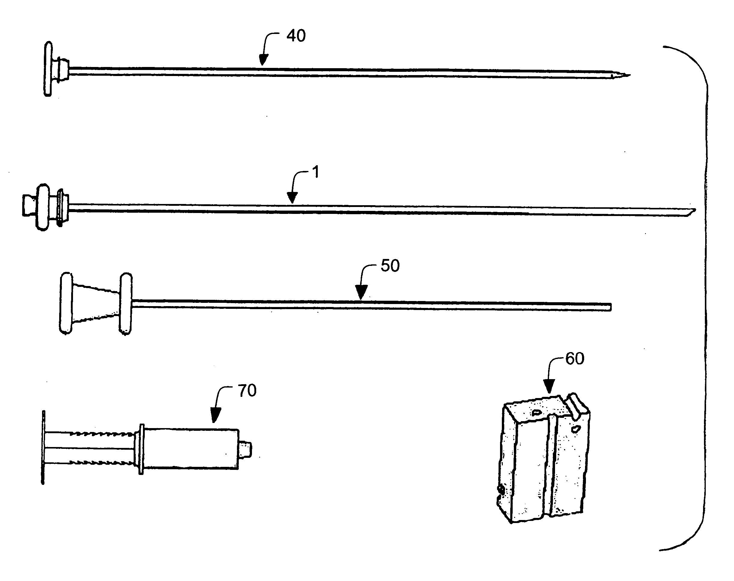
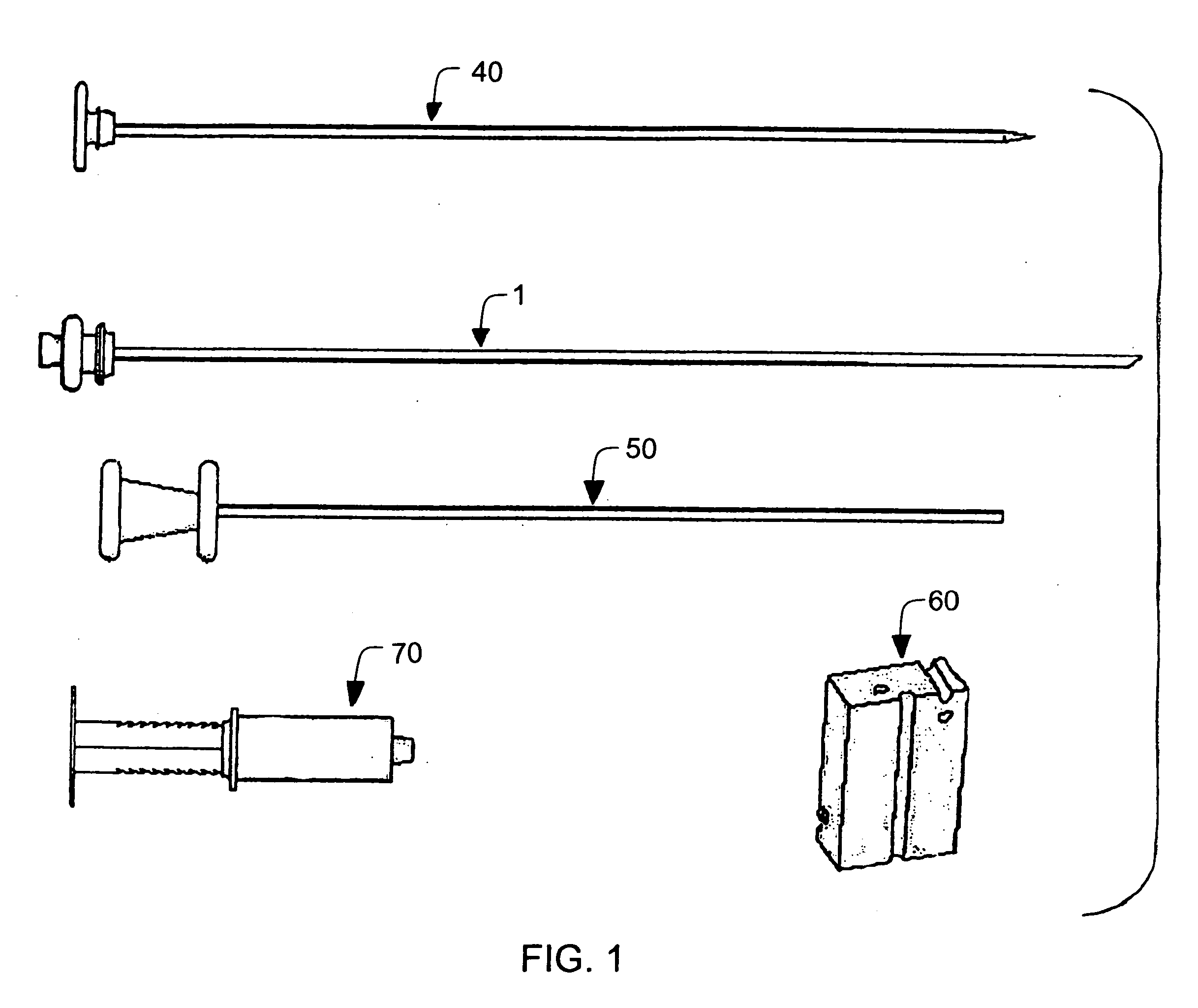
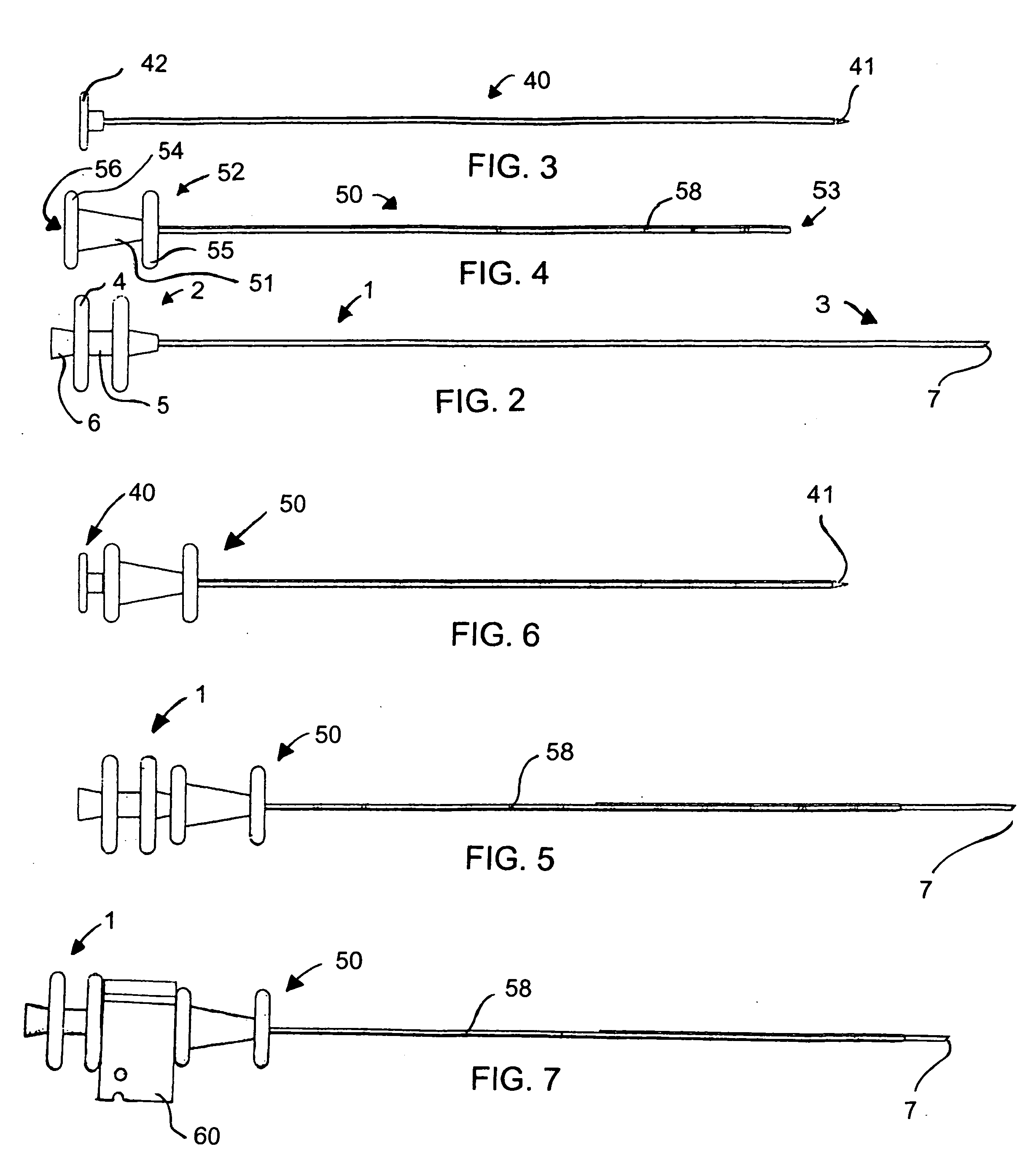
[0051] Referring now to the figures of the drawings in detail and first, particularly, to FIG. 1 thereof, there is seen a biopsy needle system according to the invention which has a biopsy needle 1, a trocar 40, a carrier 50, a controller 60 and a syringe 70. More specifically, the five-piece system includes: [0052] a. an integrated, reliable, true end-cutting, full-lumen specimen, coring, biopsy needle 1; [0053] b. an integrated, easy-insertion, biopsy needle carrier trocar 40; [0054] c. an integrated, improved-placement, high-reflectance, easy-insertion, biopsy needle carrier 50; [0055] d. an integrated, single-piece, dual-use, adjustable biopsy depth gauge and depth controller 60; and [0056] e. an integrated, dual-use, lockable, vacuum-assisted, coring and non-traumatic specimen removal syringe 70.

[0057] As is seen in FIG. 2, the biopsy needle 1 is formed as a tube-like structure, constructed of metal or other suitable material with a proximal end 2 (at the left in the figure) t...


PUM


Abstract

A biopsy needle system includes a carrier. A trocar is inserted into the carrier for percutaneous insertion to a biopsy site. A biopsy needle is inserted into the carrier, replacing the trocar, for removal of a tissue biopsy specimen. A biopsy needle and a method for obtaining a tissue biopsy specimen with the system, are also provided.

Description

BACKGROUND OF THE INVENTION [0001] 1. Field of the Invention [0002] The invention relates to a biopsy needle system, a biopsy needle and a method for obtaining a tissue biopsy specimen with the system. [0003] 2. Description of the Related Art [0004] The most critical diagnosis in medicine is the presence or absence of malignancy. Although there are many screening tests: patient awareness, physical exams, blood tests, and new imaging systems, the hallmark of the cancer diagnosis is a physical specimen, or biopsy, for microscopic analysis. [0005] Malignancy is of life or death importance, so that the quality of the biopsy is of utmost importance. Open surgical biopsy, with total control of specimen location, size, and condition, is the accepted standard of diagnostic quality and a minimally invasive, percutaneous or endoscopic biopsy must not sacrifice that quality, since a false negative may condemn the patient to an agonizing and preventable death. [0006] Obtaining tissue is more di...

Claims


Application Information

Patent Timeline
IPC IPC(8): A61B10/00
CPCA61B10/0266A61B10/0283A61B17/3403A61B10/0275A61B2019/304A61B2019/462A61B2017/32004A61B2090/034A61B2090/062
Inventor MCCLELLAN, W. THOMAS
Owner MCCLELLAN W THOMAS
Learn More