

Copolymer-bioceramic composite implantable medical devices
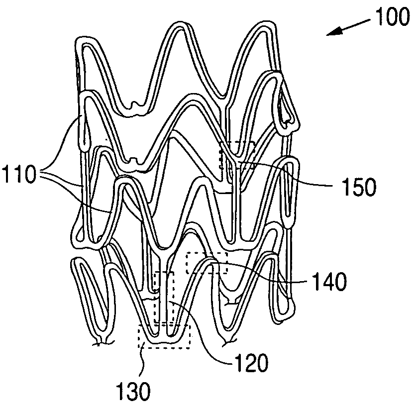
a bioceramic and composite technology, applied in the field of implantable medical devices, can solve the problems of stent pattern localized portions subjected to substantial deformation during crimping and expansion, and the struts or bar arms can crack, and the most vulnerable to failur
- Summary
- Abstract
- Description
- Claims
- Application Information
AI Technical Summary
Problems solved by technology
Method used
Image

Examples
example 1
Prophetic Example of Solution Blending of Polymer and Bioceramic Particles
[0135]Step 1: Add bioceramic particles into suitable solvent, such as chloroform, acetone, etc. and stir to form a bioceramic particle suspension solution.
[0136]Step 2: Slowly add a polymer such as PLLA, PDLLA, PLGA into suspension solution and stir until polymer dissolves completely. In this step, the solution may still have a relatively low viscosity. However, the bioceramic particles should be well dispersed while stirring.
[0137]Step 3: Slowly add the polymer into solution again to gradually increase solution viscosity. Repeat this step as needed until the polymer is completely dissolved and reasonable solution viscosity is developed.
[0138]Step 4: Apply ultrasonic mixing to suspension solution for 15-30 min to further disperse all the HAP uniformly into the PLLA solution.
[0139]Step 6: Add suspension solution to 1 L methanol to precipitate polymer and particles.
example 2
[0140]Solution blending of PLLA / HAP Composite (100:1 wt / wt)
[0141]Step 1: Added 50 mg HAP particles into 300 mL of chloroform and stirred for 10-30 minutes to form bioceramic particle suspension solution.
[0142]Step 2: Slowly added 5 g PLLA into suspension solution and stirred about 8 hours to dissolve all polymer.
[0143]Step 3: Applied ultrasonic mixing to suspension solution for 15-30 min to further disperse HAP particles into PLLA solution.
[0144]Step 4: Added suspension solution to 1 L methanol to precipitate polymer and particles.
[0145]Step 5: Filtered the precipitate and dried about 8 hours in vacuum oven at 60° C. End product is PLLA / HAP composite. Composites were also made with 2 wt % and 5 wt % HAP.
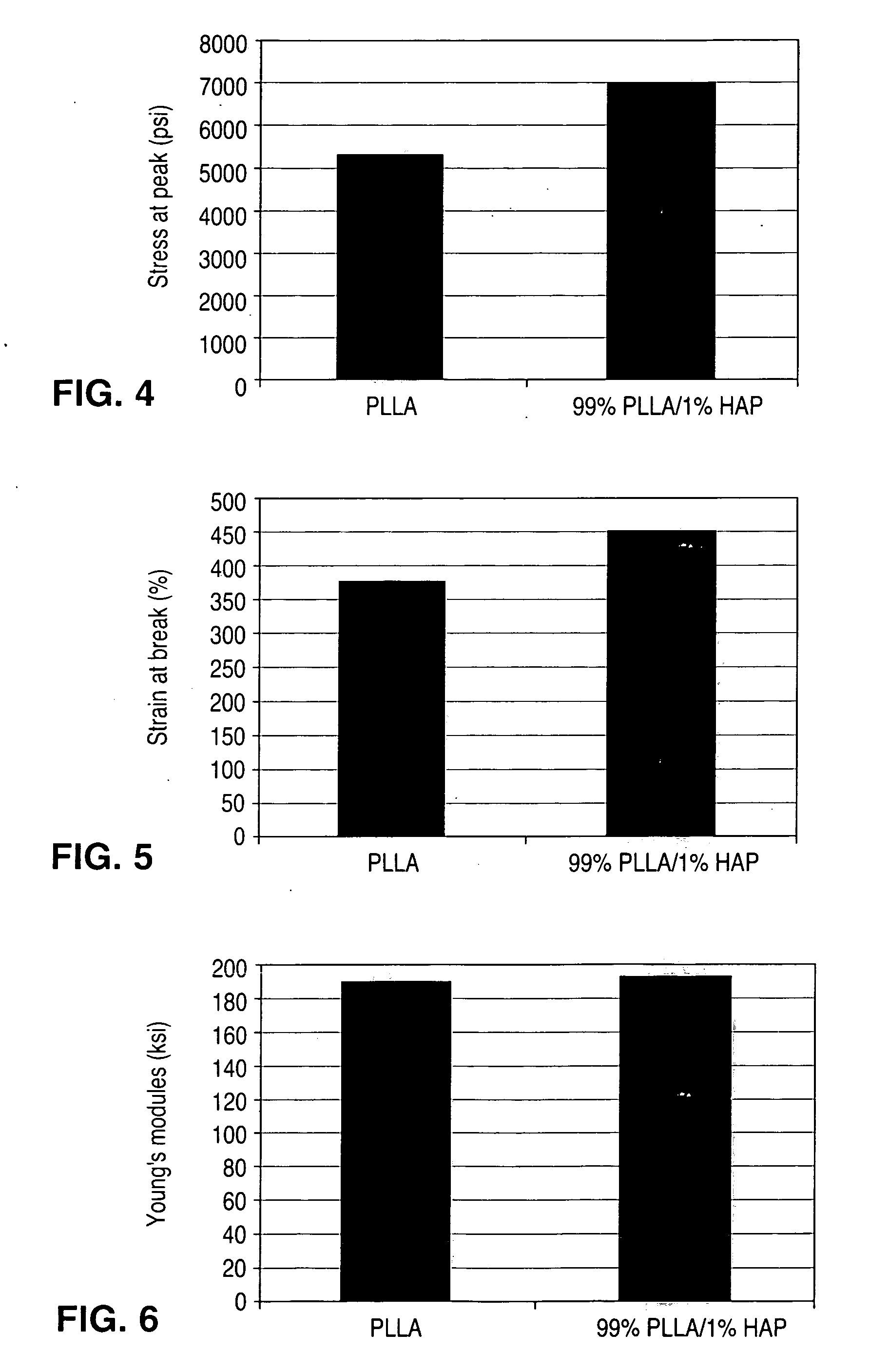
[0146]Mechanical Properties and Morphology of PLLA / HAP Composite (100:1 wt / wt)
[0147]Tensile testing of the composite samples and a pure PLLA were performed using an Instron tensile tester obtained from Instron in Canton, Mass. Test samples were prepared by hot pressing the PLLA / HAP c...
example 3
[0149]Solution Blending of PLLA / HAP Composite (2:1 wt / wt) as HAP Intermedium Mixture
[0150]Step 1: Added 25 g HAP particles to 3L chloroform and stirred for 10-30 minutes to form bioceramic particle suspension solution.
[0151]Step 2: Slowly added 50 g PLLA into suspension solution and stirred about 8 hours to dissolve all polymer.
[0152]Step 3: Applied ultrasonic mixing for 15-30 min to further disperse HAP particles into PLLA solution.
[0153]Step 4: Added suspension solution to 9 L methanol to precipitate particles and polymer.
[0154]Step 5: Filtered the precipitate and dried about 8 hours in vacuum oven at 60° C. End product is PLLA / HAP composite.
[0155]Extrusion of Precipitated PLLA / HAP (2:1 wt / wt) with PLLA
[0156]Step 1: Broke 2:1 wt / wt composite into small pieces
[0157]Step 2: Mixed 24 g of broken up composite and 376 g PLLA.
[0158]Step 3: Extruded mixture at 216° C.
PUM
| Property | Measurement | Unit |
|---|---|---|
| wt % | aaaaa | aaaaa |
| Tg | aaaaa | aaaaa |
| diameter | aaaaa | aaaaa |
Abstract
Description
Claims
Application Information