Preclinical SPECT system using multi-pinhole collimation

a spectroscopic system and multi-pinhole technology, applied in the field of nuclear medicine, can solve the problems of not being able to increase the intrinsic spatial resolution of the gamma camera, the gantry system is relatively expensive subsystem of the diagnostic tool, and the actual unit rotation of the gantry requires a large degree of spa

Inactive Publication Date: 2008-01-03
SIEMENS MEDICAL SOLUTIONS USA INC
View PDF4 Cites 23 Cited by
  • Summary
  • Abstract
  • Description
  • Claims
  • Application Information

AI Technical Summary

Benefits of technology

[0019]In another embodiment, a method of performing a preclinical scan of a test subject employs a preclinical nuclear imaging detector system including using a pinhole aperture that is positionable to effect a desired spatial resolution. In yet another embodiment, the method of performing a preclinical scan of a test subject employs a preclinical nuclear imaging detector system having a pinhole aperture that is positionable to effect a desired spatial sensitivity.

Problems solved by technology

In most clinical imaging studies, the predominant spatial resolution achieved is determined by the geometric resolution of the collimator, and thus there has not been a strong incentive to increase the intrinsic spatial resolution of the gamma camera.
One drawback associated with this requirement however, is that such gantry systems are relatively expensive subsystems of the diagnostic tool because they must be capable of providing rapid rotation of the large and heavy camera heads through very precise orbits about the patient.
Further, rotating gantries require a large degree of space for the actual unit as well as for full operational ability.
This is especially problematic with preclinical SPECT wherein lab space limitations are more prevalent.

Method used


Image

  • Preclinical SPECT system using multi-pinhole collimation
  • Preclinical SPECT system using multi-pinhole collimation
  • Preclinical SPECT system using multi-pinhole collimation

Examples


Embodiment Construction

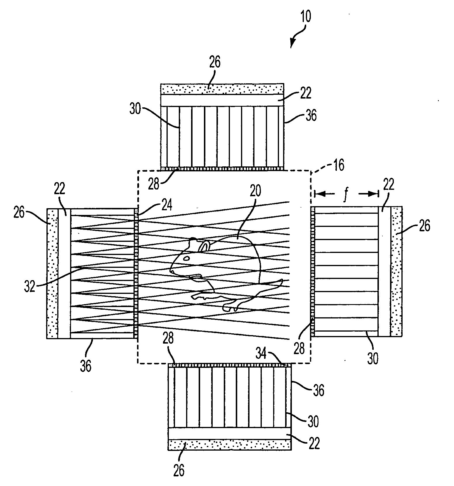
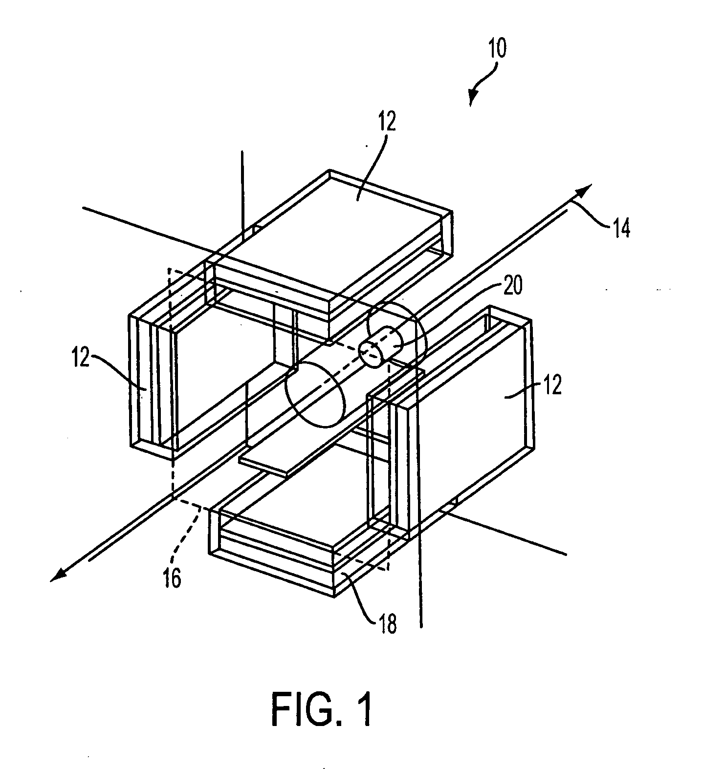
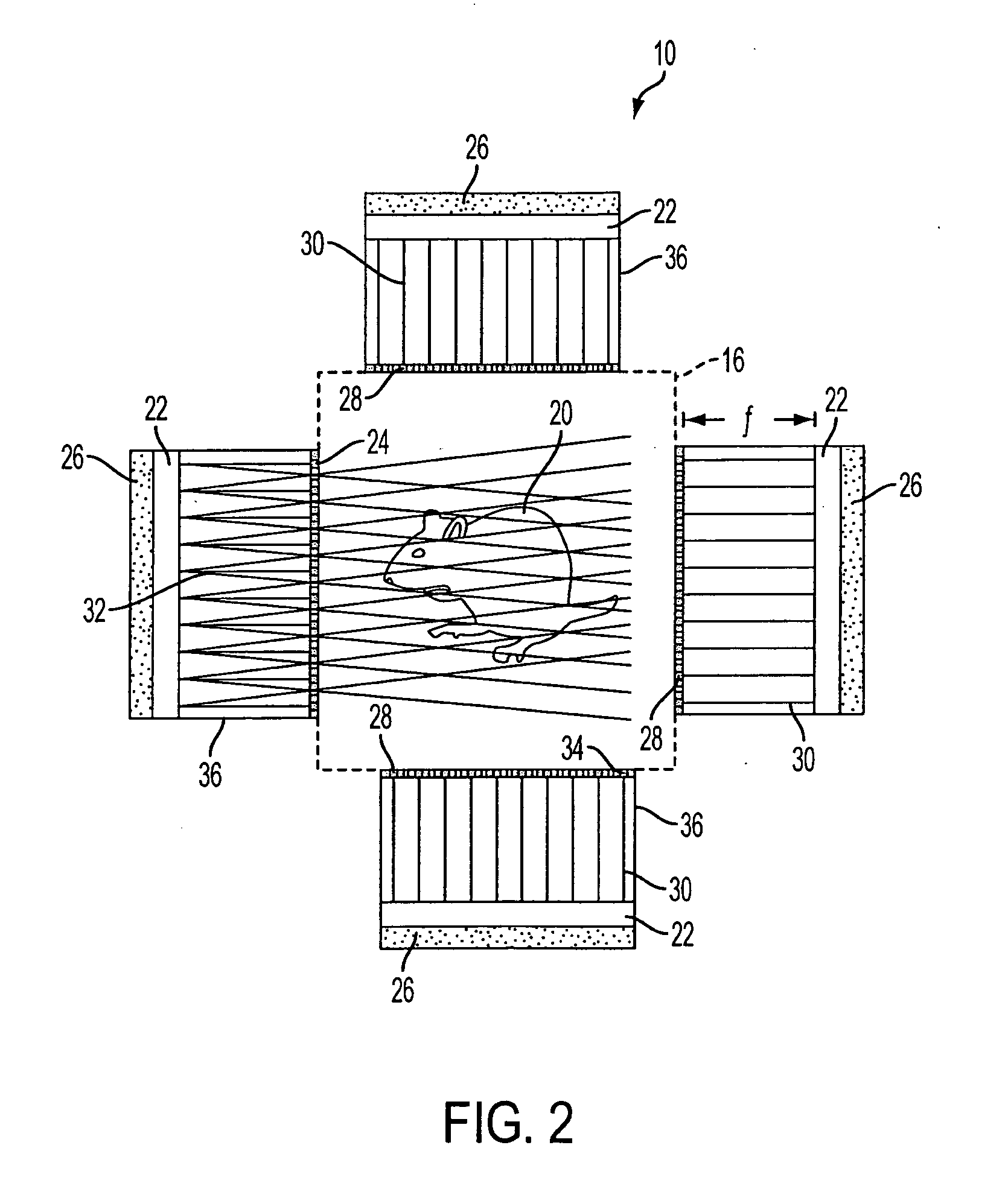
[0034]Referring now to the figures, FIGS. 1 and 6 disclose example embodiments of preclinical nuclear imaging detector systems according to the instant invention. The detectors of FIG. 1 are generally disposed in a rectangular or square configuration, whereas the detectors of FIG. 6 are generally disposed in an arcuate configuration. Generally, a preclinical nuclear imaging detector system 10 comprises one or more detector assemblies 12 secured to a gantry and / or support structure 11. Detector assemblies 12 are radially disposed about an axis 14 to thereby describe a perimeter 16 or a portion thereof and are optionally fixed relative to gantry and / or support structure 11. In the embodiment illustrated in FIG. 1, preclinical nuclear imaging detector system 10 further includes an object table 18 which is further shown supporting object 20, e.g., a phantom or test object such as a laboratory animal. Referring now to FIGS. 2 to 5, detector assembly 12 is shown as comprising a scintillat...


PUM


Abstract

A preclinical nuclear imaging detector system, including a gantry and one or more detector assemblies, each including a scintillator configured to interact with radiation emanating from a target test object being imaged and at least one pinhole collimator, having one or more pinhole apertures formed therein. The pinhole collimator is disposed between the target object and the scintillator, wherein a distance between the pinhole aperture and the scintillator is selected as a function of the number of pinhole apertures provided in the collimator and to optimize one of sensitivity or spatial resolution, such that the one or more pinhole apertures collectively project a unitary minified radiation image of the target object onto the scintillator. Further, one or more photosensors are optically coupled to the scintillator to receive interaction events from the scintillator.

Description

FIELD OF THE INVENTION[0001]The instant invention generally relates to nuclear medicine, and systems for obtaining nuclear medical images of interest. In particular, the instant invention relates to a novel detector configuration for preclinical single photon imaging including single photon emission computed tomography (SPECT) or planar imagingBACKGROUND OF THE INVENTION[0002]Nuclear imaging is a unique specialty wherein radiation is used to acquire images that show the function and anatomy of organs, bones or tissues of the body. Radiopharmaceuticals are introduced into the subject, either by injection or ingestion, and are attracted to specific organs, bones or tissues of interest. Such radiopharmaceuticals produce gamma photon emissions that emanate from the body. One or more detectors are used to detect the emitted gamma photons, and the information collected from the detector(s) is processed to calculate the position of origin of the emitted photon from the source (i.e., the bo...

Claims


Application Information

Patent Timeline
Patent Type & Authority Applications(United States)
IPC IPC(8): G01T1/20
CPCG01T1/1644A61B6/508A61B6/037
Inventor JOUNG, JINHUN
Owner SIEMENS MEDICAL SOLUTIONS USA INC
Why Eureka
Try Eureka