Ultrasonograph and Ultrasonograph Control Method

a control method and ultrasonograph technology, applied in the field of ultrasonic diagnostic equipment, can solve the problems of difficult repair of the hardened artery completely, affecting the accuracy of measurement, and losing the elasticity of the artery, so as to reduce the noise and other disturbances, and achieve accurate measurement.

Inactive Publication Date: 2008-01-24
KONICA MINOLTA INC
View PDF7 Cites 24 Cited by
  • Summary
  • Abstract
  • Description
  • Claims
  • Application Information

AI Technical Summary

Benefits of technology

[0103] According to the present invention, the maximum and minimum values of thicknesses or thickness variations are found during a maximum value finding period and a minimum value finding period, which are defined as respective partial periods of one cardiac cycle. As a result, the unwanted effects of noise and other disturbances can be reduced in the remaining periods other than the maximum and minimum value finding periods and measurements can be done accurately.
[0104] In addition, the accuracy of the greatest thickness difference, strain or elastic property is checked at the points in time when the maximum and minimum values are obtained. Thus, by considering the degrees of accuracy of the measuring results, inaccurate measuring results are much less likely to be taken for accurate ones erroneously and high-reliability diagnosis can be made based on the measuring results.
[0105] On top of that, a go / no-go test is carried out to determine whether each shape measured value or attribute property value is a correct one or not, a go / no-go ratio is calculated based on the results of the go / no-go testing, and the shape measured and attribute property values are presented depending on the go / no-go ratio. Consequently, high-reliability diagnosis can be made based on the measuring results.

Problems solved by technology

More specifically, if an atheroma is created on the arterial wall or if no arterial cells are produced anymore due to various factors such as elevated blood pressure, then the artery loses its elasticity to become hard and fragile.
However, this diagnosis needs to be carried out with a vascular catheter inserted into the blood vessel of a testee, thus imposing a heavy load on him or her.
However, it is said that once the arterial sclerosis has advanced to a certain degree, the farther advancement of that disease can be checked with the administration of medicine but it is difficult to repair the hardened artery completely.

Method used


Image

  • Ultrasonograph and Ultrasonograph Control Method
  • Ultrasonograph and Ultrasonograph Control Method
  • Ultrasonograph and Ultrasonograph Control Method

Examples


embodiment 1

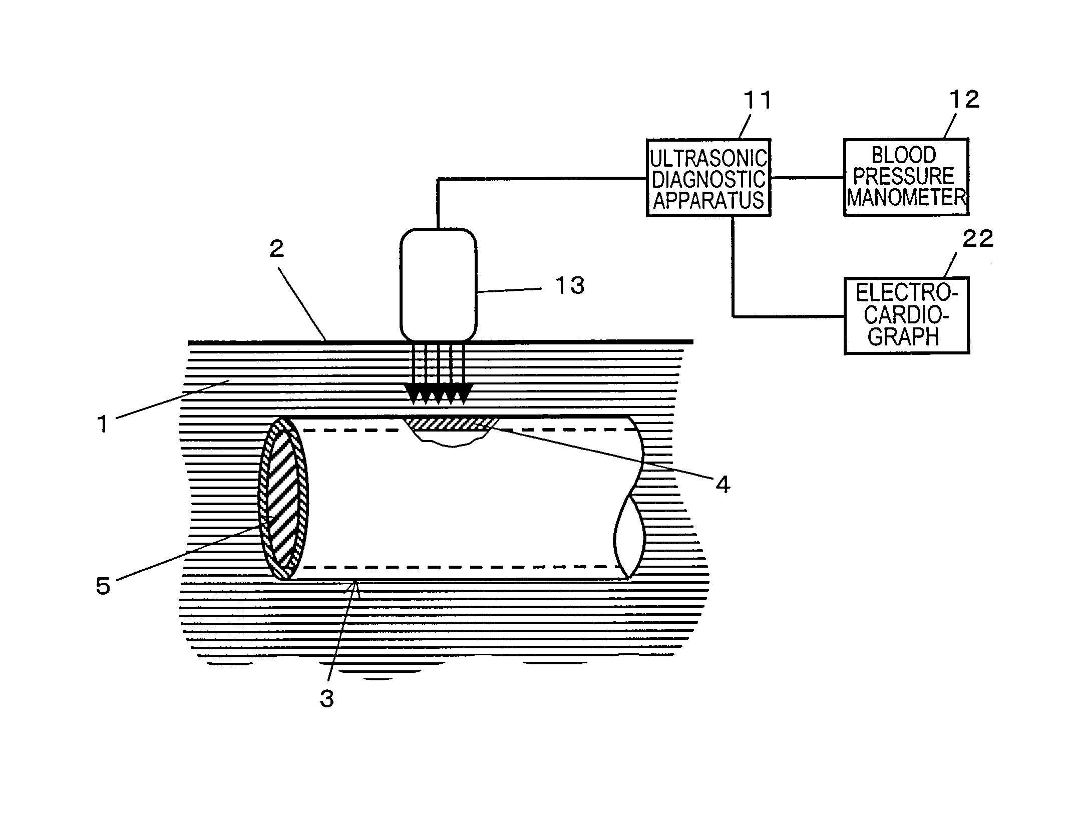
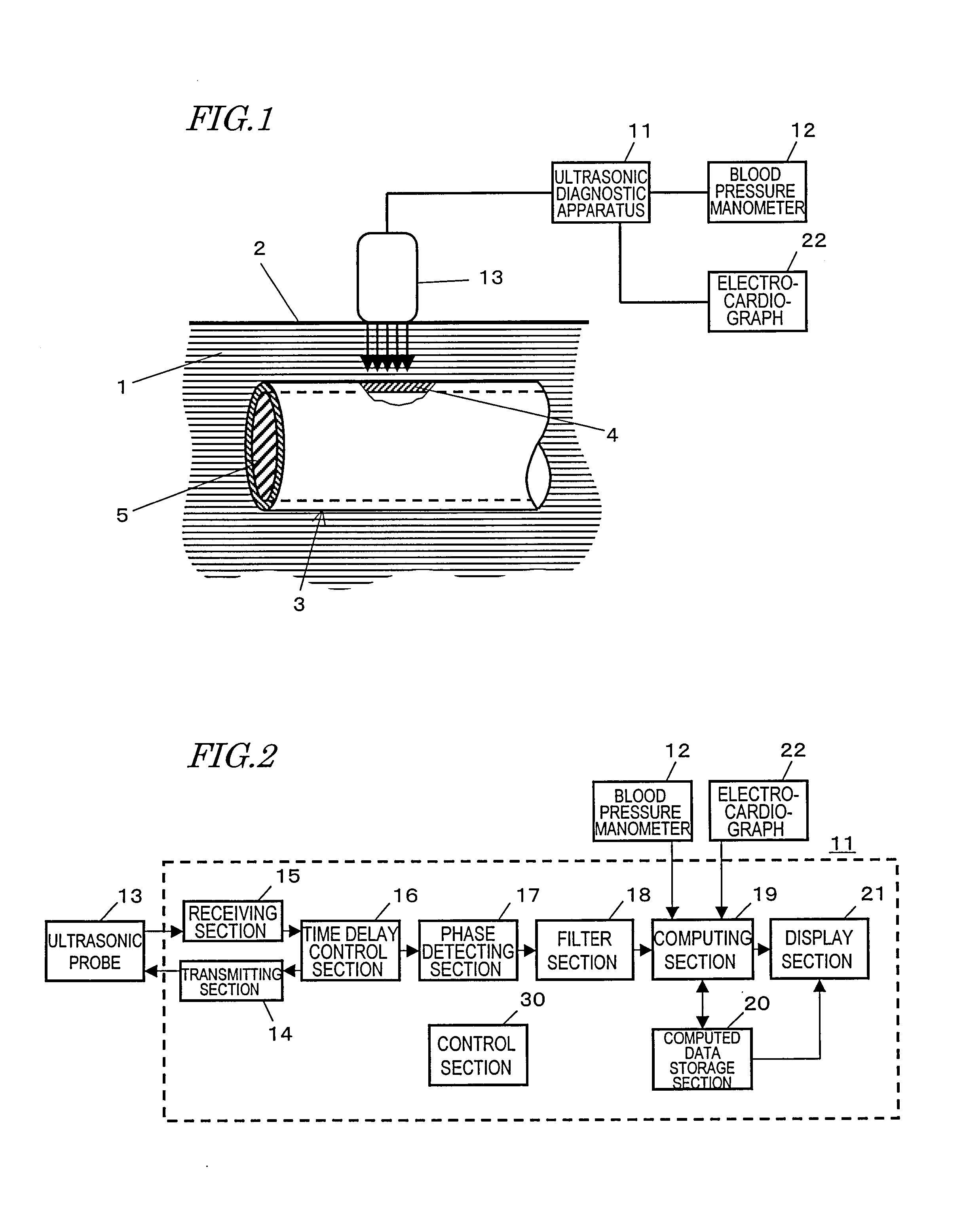
[0176] Hereinafter, a First Preferred Embodiment of an ultrasonic diagnostic apparatus according to the present invention will be described. FIG. 2 is a block diagram showing a configuration for the ultrasonic diagnostic apparatus 11. The ultrasonic diagnostic apparatus 11 includes a transmitting section 14, a receiving section 15, a time delay control section 16, a phase detecting section 17, a filter section 18, a computing section 19, a computed data storage section 20, and a display section 21. The ultrasonic diagnostic apparatus 11 further includes a control section 30 (implemented as a microcomputer, for example) for performing an overall control on all of these sections.

[0177] The transmitting section 14 generates a predetermined drive pulse signal and outputs it to the ultrasonic probe 13. An ultrasonic transmitted wave, transmitted by the ultrasonic probe 13 in response to the drive pulse signal, is reflected and scattered by a body tissue such as the wall of the blood ves...

embodiment 2

[0241] Hereinafter, a second preferred embodiment of an ultrasonic diagnostic apparatus according to the present invention will be described. FIG. 16 is a block diagram showing the configuration of core sections of the ultrasonic diagnostic apparatus of the second preferred embodiment. Although not shown in FIG. 16, the ultrasonic diagnostic apparatus of the second preferred embodiment also includes the transmitting section 14, receiving section 15, time delay control section 16, phase detecting section 17, filter section 18 and control section 30 as in the first preferred embodiment. All of these sections operate just as already described for the first preferred embodiment.

[0242] The ultrasonic diagnostic apparatus of this preferred embodiment also finds the maximum and minimum values of the thickness variations either during one cardiac cycle or during respective partial periods of one cardiac cycle and checks the degrees of accuracy of the greatest thickness difference, strain a...

embodiment 3

[0272] Hereinafter, an ultrasonic diagnostic apparatus according to a third preferred embodiment of the present invention will be described. FIG. 23 is a block diagram showing a configuration for the core sections of the ultrasonic diagnostic apparatus of this third preferred embodiment. Although not shown in FIG. 23, the ultrasonic diagnostic apparatus of the third preferred embodiment also includes the transmitting section 14, receiving section 15, time delay control section 16, phase detecting section 17, filter section 18 and control section 30 just like the counterpart of the first preferred embodiment. All of these sections operate just as already described for the first preferred embodiment.

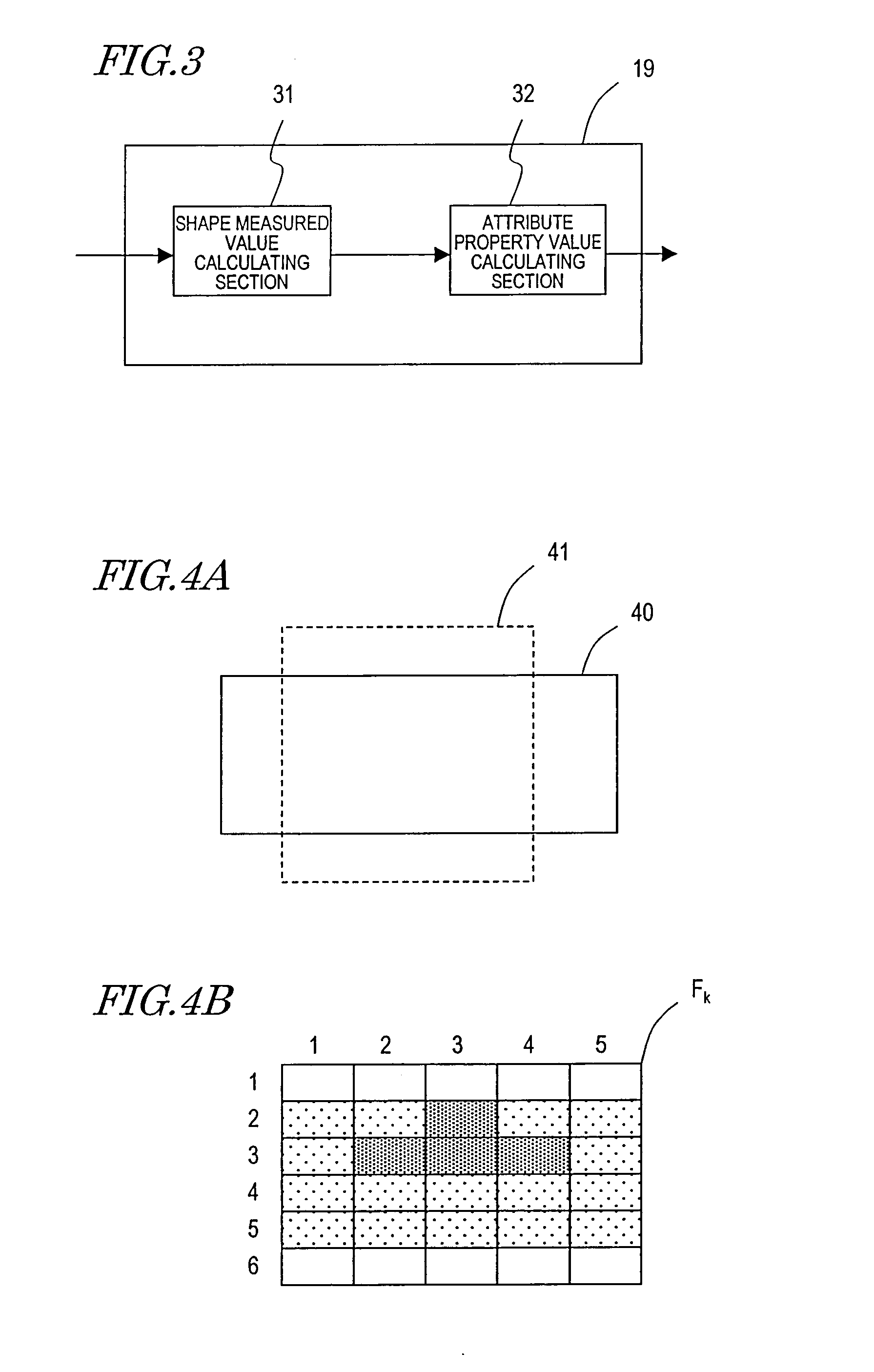
[0273] The ultrasonic diagnostic apparatus of this preferred embodiment determines whether or not the greatest thickness difference, strain or elastic property, obtained every cardiac cycle, is a highly reliable one overall. For that purpose, the computing section 19 includes a shape meas...


PUM


Abstract

An ultrasonic diagnostic apparatus according to the present invention includes: a transmitting section 14 for driving an ultrasonic probe 13 sending out an ultrasonic transmitted wave toward a tissue of an organism; a receiving section 15 for receiving an ultrasonic reflected wave, produced by getting the ultrasonic transmitted wave reflected by the tissue of the organism, through the ultrasonic probe to generate a received signal; a phase detecting section 17 for detecting the phase of the received signal to generate a phase detected signal; a positional displacement calculating section 31a for calculating the magnitudes of positional displacements at multiple measuring points, set on the tissue of the organism, based on the phase detected signal; a thickness variation calculating section 31b for calculating thicknesses or thickness variations, each measured between two arbitrary points set with respect to the measuring points, based on the magnitudes of positional displacements; and a maximum/minimum value calculating section 31c for finding the maximum and minimum thicknesses or thickness variations during a maximum value finding period and a minimum value finding period, which are defined as respective partial periods of one cardiac cycle of the organism. At least one of the greatest thickness difference, strain and elastic property is calculated based on a difference between the maximum and minimum thicknesses or thickness variations.

Description

TECHNICAL FIELD [0001] The present invention relates to an ultrasonic diagnostic apparatus. More particularly, the present invention relates to an ultrasonic diagnostic apparatus for measuring the attribute property of a tissue in an organism and also relates to a method for controlling such an ultrasonic diagnostic apparatus. BACKGROUND ART [0002] Recently, the number of people suffering from various circulatory system diseases, including heart infarction and brain infarction, has been on the rise, thus making it more and more urgent to prevent and treat these diseases. [0003] The pathopoiesis of heart or brain infarction is closely correlated to arterial sclerosis. More specifically, if an atheroma is created on the arterial wall or if no arterial cells are produced anymore due to various factors such as elevated blood pressure, then the artery loses its elasticity to become hard and fragile. Also, if the blood vessel is clogged up where the atheroma has been created or if a vascu...

Claims


Application Information

Patent Timeline
Patent Type & Authority Applications(United States)
IPC IPC(8): A61B8/00
CPCA61B5/02007A61B5/1075A61B8/08A61B8/0858G01S15/8906A61B8/485A61B8/543G01S7/52042A61B8/0891
Inventor KATO, MAKOTOHAGIWARA, HISASHISUNAGAWA, KAZUHIROTANNAKA, YOSHINAO
Owner KONICA MINOLTA INC
Why Eureka
Try Eureka