
Tympanic membrane drain tube

a tympanic membrane and drain tube technology, applied in the field of drain tubes, can solve the problems of recurrence of the former symptom, difficult to transmit sound into the internal ear through the external ear canal, and worsening of the tympanic membrane and auditory ossicle movement, etc., and achieves the effects of convenient intubation, simple structure, and convenient treatmen

Inactive Publication Date: 2008-03-06
FUJIWARA HISAO
View PDF4 Cites 15 Cited by
  • Summary
  • Abstract
  • Description
  • Claims
  • Application Information

AI Technical Summary

Benefits of technology

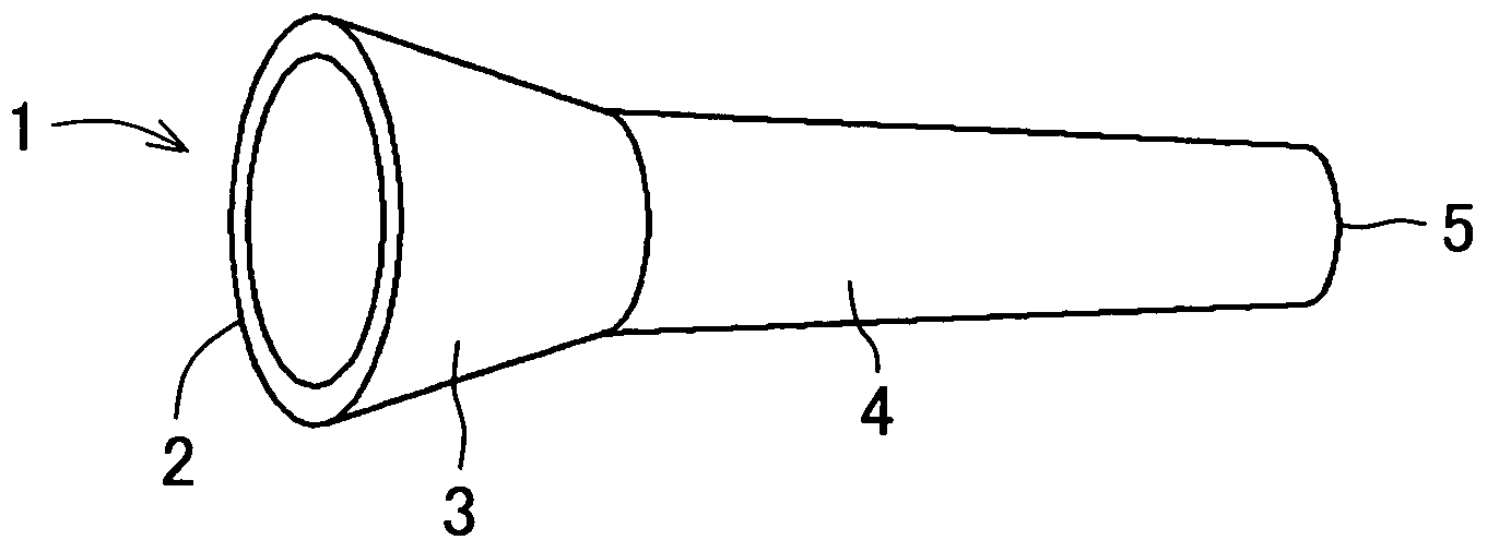
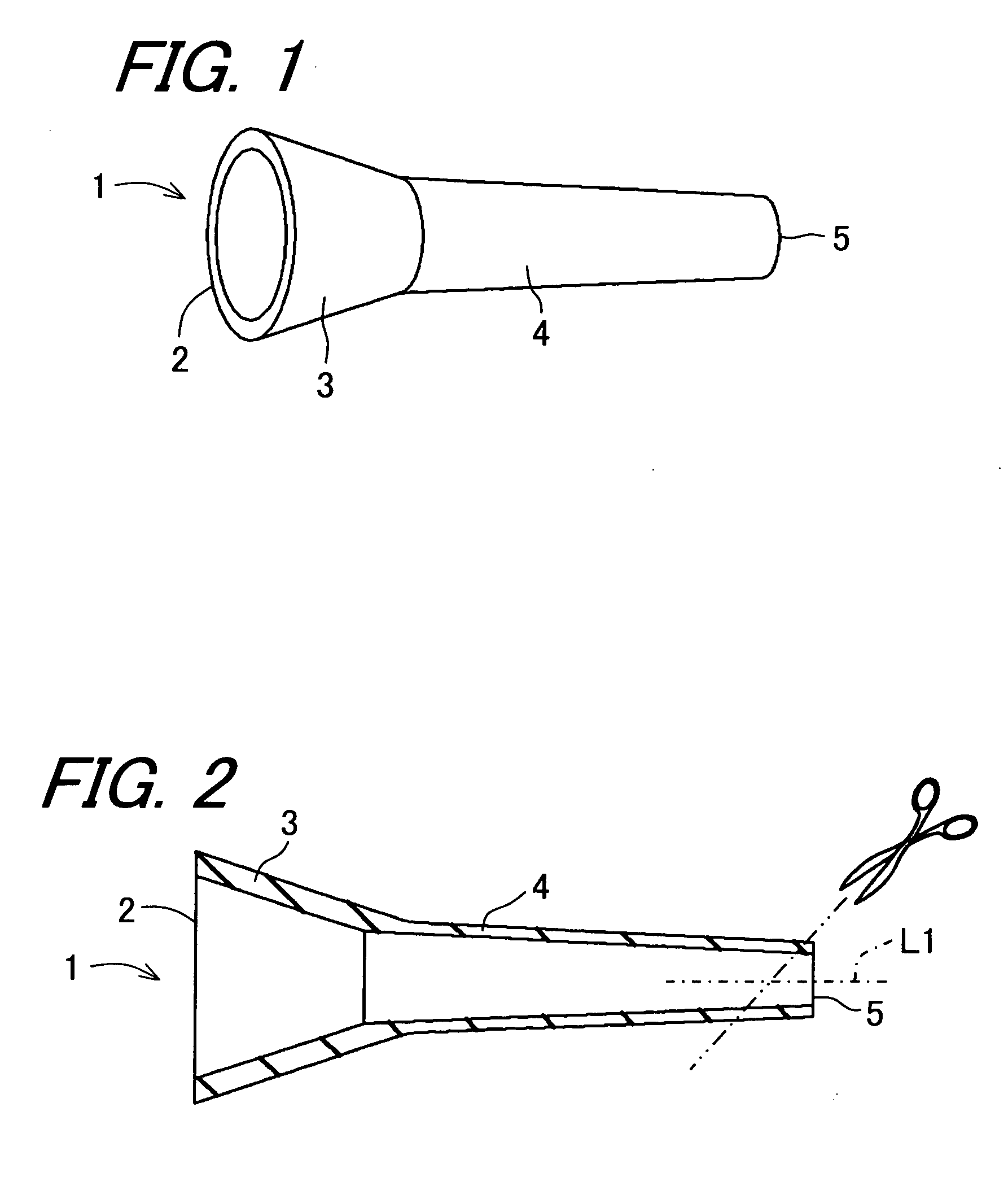
[0021]The tympanic membrane drain tube according to the invention is formed into a shape in which the tympanic membrane drain tube can be easily pinched with a dressing forceps or the like. That is to say, the tympanic membrane drain tube has a smaller flange-like portion which is, on the external ear side, formed into an annular shape that less irritates the tympanic membrane and has a smaller contact area therewith. The tympanic membrane drain tube is formed so that a tip end thereof turns slightly upward to allow visualization of the tip and an incised portion of the tympanic membrane when the drain tube is pinched with a dressing forceps or the like.
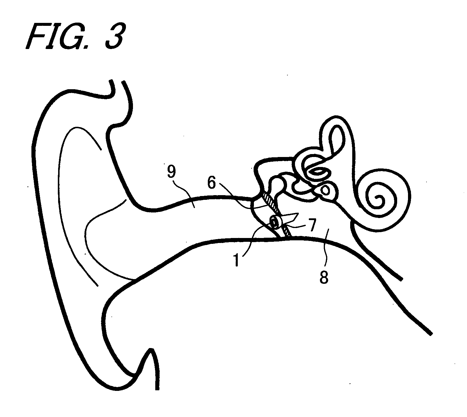
[0035]wherein the hollow tube can be safely inserted into a stoma formed in a tympanic membrane under clear vision.

Problems solved by technology

When a person contracts exudative otitis media or the like disease, the person has the middle ear falling into a hypoxic condition, with the result that an exudate accumulates in the middle ear cavity, movements of the tympanic membrane and auditory ossicle become worse, and sound entering through the external ear canal becomes hard to transmit into the internal ear.
However, an opening formed in the tympanic membrane spontaneously closes in a few days and therefore, in a case where the patient has not recovered completely, it often happens that an exudate accumulates again in the middle ear cavity which has been spontaneously closed, resulting in return to the former symptom.
It is true that the flange-like portions are effective for preventing the drain tube from coming off, but various problems arise in inserting the drain tube.
Further, since the conventional drain tube is usually made of a hard material, it is difficult to process the drain tube in accordance with the symptom of the patient.
Accordingly, it takes a lot of trouble to perform the operation, and the operation inevitably takes a long time, with the result that the operation requires considerable experience and skill therefore.
Furthermore, the patient is forced to bear a considerably level of invasion that involves a pain.
As a result, the patient needs to stay in hospital, and thus in many cases, the operation cannot be performed in the doctor's office.
Furthermore, a mucus plug is liable to be formed in the tube immediately after the operation, which may cause a loss of the ventilation effect.
Besides, a foreign-body reaction arising between the tube and the tympanic membrane causes inflammation which results in granulation and crust formation.
On the other hand, when the symptom subsides and the drain tube thus becomes unnecessary, the removal of the drain tube imposes a heavy burden including pain on the patient.

Method used


Image

  • Tympanic membrane drain tube
  • Tympanic membrane drain tube
  • Tympanic membrane drain tube

Examples


second embodiment

[0068]FIG. 5 is a front view illustrating a tympanic membrane drain tube 1A according to the invention. The tympanic membrane drain tube 1A illustrated in FIG. 5 has the rear end 2 provided with an annular flange portion 11. In a case where the tympanic membrane drain tube 1A in this form is inserted into the tympanic membrane 6, it does not happen that the tympanic membrane drain tube 1A is excessively inserted by mistake, even if the stoma 7 is too large.

third embodiment

[0069]FIG. 6 is a front view illustrating a tympanic membrane drain tube 1B according to the invention. The tympanic membrane drain tube 1B illustrated in FIG. 6 has the hollow tube 4 provided with a plurality of fine perforations 12. In a case where the tympanic membrane drain tube 1b in this form is attached to the tympanic membrane 6, a larger amount of air circulates from the external ear canal 9 to the middle ear cavity 8, whereby it is possible to further promote an effect of remedy. That is to say, the plurality of fine perforations 12 formed in the hollow tube 4 allows increase in amount of air passing through the tympanic membrane drain tube 1B without the need of increase in outer and inner diameters of the hollow tube 4.

fourth embodiment

[0070]FIG. 7 is a front view illustrating a tympanic membrane drain tube 1C according to the invention. A tongue-like assist tool 13 is attached to the rear end 2 of the hollow tube 4 in the tympanic membrane drain tube 1C illustrated in FIG. 7 so that the tympanic membrane drain tube 1c can be pinched easily with an instrument at the time of its removal. Consequently, it becomes possible to shorten a length of time to perform an operation for the removal of the tympanic membrane drain tube 1C.


PUM


Abstract

A tympanic membrane drain tube is formed so that it is possible to freely trim a tip end of a hollow tube which has a large diameter at a rear end on the external ear side and tapers toward a front end, whereby the drain tube can be inserted into a stoma formed in the tympanic membrane under clear vision, and can naturally shed.

Description

BACKGROUND OF THE INVENTION[0001]1. Field of the Invention[0002]The present invention relates to a drain tube for use in otologic surgical treatment, of which drain tube ventilation effect causes an improvement in hypoxic condition attributable to exudative otitis media, chronic ottitis media, and the like disease so that middle ear mucosa is normalized to thereby drain an accumulated middle ear exudate to outside of a middle ear cavity.[0003]2. Description of the Related Art[0004]When a person contracts exudative otitis media or the like disease, the person has the middle ear falling into a hypoxic condition, with the result that an exudate accumulates in the middle ear cavity, movements of the tympanic membrane and auditory ossicle become worse, and sound entering through the external ear canal becomes hard to transmit into the internal ear.[0005]Therefore, when an exudate has accumulated in the middle ear cavity, it is necessary to incise the tympanic membrane and forcedly aspira...

Claims


Application Information

Patent Timeline
IPC IPC(8): A61F11/00
CPCA61F11/002A61F11/202
Inventor FUJIWARA, HISAO
Owner FUJIWARA HISAO
Learn More