System and method for in vivo imaging of blood vessel walls to detect microcalcifications

a blood vessel wall and in vivo imaging technology, applied in the field of systems for imaging blood vessel walls, can solve the problems of many different possible acute health problems, and the risk of rupture of atheroma is often not known

Inactive Publication Date: 2008-04-17
COLUMBIA UNIV (US) +1
View PDF81 Cites 5 Cited by
  • Summary
  • Abstract
  • Description
  • Claims
  • Application Information

AI Technical Summary

Problems solved by technology

Blood vessel occlusion may lead to morbidity and death of tissues left undernourished and / or oxygen starved.
Consequently, many different possible acute health problems may arise, depending on the location of the occlusion, including some life-threatening conditions, such as ischemic stroke, myocardial infarction, and other cardiovascular diseases.
Unfortunately, the risk of rupture of an atheroma is often not known.
Additionally, atheromas can remain asymptomatic for many years and may remain asymptomatic until a blood vessel has already become occluded.

Method used


Image

  • System and method for in vivo imaging of blood vessel walls to detect microcalcifications
  • System and method for in vivo imaging of blood vessel walls to detect microcalcifications
  • System and method for in vivo imaging of blood vessel walls to detect microcalcifications

Examples


Embodiment Construction

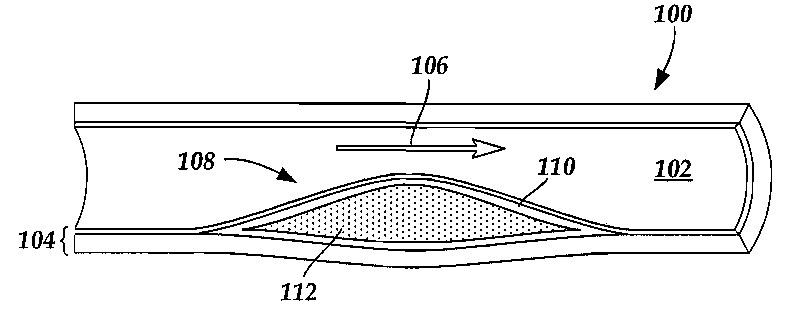
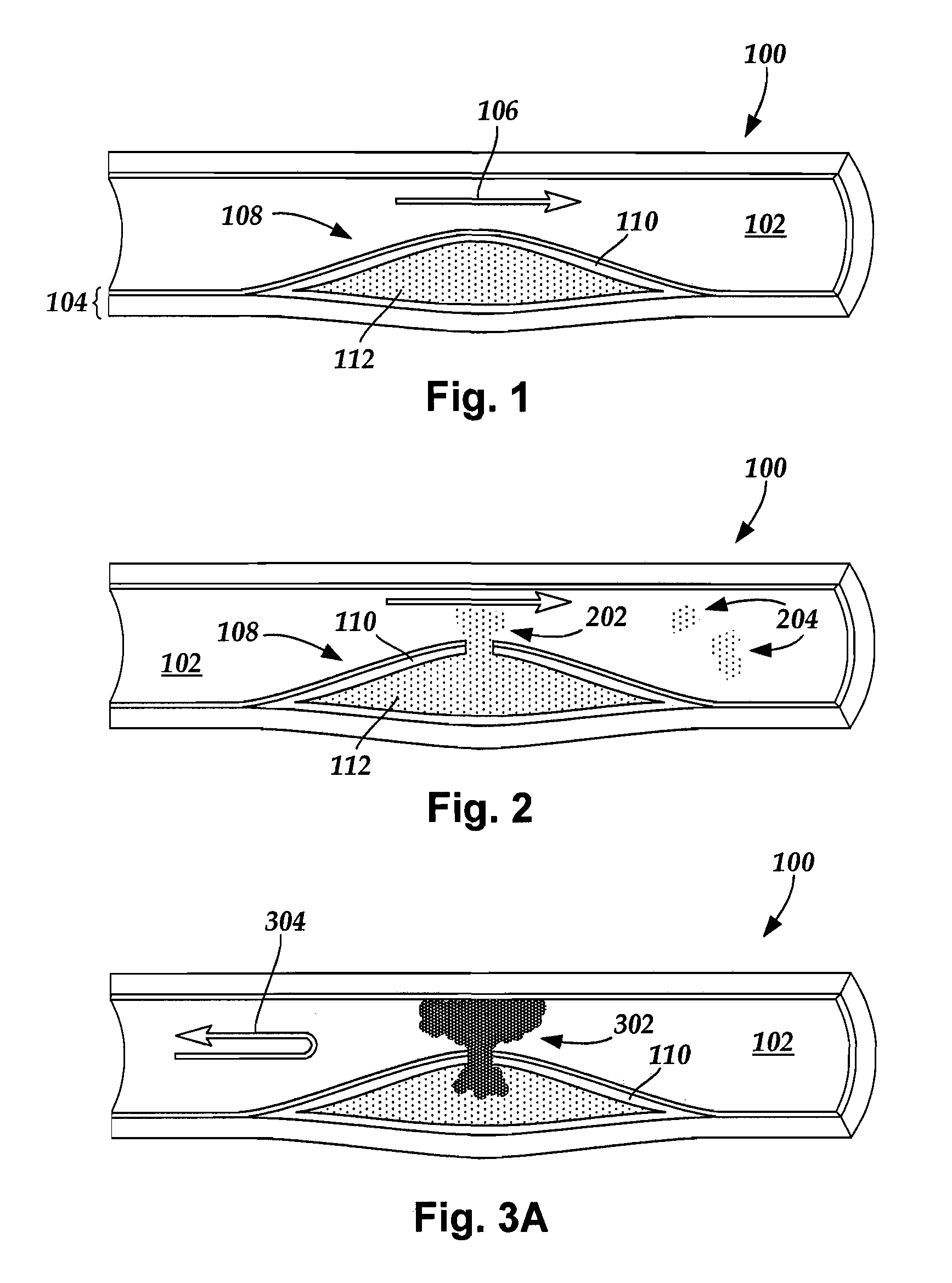
[0018] The present invention is directed to systems for imaging blood vessel walls to predict the risk of fibrous cap rupture and methods of using the systems. The present invention is also directed to systems that include imaging blood vessel walls to identify microcalcifications, as well as methods of using the systems.

[0019] Suitable medical diagnostic systems include, but are not limited to, in vivo imaging techniques for imaging a portion of a blood vessel of a patient to observe calcified inclusions (“microcalcifications”) in a fibrous cap (“cap”) of an atheroma. The term “microcalcification” refers to a calcified inclusion with a diameter of fifty micrometers or less. For example, a microcalcification diameter may be up to fifty micrometers, up to forty micrometers, up to twenty-five micrometers, up to twenty micrometers, between five micrometers and fifty micrometers, between five micrometers and forty micrometers, between five micrometers and twenty-five micrometers, and b...


PUM


Abstract

A system includes an in vivo imaging device for imaging a blood vessel with a resolution level of at least fifty micrometers. The in vivo imaging device is capable of detecting a microcalcification in a fibrous cap of an atheroma. The system also includes a processor for receiving an image of the blood vessel from the in vivo imaging device. The processor uses the image to determine whether the blood vessel contains at least one microcalcification within the fibrous cap. In some embodiments, the processor is configured and arranged to predict a risk of rupture of the fibrous cap based, at least in part, on the presence of the at least one microcalcification. In some embodiments, treatment of a patient is based on the determination from the imaging whether the blood vessel includes at least one microcalcification within the fibrous cap of the atheroma.

Description

CROSS-REFERENCE TO RELATED APPLICATIONS [0001] This application claims priority from U.S. Ser. No. 60 / 848,027, filed Sep. 28, 2006, which is hereby incorporated by reference in its entirety.TECHNICAL FIELD [0002] The present invention is directed to systems for imaging blood vessel walls to predict the risk of fibrous cap rupture and methods of using the systems. The present invention is also directed to systems that include imaging blood vessel walls to identify microcalcifications, as well as methods of using the systems. BACKGROUND OF THE INVENTION [0003] Atherosclerosis is a disease affecting arterial blood vessels. Over time, arterial walls may respond to the deposition of lipoproteins by undergoing an inflammatory response and forming plaques, or atheromas. Atheromas can eventually lead to the occlusion of blood vessels by either swelling to a sufficient size to occlude a blood vessel or by rupturing and causing a downstream occlusion. Blood vessel occlusion may lead to morbid...

Claims


Application Information

Patent Timeline
Patent Type & Authority Applications(United States)
IPC IPC(8): A61B8/13
CPCA61B5/0066A61B5/0068A61B5/0073A61B5/0075A61B6/583A61B5/055A61B6/03A61B8/12A61B5/0084
Inventor WEINBAUM, SHELDONVENGRENYUK, YULIYACARDOSO, LUISPARRA, LUCASCARLIER, STEPHANEXANTHOS, SAVVAS
Owner COLUMBIA UNIV (US)
Why Eureka
Try Eureka