Methods and systems for attenuation correction in medical imaging

a technology of attenuation correction and medical imaging, applied in the field of medical imaging systems, can solve the problems of indeterminate or incorrect diagnosis of nodules, inability to accurately detect nodules, and high influence on image quality of at least some known pet and ct systems by physiological patient movemen

Active Publication Date: 2008-05-08
GENERAL ELECTRIC CO
View PDF1 Cites 56 Cited by
  • Summary
  • Abstract
  • Description
  • Claims
  • Application Information

AI Technical Summary

Problems solved by technology

However, due to the comparatively lower resolution of PET images as compared to CT images, and due to the effects of respiratory or patient motion during a PET scan, nodule activity can be blurred in the PET scan.
Consequently, it can be difficult to quantify the nodule activity with a PET scan alone, which may result in an indeterminate or incorrect outcome of the diagnosis of the nodule.
More particularly, the image quality of at least some known PET and CT systems is highly affected by physiological patient movement.
Such image quality may affect diagnosis.
Further, misalignment of a CT attenuation map and the PET emission image that is due to respiratory motion may cause errors in attenuation correction (AC) factors and may produce artifacts in the final reconstructed AC PET image.
For instance, “under attenuation correction” may have the potential of introducing artifacts that resemble artificial myocardial perfusion defects in cardiac PET.
Thus, in the case of cardiac PET, wherein helical CT data is used, attenuation artifacts may result in areas having artificially reduced tracer uptake in the myocardial wall that may be incorrectly interpreted as perfusion defects by utilizing helical CT attenuation correction (CTAC) data.

Method used


Image

  • Methods and systems for attenuation correction in medical imaging
  • Methods and systems for attenuation correction in medical imaging
  • Methods and systems for attenuation correction in medical imaging

Examples


Embodiment Construction

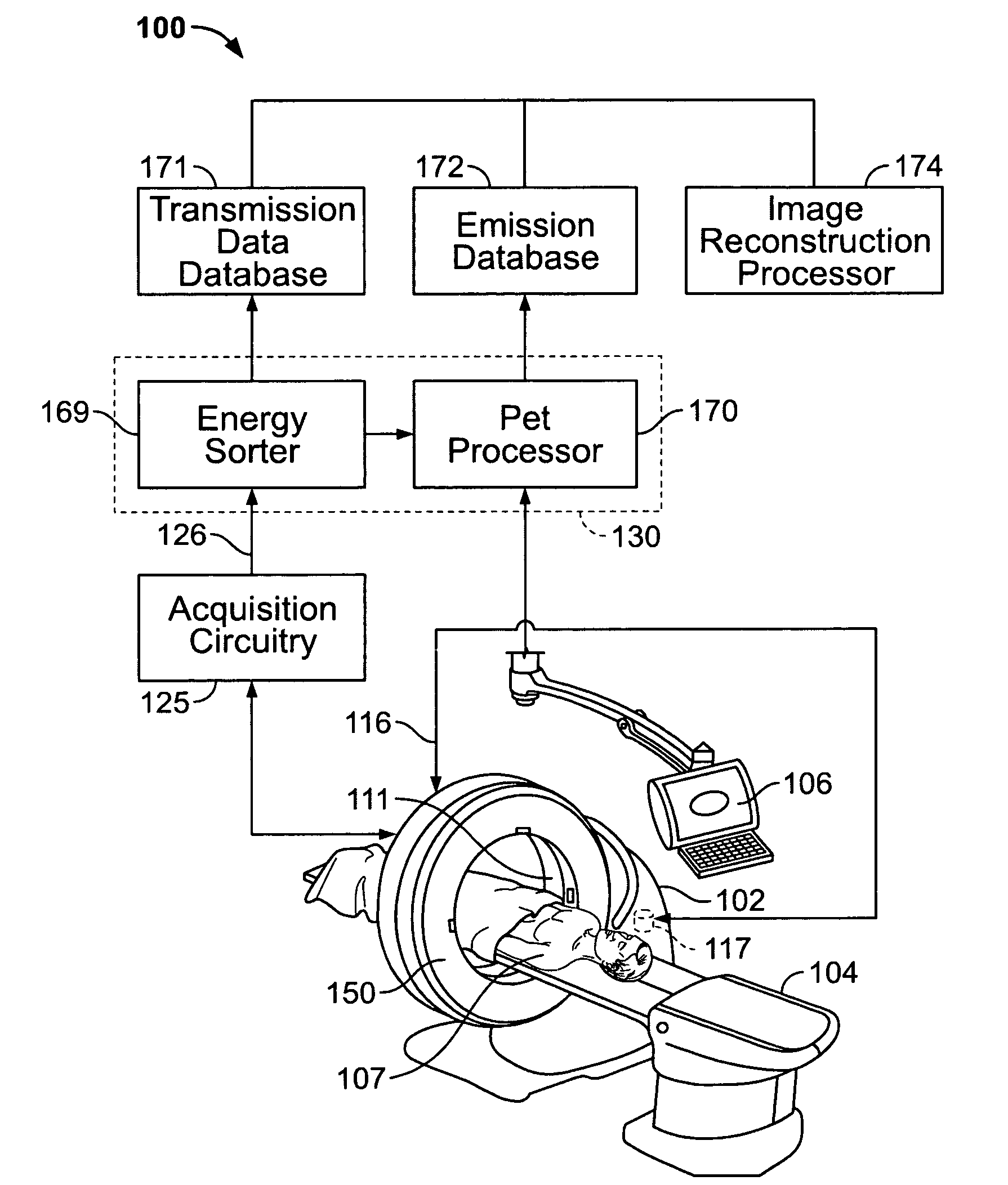
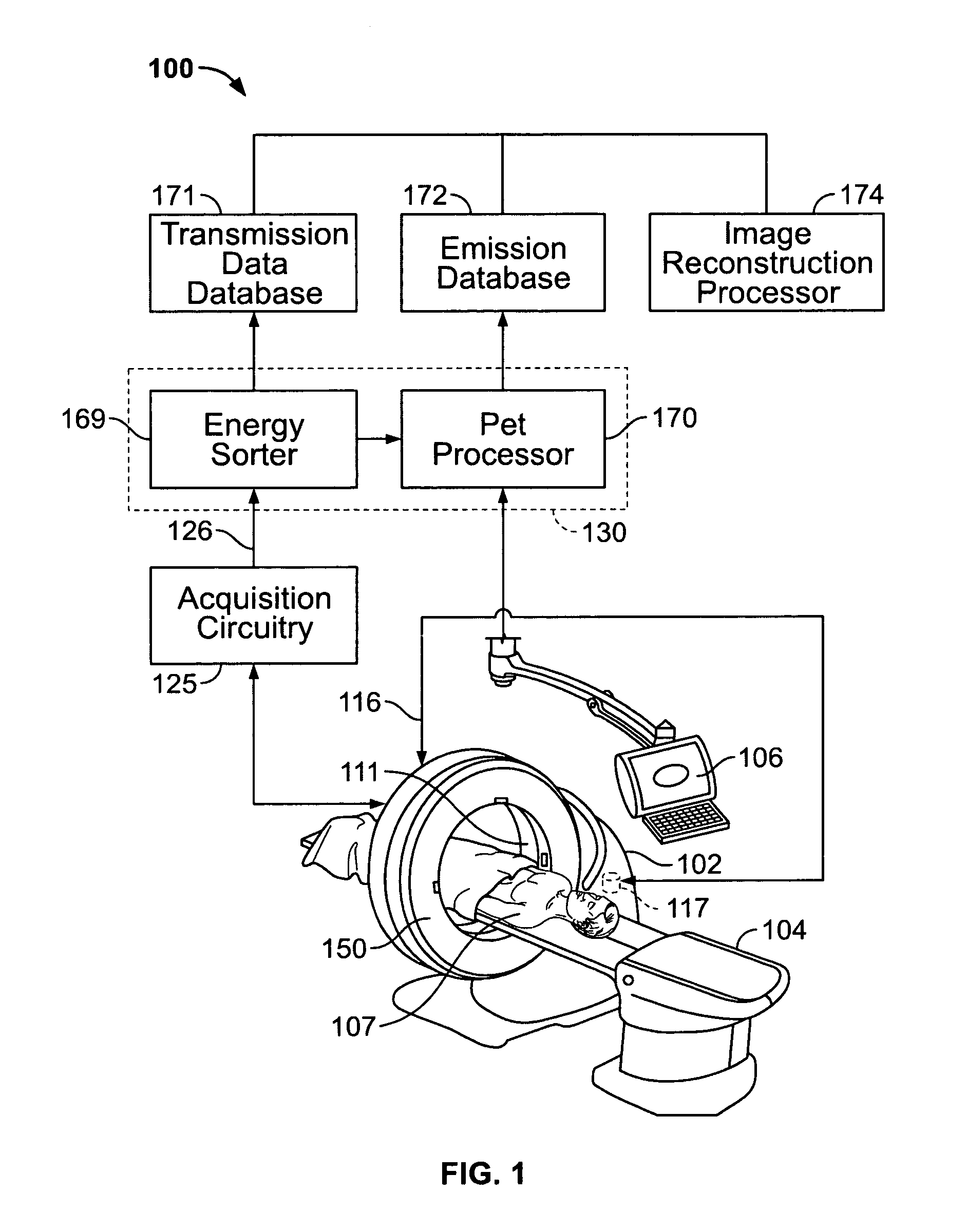
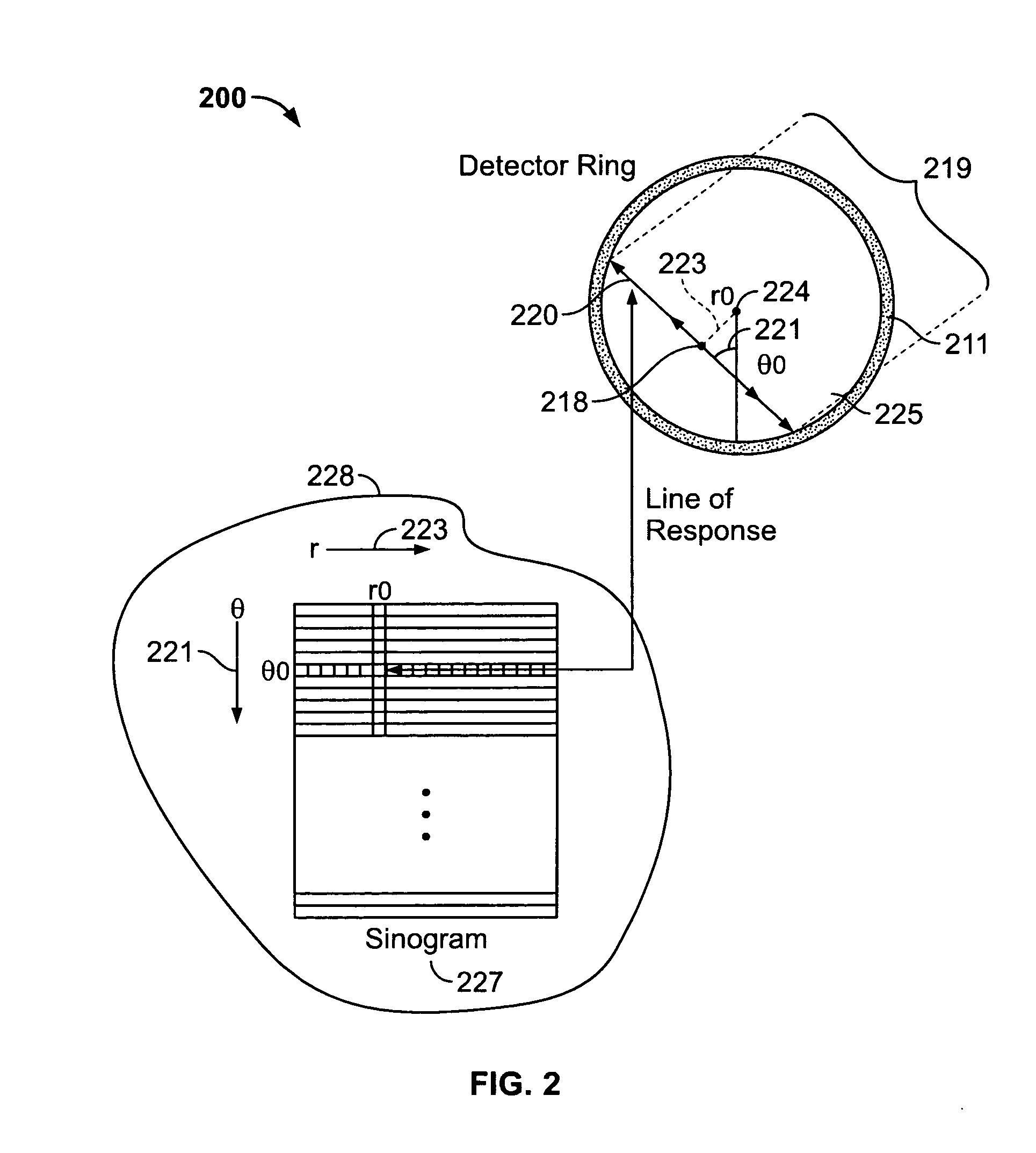
[0012]In the following detailed description, reference is made to the accompanying drawings which form a part hereof, and in which is shown by way of illustration specific embodiments in which the present invention may be practiced. These embodiments, which are also referred to herein as “examples,” may be combined, or other embodiments may be utilized and structural, logical and electrical changes may be made without departing from the scope of the various embodiments of the present invention. The following detailed description is, therefore, not to be taken in a limiting sense, and the scope of the various embodiments of the present invention is defined by the appended claims and their equivalents.

[0013]In this document, the terms “a” or “an” are used, to include one or more than one. In this document, the term “or” is used to refer to a nonexclusive or, unless otherwise indicated. In addition, as used herein, the phrase “pixel” also includes embodiments of the present invention w...


PUM


Abstract

Methods and systems for imaging a patient are provided. The method includes scanning a patient and acquiring a plurality of frames of cine computed tomography (CT) images during one complete respiratory cycle. In one embodiment, a method is provided that includes selecting an organ of interest in the cine CT data and selecting a value for each pixel in the organ of interest that represents the maximum density measurement. An attenuation corrected positron emission tomography (PET) image is constructed based on the maximization of the pixel intensity of the organ of interest in the CT attenuation correction map. Incorrect attenuation correction values for PET images can be avoided by utilizing the CT attenuation correction map.

Description

BACKGROUND OF THE INVENTION[0001]This invention relates generally to medical imaging systems, and more particularly to attenuation correction for medical imaging.[0002]A nodule found during a computed tomography (CT) scan often requires a patient to return many months later and obtain another CT scan to determine malignancy based on a nodule doubling time. Positron emission tomography (PET) scans may be helpful in diagnosis due to increased metabolic activity in the region of the nodule. However, due to the comparatively lower resolution of PET images as compared to CT images, and due to the effects of respiratory or patient motion during a PET scan, nodule activity can be blurred in the PET scan. Consequently, it can be difficult to quantify the nodule activity with a PET scan alone, which may result in an indeterminate or incorrect outcome of the diagnosis of the nodule.[0003]More particularly, the image quality of at least some known PET and CT systems is highly affected by physi...

Claims


Application Information

Patent Timeline
Patent Type & Authority Applications(United States)
IPC IPC(8): A61B6/00G01D18/00G01T1/16
CPCA61B6/032A61B6/469A61B6/037A61B6/5235A61B6/488
Inventor THOMAS, ALICIA MARIAO'DAY, PATRICK JOSEPH
Owner GENERAL ELECTRIC CO
Why Eureka
Try Eureka