Solid type microneedle and methods for preparing it

a microneedle and solid type technology, applied in the field of solid microneedles, can solve the problems of pain in inability to achieve painless skin penetration, and inability to reliably deliver drugs or cosmetic components to the target site, so as to minimize the pain of skin penetration of microneedles

Inactive Publication Date: 2008-05-08
IND ACADEMIC CORP FOUND YONSEI UNIV
View PDF5 Cites 55 Cited by
  • Summary
  • Abstract
  • Description
  • Claims
  • Application Information

AI Technical Summary

Benefits of technology

[0012] It is important that microneedles should have a structure, which is thin and long enough to minimize not only pain in their penetration into the skin, but also foreign matter sensation after their insertion into the skin. According to the present invention, the solid microneedles can be fabricated to have the desired diameter and length without any particular limitation. Preferably, the solid microneedles can be fabricated to have an upper end diameter of 5-40 μm and an effective length of 500-2,000 μm.

Problems solved by technology

However, the solid silicon microneedle according to this method has a diameter of 50-100 μm and a length of 500 μm, and thus it has problems that it is impossible to realize painless skin penetration and that in-vivo delivery of a drug or a cosmetic component to the target site is not reliably achieved.
Said Japanese Patent suggests the fabrication of transdermal microneedles and the transdermal delivery of drugs through the fabricated microneedles, but the skin penetration of the microneedles involves pain.
Due to the technical limitation in the fabrication of a mold, it is impossible to fabricate a microneedle, which has the length required for effective drug delivery, that is, a length of 1 mm or more, and, at the same time, an appropriate upper end diameter which causes no pain.
For this reason, it is limited in its ability to allow a drug or a beauty component to permeate deep into the skin.
In the prior solid microneedles, the raw material thereof was limited to materials such as silicon, polymers, metal, glass or the like, due to the limitation on the fabrication methods thereof, and it was not easy to achieve the desired effects, because they were fabricated to have a diameter of 50-100 μm at the upper end part and a length of 500 μm.

Method used


Image

  • Solid type microneedle and methods for preparing it
  • Solid type microneedle and methods for preparing it
  • Solid type microneedle and methods for preparing it

Examples


Embodiment Construction

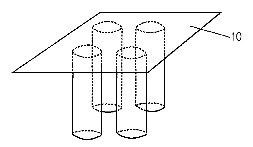
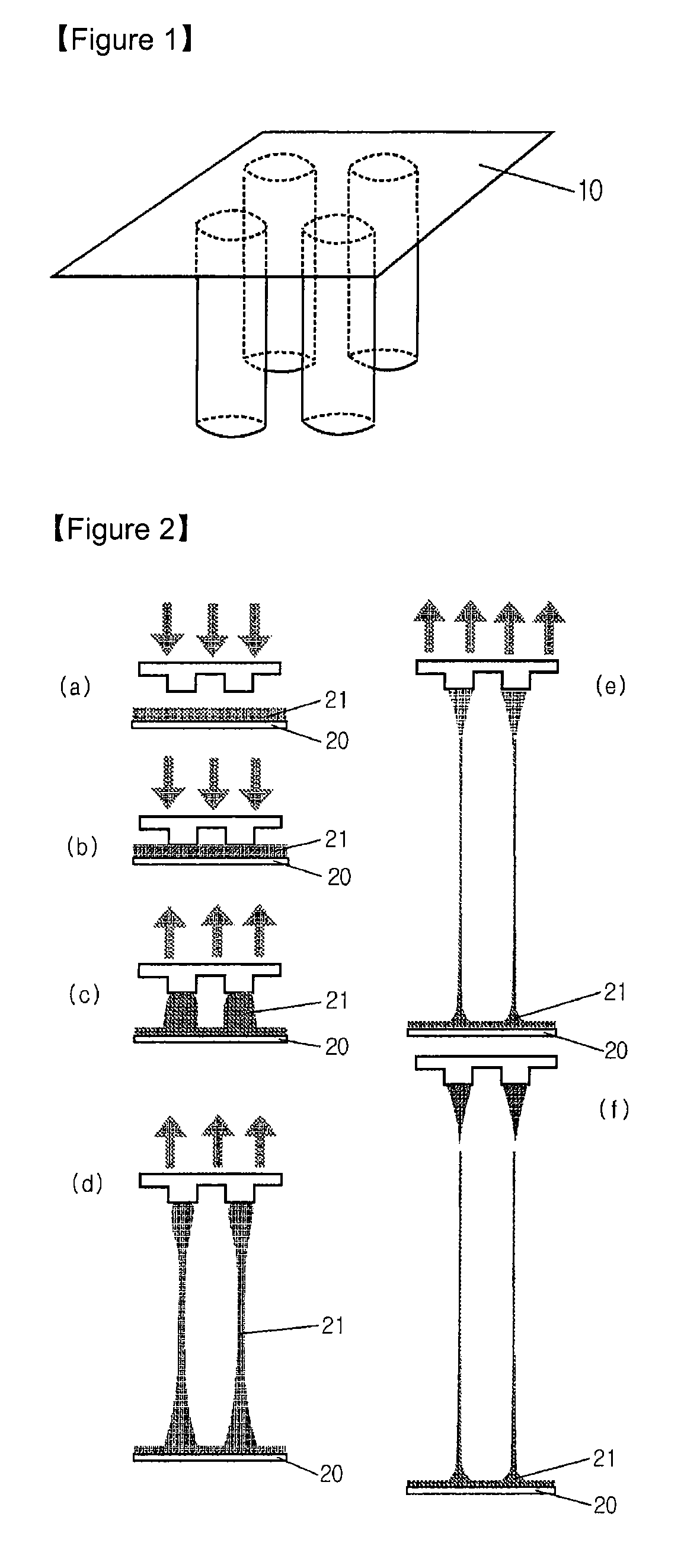
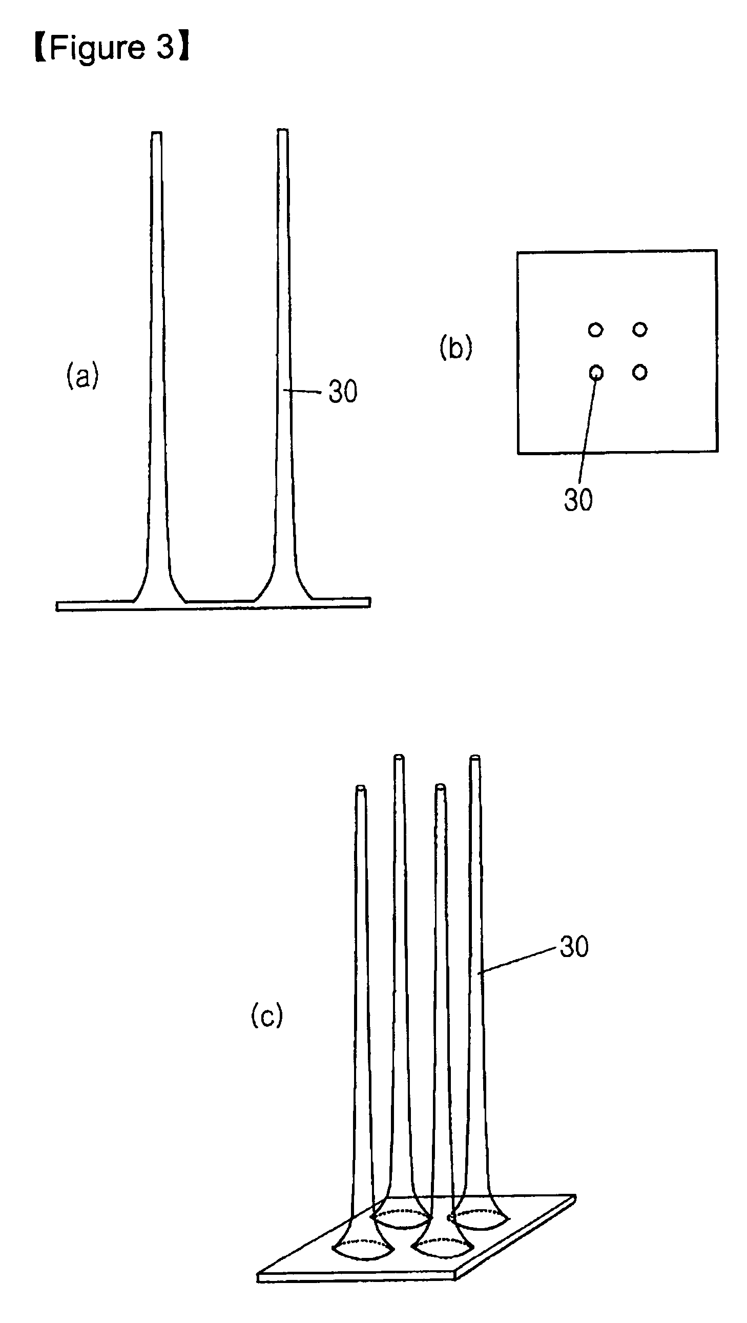
[0024] SU-8 2050 photoresist (commercially purchased from Microchem) having a viscosity of 14,000 cSt was used to fabricate solid microneedles. For this purpose, SU-8 2050 was coated on a flat glass panel to a certain thickness, and it was maintained at 120° C. for 5 minutes to maintain its flowing properties. Then, the coated material was brought into contact with a frame having 2×2 pillar patterns formed thereon, each pillar having a diameter of 200 μm (See FIG. 1). The temperature of the glass panel was slowly lowered to 90-95° C. over about 5 minutes to solidify the coated SU-8 2050 and to increase the adhesion between the frame and the SU-8. Then, while the temperature was slowly lowered from 90-95° C., the coated SU-82050 was drawn at the speed of 1 μm / s for 60 minutes using the frame which adhered to the coated SU-82050 (See FIG. 2). After 60 minutes of drawing, solid microneedles, each having a length of about 3,600 μm, were formed. Subsequently, the solid microneedles were ...


PUM


Abstract

Disclosed herein are biodegradable solid microneedles and a fabrication method thereof. The microneedles are small in diameter and are long and hard enough to pass through the stratum corneum. Thus, the biodegradable solid microneedles can be used for painless transdermal drug delivery, the detection of biological samples such as blood, and biopsy.

Description

TECHNICAL FIELD [0001] The present invention relates to solid microneedles and a fabrication method thereof. Furthermore, the present invention relates to in-vivo delivery of a drug or a cosmetic component through solid microneedles. BACKGROUND ART [0002] Generally, microneedles are used in in-vivo drug delivery, the detection of biological samples, and biopsy. Drug delivery with microneedles aims to deliver a drug through the skin rather than biological circulatory systems such as blood vessels or lymphatic vessels. Accordingly, the microneedles should not cause pain when they penetrate the skin, and should have sufficient length such that they can deliver drugs to the target site. In addition, the microneedles should have excellent physical hardness such that they can penetrate the stratum corneum having a thickness of 10-20 μm. Since in-plane microneedles were suggested (“Silicon-processed Microneedles”, Journal of microelectrochemical systems Vol. 8, Nol, March 1999), various ty...

Claims


Application Information

Patent Timeline
Patent Type & Authority Applications(United States)
IPC IPC(8): A61M5/32
CPCA61M37/0015A61M2037/0061A61M2037/0053A61M37/00
Inventor JUNG, HYUNG ILLEE, KWANG
Owner IND ACADEMIC CORP FOUND YONSEI UNIV
Why Eureka
Try Eureka