Methods and systems for optimizing high resolution image reconstruction

a high-resolution image and reconstruction technology, applied in tomography, instruments, applications, etc., can solve the problems of insufficient resolution for accurate measurement of lungs, current clinical image acquisition techniques for lungs that do not provide enough resolution for reconstruction parameters used clinically for diagnostic reading of lung images, and inability to quantitatively analyze reconstruction parameters, etc., to facilitate quantification of image structures and facilitate quantification

Active Publication Date: 2008-05-22
GENERAL ELECTRIC CO
View PDF17 Cites 32 Cited by
  • Summary
  • Abstract
  • Description
  • Claims
  • Application Information

AI Technical Summary

Benefits of technology

[0004]In one embodiment, a method for reconstructing a multi-resolution image of an object includes reconstructing a first three-dimensional image at a first resolution, determining at least one volume of interest in the generated image, and reconstructing a second three-dimensional image of the determined at least one volume of interest at a second resolution, the second resolution being higher than the first resolution such that a quantification of image structures is facilitated.
[0005]In another embodiment, an imaging system includes a stationary member, a rotating member rotatably coupled to the stationary member wherein the rotating member includes an opened area proximate an axis about which the rotating member rotates. The imaging system also includes an x-ray source provided on the rotating member, and an x-ray detector disposed on the rotating member and configured to receive x-rays from the x-ray source. The system further includes a processor communicatively coupled to at least one of the x-ray source and the x-ray detector wherein the processor is configure to receive image data relating to an object and then reconstruct a first image at a first resolution using the received image data, determine at least one volume of interest in the generated image, and reconstruct a second image of the determined at least one volume of interest at a second resolution, the second resolution being greater than the first resolution such that a quantification of image structures is facilitated.

Problems solved by technology

However, because of limitations of storage space and workflow, acquisition of images at the highest resolution available on a CT system may not be possible.
Current clinical image acquisition techniques for lungs do not provide enough resolution for an accurate measurement of these airways.
High-resolution reconstruction can help the accuracy but current clinical protocols do not use these because of the high number of images required to cover an entire field of view at such a reconstruction resolution.
Moreover, some of the reconstruction parameters used clinically for diagnostic reading of lung images have been found to be not suitable for quantitative analysis.

Method used


Image

  • Methods and systems for optimizing high resolution image reconstruction
  • Methods and systems for optimizing high resolution image reconstruction
  • Methods and systems for optimizing high resolution image reconstruction

Examples


Embodiment Construction

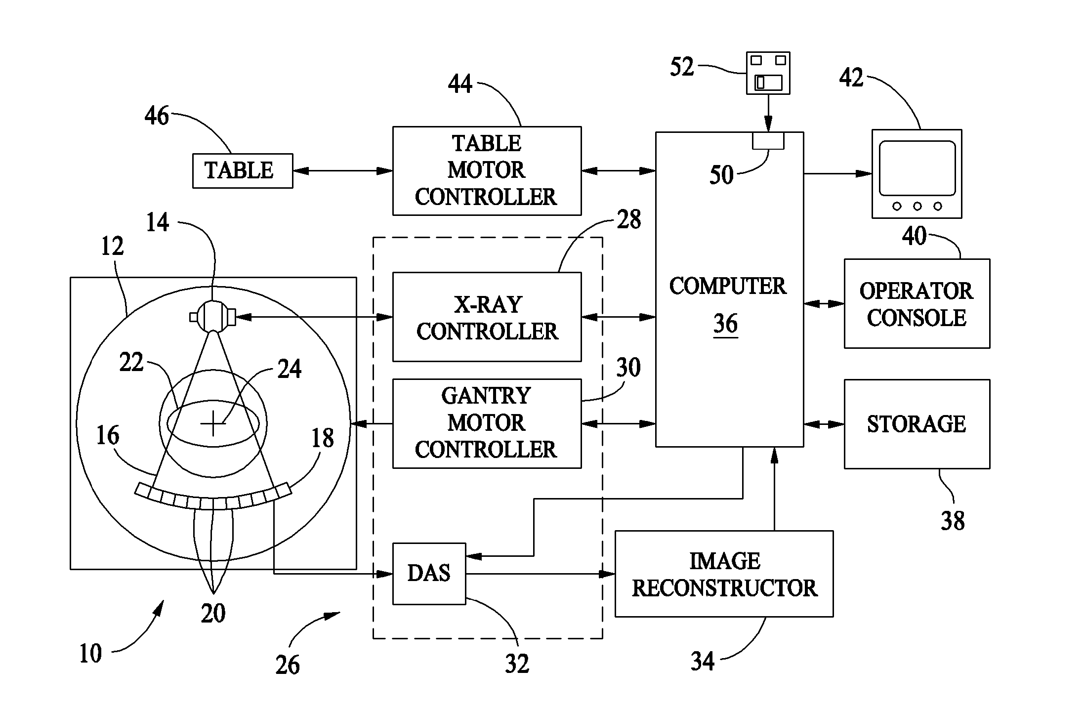
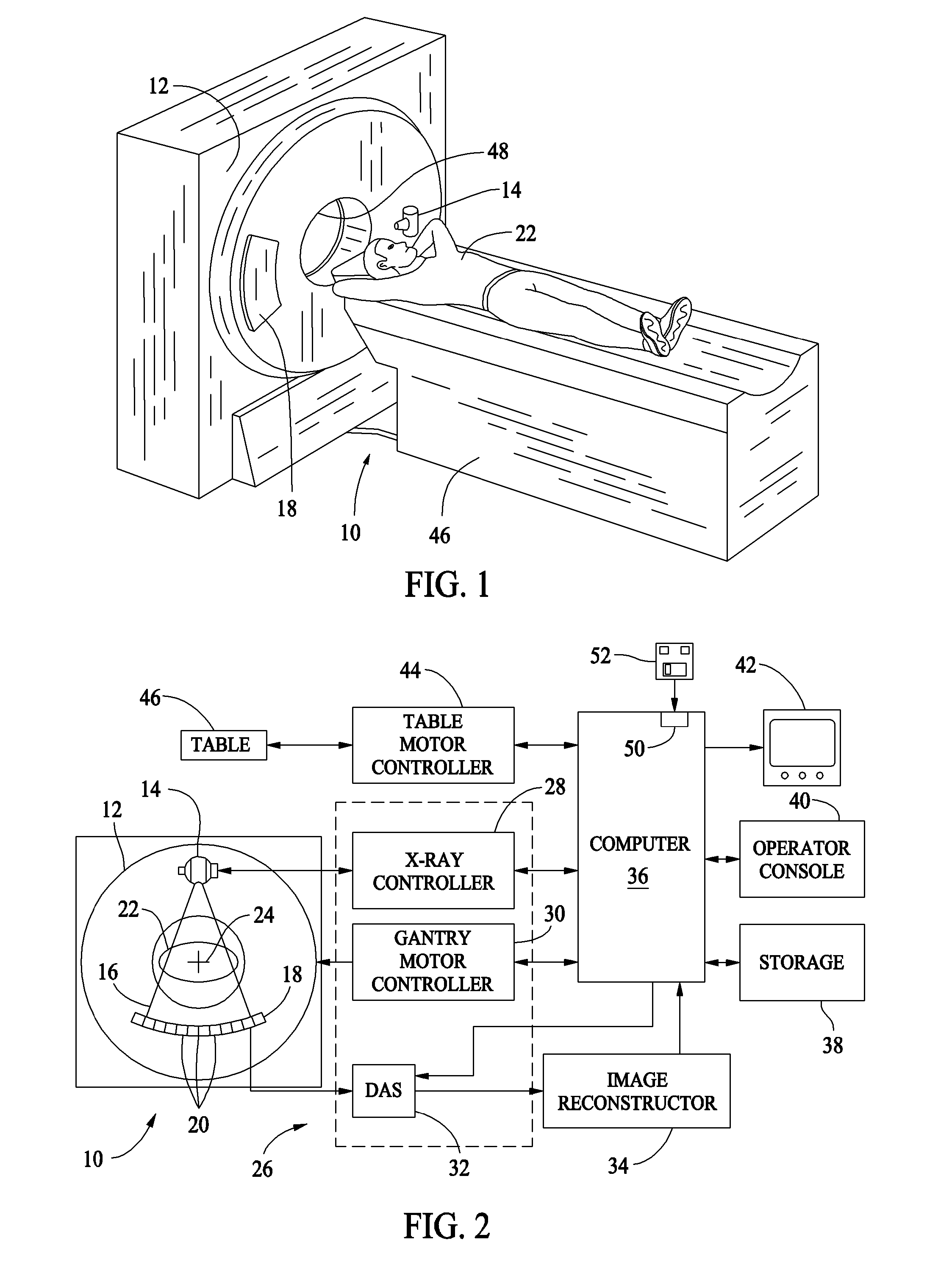
[0012]As used herein, an element or step recited in the singular and proceeded with the word “a” or “an” should be understood as not excluding plural said elements or steps, unless such exclusion is explicitly stated. Furthermore, references to “one embodiment” of the present invention are not intended to be interpreted as excluding the existence of additional embodiments that also incorporate the recited features. Moreover, unless explicitly stated to the contrary, embodiments “comprising” or “having” an element or a plurality of elements having a particular property may include additional such elements not having that property. For example, CT imaging apparatus embodiments may be described herein as having a plurality of detector rows that are used in a certain process. Such embodiments are not restricted from having other detector rows that are not used in that process.

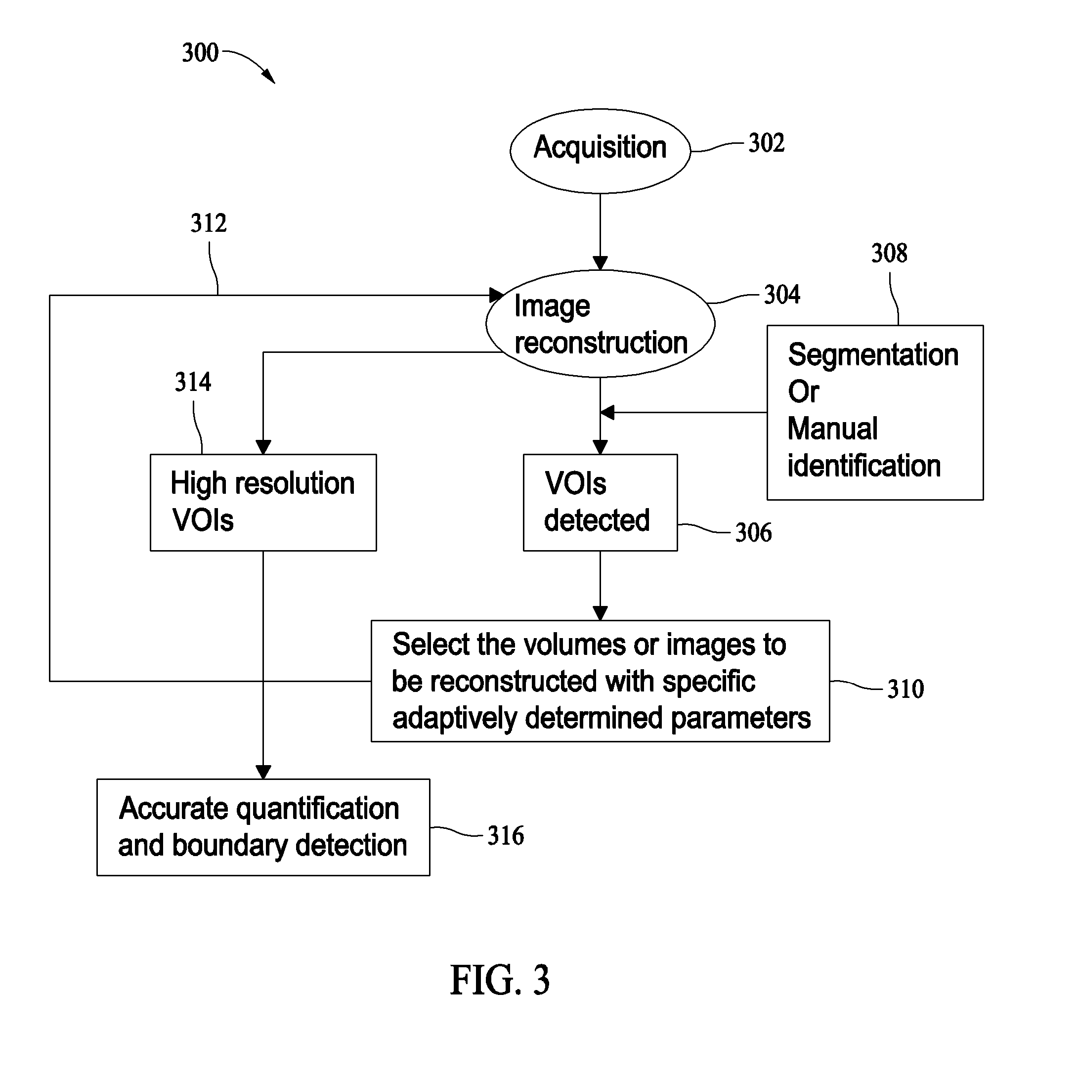
[0013]Also as used herein, the phrase “reconstructing an image” is not intended to exclude embodiments of the pr...


PUM


Abstract

Methods and apparatus for reconstructing a multiple resolution images of an object are provided. The method includes reconstructing a first three-dimensional image at a first resolution, determining at least one volume of interest in the generated image, and reconstructing a second three-dimensional image of the determined at least one volume of interest at a second resolution, the second resolution being higher than the first resolution such that a quantification of image structures is facilitated.

Description

BACKGROUND OF THE INVENTION[0001]This invention generally relates to rotating imaging scanner systems and more particularly, to methods and systems for optimizing high resolution image reconstruction.[0002]Detecting an accurate shape and size of small objects in CT images for example, a volume of soft plaque in a coronary vessel is affected by the resolution of the images that are being analyzed. To achieve the most accurate estimation of small volumes, the images of the object should be viewed at the highest possible resolution. However, because of limitations of storage space and workflow, acquisition of images at the highest resolution available on a CT system may not be possible.[0003]Small airways in lungs are the early precursors of thoracic diseases for example, but not limited to chronic obstructive pulmonary disease (COPD), asthma, and bronchitis. Changes in the wall thickness and lumen of these small airways (<2 mm) facilitate indicating the progression of the disease a...

Claims


Application Information

Patent Timeline
Patent Type & Authority Applications(United States)
IPC IPC(8): A61B6/03G06K9/00
CPCA61B6/032A61B6/583G06T11/006
Inventor DUTTA, SANDEEPSIROHEY, SAAD AHMEDAVINASH, GOPAL B.SKINNER, JOHN V.NEZET, PATRICIA LEHAAS, DEANN MARIEGAUTHAM, SARDAR MALJOSHI, MUKTA C.
Owner GENERAL ELECTRIC CO
Why Eureka
Try Eureka