Endoscopic System for In-Vivo Procedures

a tissue characterization and endoscope technology, applied in the field of in-vivo tissue characterization endoscope, can solve the problems of unfulfilled medical goals, untreated, undetected and untreated, and single colonoscopy is often not enough to identify the source of colorectal cancer

Inactive Publication Date: 2008-06-26
DUNE MEDICAL DEVICES
View PDF93 Cites 140 Cited by
  • Summary
  • Abstract
  • Description
  • Claims
  • Application Information

AI Technical Summary

Problems solved by technology

In spite of enormous expenditures of financial and human resources, early detection of malignant tumors remains an unfulfilled medical goal.
While it is known that a number of cancers are treatable if detected at an early stage, lack of reliable screening procedures results in their being undetected and untreated.
However, a single colonoscopy is often not sufficient to identify the source of colorectal bleeding which is typically sporadic and in many cases would be best located by observing the entire colon over a period of time.
The aforementioned endoscopes, while providing means to access and visualize portions of the gastrointestinal track, do not provide means of detecting gastrointestinal pathologies, which are not clearly visible.
In particular, they do not provide means for localization and differentiation of occult tumors.
Similarly, lung cancer is the leading cause of cancer death in both men and women in Western society.
However, detection at an early stage is rare.
However, biopsy diagnosis performed in a laboratory and follow up procedures based on laboratory biopsy suffer from inherent disadvantages, as follows:
ii. it may happen that the biopsy is taken from a region near the tumor, and not the tumor itself, leading to erroneous false negative results
iii. the exact location from which the biopsy was taken, may be difficult to reproduce; an

Method used


Image

  • Endoscopic System for In-Vivo Procedures
  • Endoscopic System for In-Vivo Procedures
  • Endoscopic System for In-Vivo Procedures

Examples


Embodiment Construction

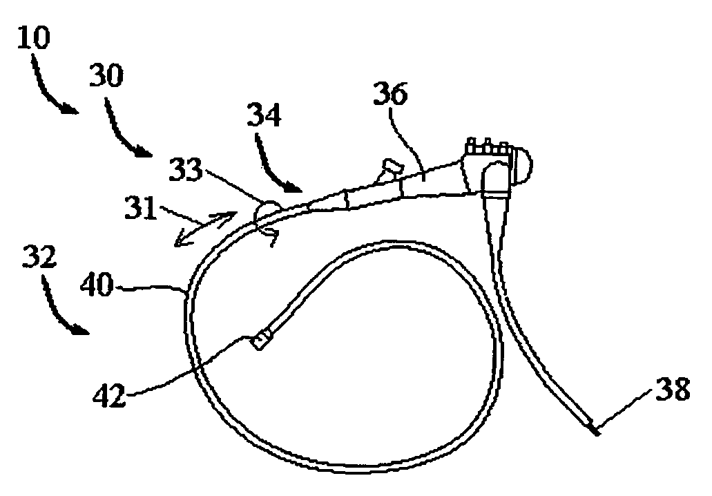
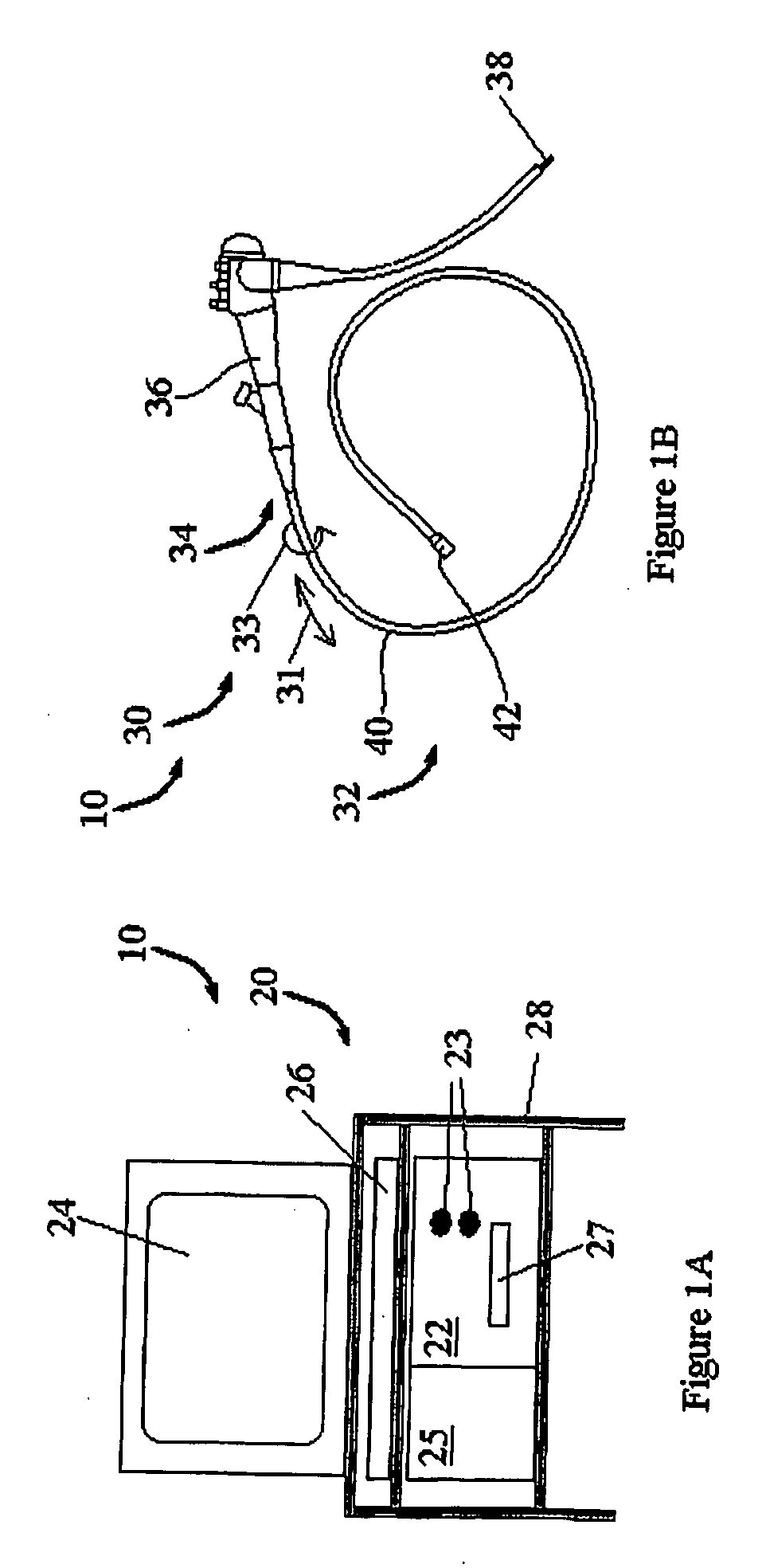
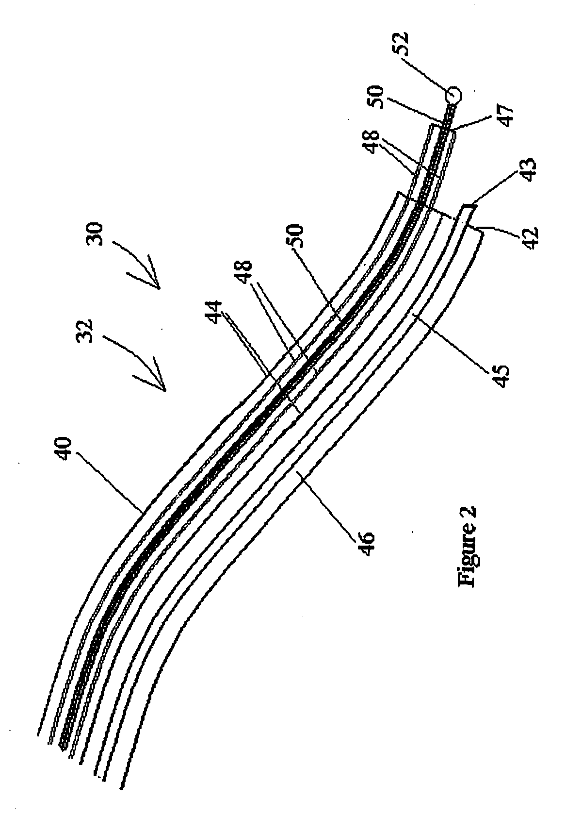
[0083]The present invention relates to an endoscopic system for in-vivo tissue characterization, using a nonirradiative electromagnetic sensor. The endoscopic system is further configured to employ several follow-up procedures, for example, biopsy sampling, localized surgery, dispensing a medicament, and the like, so that on the whole, the endoscopic system provides for the early detection of cancerous and pre-cancerous tissue, in vivo, and for the application of immediate follow-up procedures to any such tissue.

[0084]The principles and operation of the device and method according to embodiments of the present invention may be better understood with reference to the drawings and accompanying descriptions.

[0085]Before explaining at least one embodiment of the invention in detail, it is to be understood that the invention is not limited in its application to the details of construction and the arrangement of the components set forth in the following description or illustrated in the d...


PUM


Abstract

An endoscopic system for in-vivo tissue characterization, using a nonirradiative electromagnetic sensor, is described. The endoscopic system is further configured to employ several follow-up procedures, for example, biopsy sampling, localized surgery, dispensing a medicament, and the like, so that on the whole, the endoscopic system provides for the early detection of cancerous and pre-cancerous tissue, in vivo, and for the application of immediate follow-up procedures to any such tissue.

Description

FIELD AND BACKGROUND OF THE INVENTION[0001]The present invention relates to an endoscopic system for in-vivo tissue characterization, employing a nonirradiative electromagnetic sensor.[0002]The impact of cancer is great. In spite of enormous expenditures of financial and human resources, early detection of malignant tumors remains an unfulfilled medical goal. While it is known that a number of cancers are treatable if detected at an early stage, lack of reliable screening procedures results in their being undetected and untreated.[0003]Various forms of endoscopes are currently in use. For example, diagnosis of different conditions of the colon generally involves using a colonoscope. A typical colonoscope includes, at its distal end, with respect to an operator, a light source, a video chip, and a suction channel. These elements are all in communication with a proximal end of the colonoscope via wires and channels housed within a flexible tube. The distal end is inserted into a patie...

Claims


Application Information

Patent Timeline
Patent Type & Authority Applications(United States)
IPC IPC(8): A61B1/018A61M25/09
CPCA61B1/018
Inventor HASHIMSHONY, DAN
Owner DUNE MEDICAL DEVICES
Why Eureka
Try Eureka