Method using three-dimensional mechanics and tissue specific imaging of blood vessels and plaques for detection

A technology of three-dimensional mechanics and tissue characteristics, which is applied in the detection of organ movement/change, medical science, and measurement devices. It can solve problems such as inaccurate blood flow velocity, reduced estimation accuracy of vessel wall shear rate, and spectral ambiguity.

Inactive Publication Date: 2015-03-11
XI AN JIAOTONG UNIV
View PDF0 Cites 46 Cited by
  • Summary
  • Abstract
  • Description
  • Claims
  • Application Information

AI Technical Summary

Problems solved by technology

However, the traditional B-ultrasound imaging mode is realized by sequentially focusing on transmitting and receiving scan lines one by one. The imaging frame rate of this imaging mode is only tens of hertz, while the pulse wave velocity of the human body can reach several meters per second. Therefore, the frame rate of traditional ultrasound imaging Rate Difficult to Capture Pulse Wave Propagation
In addition, there is a certain angle between the pulse Doppler emission beam and the blood flow in the traditional B-ultrasound, and only the flow ve

Method used


Image

  • Method using three-dimensional mechanics and tissue specific imaging of blood vessels and plaques for detection
  • Method using three-dimensional mechanics and tissue specific imaging of blood vessels and plaques for detection
  • Method using three-dimensional mechanics and tissue specific imaging of blood vessels and plaques for detection

Examples


Embodiment Construction

[0054] The present invention is described in detail below in conjunction with accompanying drawing.

[0055] The three-dimensional mechanical characteristics and tissue characteristic imaging detection method of blood vessels and plaques of the present invention, its specific implementation mode, comprises the following several steps:

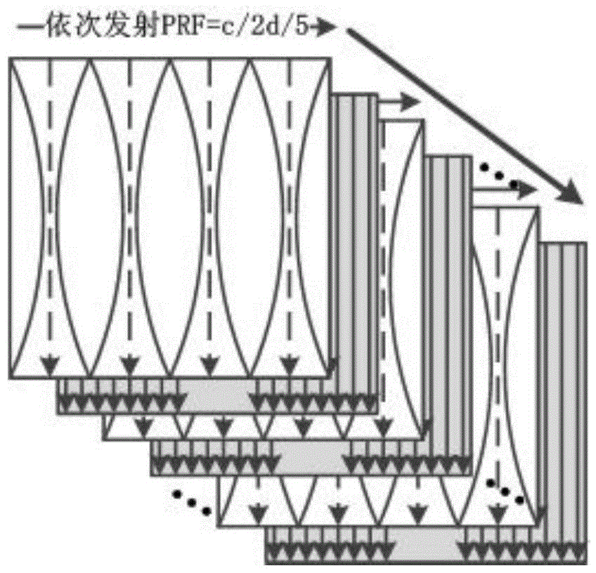
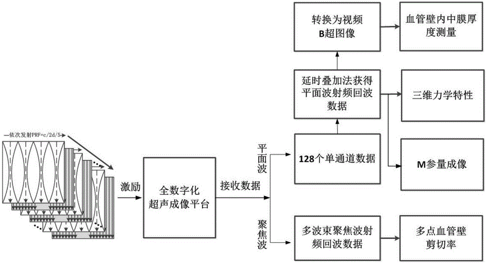
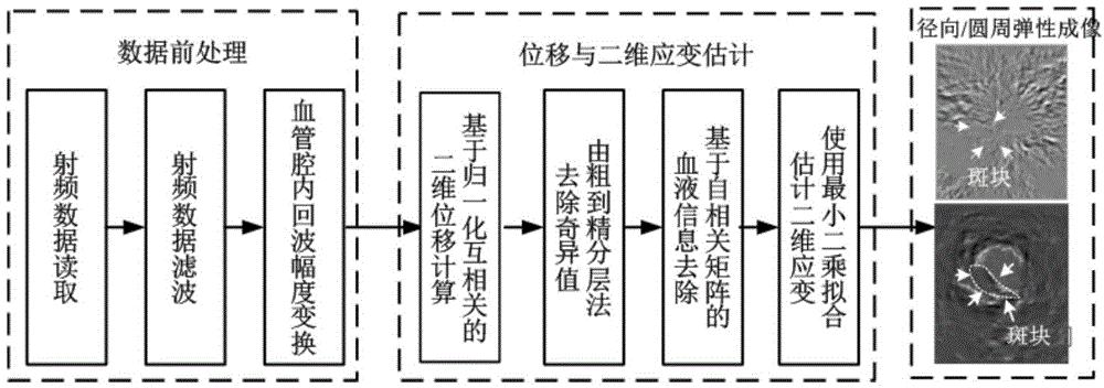
[0056] Step 1. Use multi-beam focused waves and ultra-fast plane waves to alternately transmit sequences, conduct elastography from the radial and circumferential directions of carotid vessels and plaques, obtain the local propagation velocity of pulse waves along the vessel axis, and obtain vessel radial , Circumferential and axial three-dimensional mechanical properties. Calculate the change curve of the shear rate of the vessel wall at multiple points at the plaque site with the cardiac cycle, and extract parameters such as the expansion coefficient, compliance coefficient, and hardness index of the vessel wall to characterize the degree of ...


PUM


Abstract

The invention relates to a method using three-dimensional mechanics characteristics and tissue specific imaging of blood vessels and plaques for detection. The method is on the basis of a multi-beam focus wave and superfast plane wave alternative transmitting sequence and can also be extended to a line-by-line scan imaging manner; the forms and the functions of the blood vessels and the plaques are evaluated and imaged respectively from the aspects of the radial-direction, circumferential and axial-direction three-dimensional mechanics characteristics, blood vessel wall shear rate, plaque form and tissue characterization, and the like; the new method which is provided for detection of the vulnerable plaques of a carotid artery is regarded as the improvement of the existing method.

Description

technical field [0001] The invention relates to the technical field of ultrasonic imaging, in particular to a method for imaging and detecting three-dimensional mechanical and tissue characteristics of carotid arteries and plaques based on alternating emission sequences of multi-beam focused waves and ultrafast plane waves. Background technique [0002] Atherosclerosis is a systemic systemic disease, and regular ultrasound examination of carotid arteries has become the basic method for clinical atherosclerotic plaque screening. Existing diagnostic techniques for clinical vascular plaques mainly rely on imaging methods such as B-ultrasound images, pulse Doppler, and color Doppler to determine the location, size, and stenosis of plaques. Although the degree of stenosis is often used as an important routine measurement parameter in the diagnosis and treatment of atherosclerosis, many studies have shown that the occurrence of most acute cerebrovascular events is not directly rel...

Claims


Application Information

Patent Timeline
IPC IPC(8): A61B8/00A61B8/08
CPCA61B8/0891A61B8/14A61B8/4488A61B8/5238G01S7/52042G01S7/5209G01S15/8979G01S15/8993
Inventor 万明习万锦锦胡咪咪宗瑜瑾何方莉张宇
Owner XI AN JIAOTONG UNIV
Why Eureka
Try Eureka