Real-time optoacoustic monitoring with electophysiologic catheters

a technology of electophysiologic catheters and optoacoustic monitoring, which is applied in the field of electrophysiologic catheters, can solve the problems of inability to obtain real-time information regarding the condition of the treatment site within the body, inability to provide information to the clinician, and irregular heart beating, etc., and achieves a high resolution and greater sensitivity to materials

Inactive Publication Date: 2008-06-26
BIOSENSE WEBSTER INC
View PDF26 Cites 49 Cited by
  • Summary
  • Abstract
  • Description
  • Claims
  • Application Information

AI Technical Summary

Benefits of technology

[0011]The present invention recognizes that light delivered in sufficiently short pulse widths is selectively absorbed by tissue elements and surrounding medium (blood), and is converted to heat. This heat produces an acoustic wave which can be detected by an acoustic sensor. A delay in receive time of the acoustic wave is proportional to the distance of elements generating the acoustic wave from the light delivery optics, and can be used to determine tissue thickness. To that end, optoacoustic imaging employs non-resonant acoustic frequencies which result from optical absorption properties of materials within the field of view of the light delivery optics. As such, the signal output has greater sensitivity to materials with different optical absorption properties, such as those between tissue and blood or air. It is therefore possible to obtain high resolution imaging of biological tissue through blood, with an operable range up to several centimeters (as determined by wavelength, optical absorption, and acoustic sensor size). Such imaging can be particularly advantageous during and concurrently with ablation for visualization of lesion formation.

Problems solved by technology

For certain types of minimally invasive medical procedures, real time information regarding the condition of the treatment site within the body is unavailable.
This lack of information inhibits the clinician when employing a catheter to perform a procedure.
This condition in the heart causes abnormal electrical signals to be generated in the endocardial tissue resulting in irregular beating of the heart.
The most frequent cause of cardiac arrhythmias is an abnormal routing of electricity through the cardiac tissue.
Furthermore, the ablation process can also cause undesirable charring of the tissue and localized coagulation, and can evaporate water in the blood and tissue leading to steam pops.
Clearly, post ablation evaluation is undesirable since correction requires additional medical procedures.
Biochemical differences between ablated and normal tissue can result in changes in electrical impedance between the tissue types.
Measuring impedance merely provides data as to the location of the tissue lesion but does not give qualitative data to evaluate the effectiveness of the lesion.
This technique, however, measures the success or lack thereof from each lesion, and yields no real-time information about the lesion formation.
But tissues can be acoustically homogenous and therefore undetectable by ultrasound imaging.
Similar limitations are posed by optical imaging based on time-resolved or phase resolved detection of diffusely reflected light pulses or photon density waves.
However, application of this technology in vivo, and particularly in vivo endocardial and epicardial applications, has been limited due to various factors, including space constraints and integration of the equipment to provide irradiation and detection of optoacoustic data.

Method used


Image

  • Real-time optoacoustic monitoring with electophysiologic catheters
  • Real-time optoacoustic monitoring with electophysiologic catheters
  • Real-time optoacoustic monitoring with electophysiologic catheters

Examples


Embodiment Construction

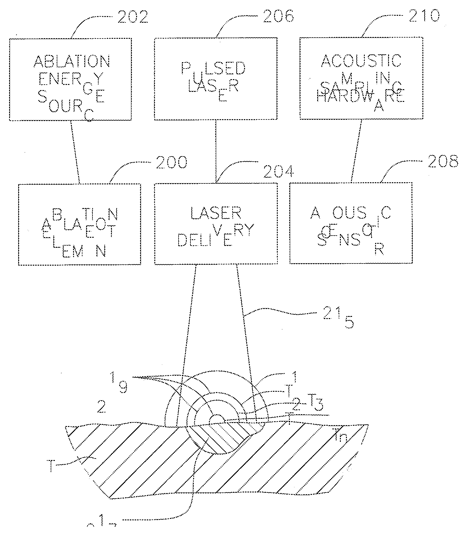
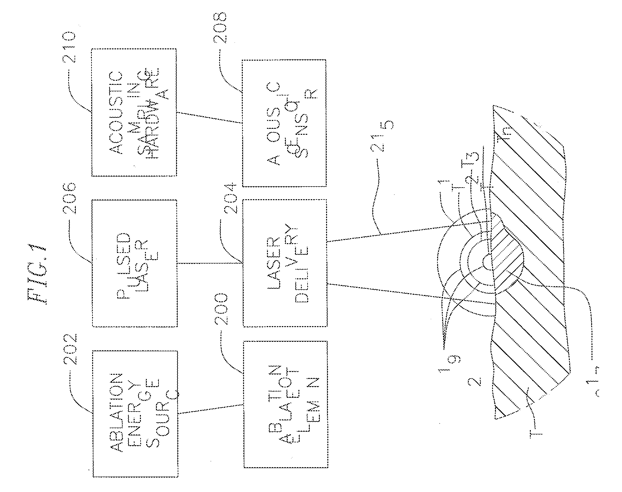
[0033]FIG. 1 illustrates an embodiment of a system S for laser optoacoustic monitoring to provide real time assessment of lesion formation, tissue status, and tissue morphology. Tissue T is subjected to RF ablation by an ablation element 200 that is energized by an ablation energy source 202 to form lesion 217. A laser delivery means 204 irradiates the lesion 217 and surrounding tissue within its field of view 215 to stimulate pressure waves 219 (with different delay times T1, T2. Tn) which are detected by acoustic transducers 208 for imaging the lesion against the surrounding tissue. The laser delivery means can include a fiber optic cable housed in a catheter that is equipped solely or primarily for irradiation, or an integrated catheter as described further below. As understood by one of ordinary skill in the art, the imaging provided by the present invention is based on contrast provided by differential absorption. To that end, a pulsed laser light source 206 drives the laser de...


PUM


Abstract

A system and method for opto-acoustic tissue and lesion assessment in real time on one or more of the following tissue characteristics: tissue thickness, lesion progression, lesion width, steam pop, and char formation, system includes an ablation element, laser delivery means, and an acoustic sensor. The invention involves irradiating tissue undergoing ablation treatment to create acoustic waves that have a temporal profile which can be recorded and analyzed by acoustic sampling hardware for reconstructing a cross-sectional aspect of the irradiated tissue. The ablation element (e.g., RF ablation), laser delivery means and acoustic sensor are configured to interact with a tissue surface from a common orientation; that is, these components are each generally facing the tissue surface such that the direction of irradiation and the direction of acoustic detection are generally opposite to each other, where the stress waves induced by the laser-induced heating of the tissue below the surface are reflected back to the tissue surface.

Description

FIELD OF INVENTION[0001]The present invention relates to electrophysiologic catheters, and in particular to laser-optoacoustic electrophysiologic catheters for monitoring tissue and lesion assessment.BACKGROUND[0002]For certain types of minimally invasive medical procedures, real time information regarding the condition of the treatment site within the body is unavailable. This lack of information inhibits the clinician when employing a catheter to perform a procedure. An example of such procedures is tumor and disease treatment in the liver and prostate. Yet another example of such a procedure is cardiac ablation used to treat atrial fibrillation. This condition in the heart causes abnormal electrical signals to be generated in the endocardial tissue resulting in irregular beating of the heart.[0003]The most frequent cause of cardiac arrhythmias is an abnormal routing of electricity through the cardiac tissue. In general, most arrhythmias are treated by ablating suspected centers o...

Claims


Application Information

Patent Timeline
Patent Type & Authority Applications(United States)
IPC IPC(8): A61B18/18
CPCA61B5/0084A61B18/1492A61B8/12A61B5/0095
Inventor SHARAREH, SHIVALIEBER, CHAD A.
Owner BIOSENSE WEBSTER INC
Why Eureka
Try Eureka