Method and system for detecting cancer regions in tissue images

a tissue image and cancer technology, applied in the field of tissue diagnosis, can solve the problems of many cases being detected late, lack of options, and insufficient prostate cancer detection technologies and methods, and achieve the effects of reducing the number of cases, and reducing the number of cancer cases

Inactive Publication Date: 2008-07-17
MEDICAL DIAGNOSTICS TECH
View PDF21 Cites 10 Cited by
  • Summary
  • Abstract
  • Description
  • Claims
  • Application Information

AI Technical Summary

Benefits of technology

[0015]The present invention provides significantly improved automatic pixel classification between a pixel representing a tissue having a given condition, such as cancer and not having the given condition.
[0016]Embodiments of the present invention provide significantly higher detection rate, and significantly lower classification error than known prior art automated ultrasound image feature detection.
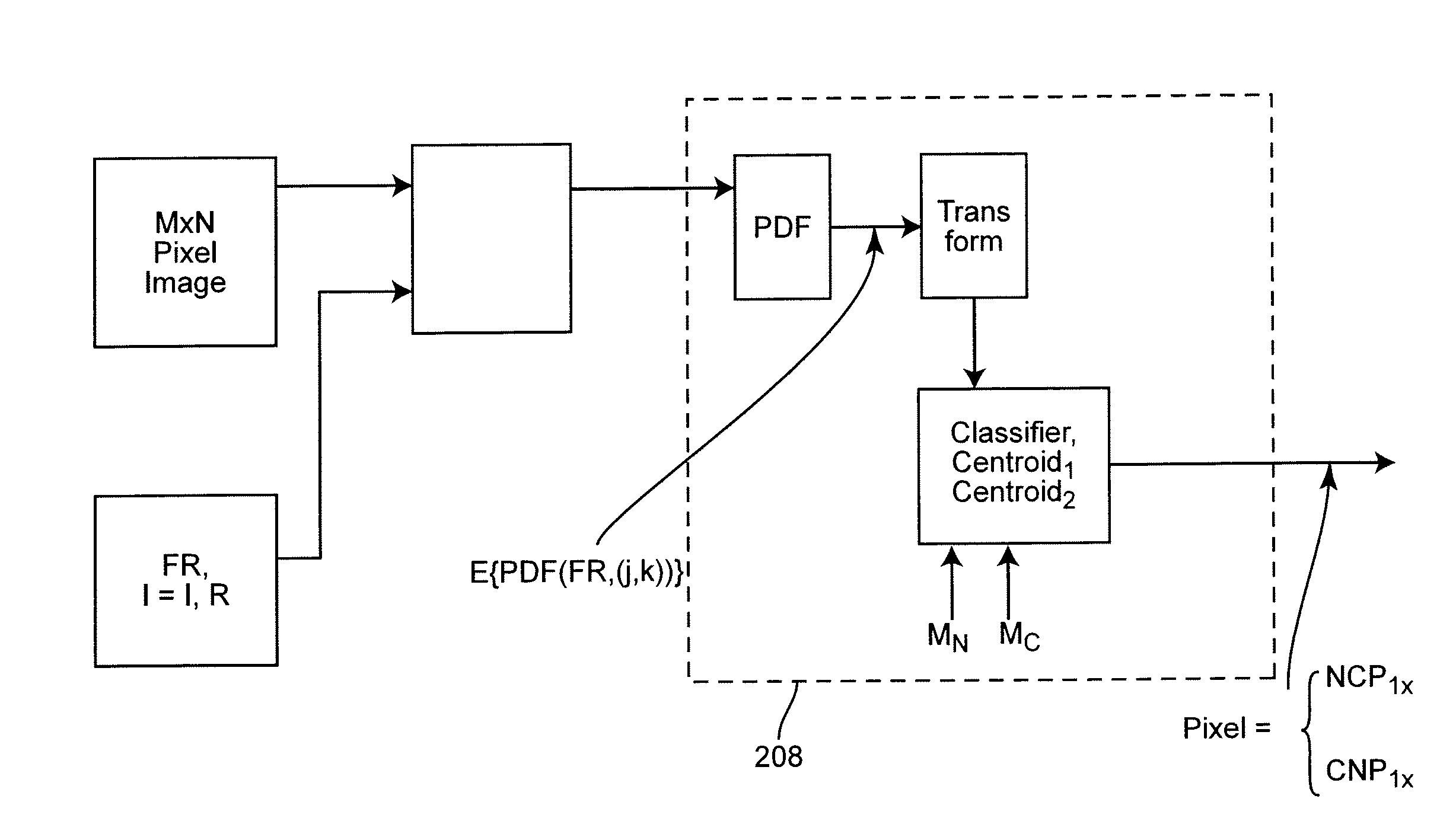
[0018]Embodiments of the present invention generate classification vectors by applying to training images having known pixel types the same basis functions that will be used to classify unknown pixels, to generate classification vectors optimized for detection sensitivity and minimal error.
[0019]Embodiments of the present invention extract further characterizing information, effectively filtering speckles and artifacts, while improving sensitivity, by estimating a probability density function for the results obtained from applying the basis functions to the subject pixels.
[0021]Embodiments of the present invention further include a multiple pass classification, providing a conditional classification by an initial pass, subject to confirmation by classifying neighbors on subsequent passes. These embodiments of the invention provide significant benefit of improved detection sensitivity, reducing false negatives, without substantial concurrent increase in false positives. These benefits and improvements may reduce the unfortunate instances of late stage initial discovery of prostate cancer.

Problems solved by technology

Unfortunately, many cases are detected late, after the prostate cancer cells have metastasized or otherwise escaped the confine of the prostate.
However, current prostate cancer detection technologies and methods are clearly inadequate, as evidenced by the still significant number of cases detected at a later stage of the cancer, where treatment options are more limited and the prognosis may be statistically unfavorable.
The two most common current screening methods are: (1) the PSA test and (2) a urologist performing digital rectal exam, and both of these methods have a significant error rate.
Digital rectal examination has a significant false negative rate because, at least in part, many prostate cancer cases may have already progressed before being detected, or being detectable by even the most skilled urologist.
Biopsy of the prostate, although one currently essential tool in the battle against prostate cancer, is invasive, is generally considered to be inconvenient, is not error free, and is expensive.
However, although there have been attempts toward improvement, current TRUS systems are known as exhibiting what is currently considered insufficient image quality for even the most skilled urologist to accurately detect cancerous regions early enough to be of practical use.

Method used


Image

  • Method and system for detecting cancer regions in tissue images
  • Method and system for detecting cancer regions in tissue images
  • Method and system for detecting cancer regions in tissue images

Examples


Embodiment Construction

[0043]The following detailed description refers to accompanying drawings that form part of this description. The description and its drawings, though, show only examples of systems and methods embodying the invention and with certain illustrative implementations. Many alternative implementations, configurations and arrangements can be readily identified by persons of ordinary skill in the pertinent arts upon reading this description.

[0044]It will be understood that like numerals appearing in different ones of the accompanying drawings, regardless of being described as the same or different embodiments of the invention, reference functional blocks or structures that are, or may be, identical or substantially identical between the different drawings.

[0045]Unless otherwise stated or clear from the description, the accompanying drawings are not necessarily drawn to represent any scale of hardware, functional importance, or relative performance of depicted blocks.

[0046]Unless otherwise s...


PUM


Abstract

A pixel of an image is classified between a first kind and a second kind by centering a sample mask on the pixel and applying each of a population of R given basis functions to the mask pixels to generate, for each basis function, a bucket of values. A probability density function is estimated for each of the bucket of values. Each of the R probability density functions is transformed to a single valued result, to generate an R-dimensional sample classification vector. The R-dimensional sample classification vector is classified against a R-dimensional first centroid vector and a R-dimensional second centroid vector, each of centroid vectors constructed in a previous training of applying the same population of R given basis functions to pixels known as being the first kind and to pixels known as being the second kind. Optionally, pixels may be conditionally classified and then finally classified based on subsequent classification of neighbor pixels.

Description

CROSS-REFERENCE TO RELATED APPLICATIONS[0001]This application claims priority to U.S. Provisional Application Ser. No. 60 / 880,310, filed Jan. 12, 2007, which is hereby incorporated by reference.FIELD OF THE INVENTION[0002]Embodiments of the invention pertain to diagnostic imaging of tissue and, in some embodiments, to mapping pixel regions to an N-dimensional space and recognizing certain tissue characteristics based on the mapping.BACKGROUND OF THE INVENTION[0003]Even though new methods for treatment and new strategies for detection have become available, prostate cancer remains the second most common cancer that kills men in the United States. Only lung cancer causes a higher number of deaths. The numbers are telling. More than 230,000 new cases of prostate cancer were diagnosed in the U.S. during 2005.[0004]The number of new cases alone is not a complete measure of the problem. Prostate cancer is a progressive disease and, generally, the earlier the stage of its progress when fir...

Claims


Application Information

Patent Timeline
Patent Type & Authority Applications(United States)
IPC IPC(8): G06K9/00
CPCG06K2209/053G06K9/522G06V10/431G06V2201/032
Inventor YFANTIS, SPYROS A.
Owner MEDICAL DIAGNOSTICS TECH
Why Eureka
Try Eureka