Multimodal Catheter for Focal Brain Monitoring and Ventriculostomy

a multi-modal catheter and brain technology, applied in the field of ventricular catheters, can solve the problems of inability to characterize the underlying brain tissue, the inability to monitor the brain, the inability to use speech and/or memory, etc., and achieve the effects of convenient connection, convenient use, and convenient us

Active Publication Date: 2008-07-17
ZAUNER ALOIS
View PDF34 Cites 26 Cited by
  • Summary
  • Abstract
  • Description
  • Claims
  • Application Information

AI Technical Summary

Benefits of technology

[0029]A further object of the invention is to provide a connector that is more durable, more sanitary, and easier to connect.
[0030]To achieve these objects, a part of a ring connector can be attached to a proximate end of the ventricular catheter. This allows the ventricular catheter to be quickly joined to a mating ring connector part on a connector to be attached to the ventricular catheter. The ring connector allows others additional connectors to be attached to the EVD catheter. The ring connection when connected is sealed to prevent pathogens from entering the EVD catheter and infecting the brain. Ring connectors are strong enough to remain connected even if the patient moves or unintentionally touches the catheter. The improved durability and sanitation extend the amount of time that the EVD catheter can remain inserted in the patient's brain. Typically, newer EVD catheters contain an antibiotic in the wall of the catheter to reduce the risk for infection. However, infection risks remain at the proximal end and along the connectors, using current technologies.
[0031]To facilitate quick connection of the ventricular catheter to a connector, each half of the ring connector can have an alignment mark made on it. The alignment marks are drawn to align with each other when the various leads (i.e. conduits, probes, ICP sensor, and thermocouple) align with leads in the connector. The use of alignment marks promotes quick connection with a reliable connection between the sensor and the respective monitor.

Problems solved by technology

Measurements made by various sensors or catheters in the tissue having microhemorrhages have been found to be inaccurate and not characterize the underlying brain tissue.
Insertion catheters into both hemispheres of the brain can lead to loss of speech and / or memory.
Another problem with the prior art is the connectors for the sensors.
The bolts are bulky (approximately, 5 cm by 4 cm by 1 cm) and have no means for connecting to the EVD catheter.
Patients often move, brush, or even remove the bolt accidentally.
In addition, the bolt's weak connection fails to prevent infection.
Another problem with the prior-art EVD systems is that the sensor cannot be added subsequent to the insertion of the EVD catheter.
Therefore, once the EVD catheter is inserted, additional sensors cannot be added without removing the EVD catheter and inserting a replacement.
In addition, surgeons avoid using all of the possible sensors with every EVD catheter because the sensors are often very expensive so using them without a specific reason is prohibitive.
Manipulating the EVD catheter and / or sensors once they are placed requires reopening of the incision and this significantly increases the infection risk.
A related problem is that the sensors are only approved to be inserted for four to seven days.
An additional problem with the prior art is that the sensor is added coaxially along with the EVD catheter.

Method used


Image

  • Multimodal Catheter for Focal Brain Monitoring and Ventriculostomy
  • Multimodal Catheter for Focal Brain Monitoring and Ventriculostomy
  • Multimodal Catheter for Focal Brain Monitoring and Ventriculostomy

Examples


Embodiment Construction

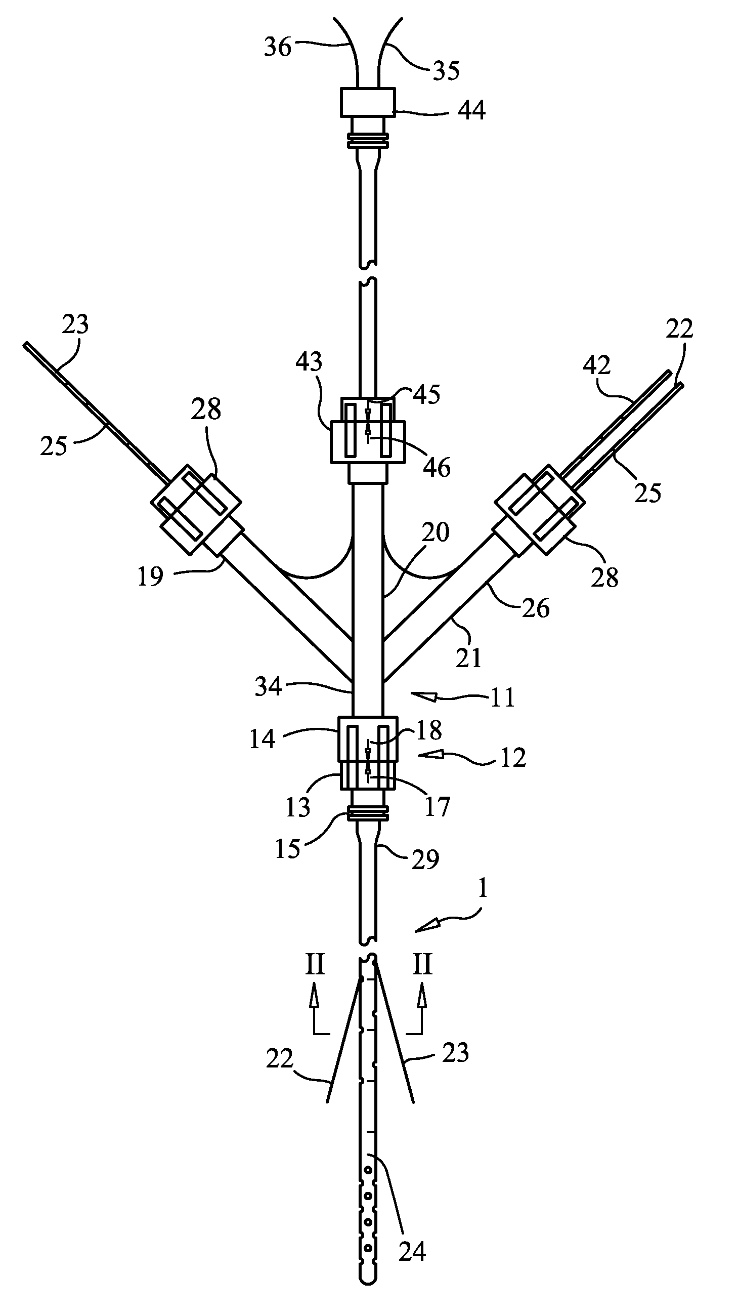
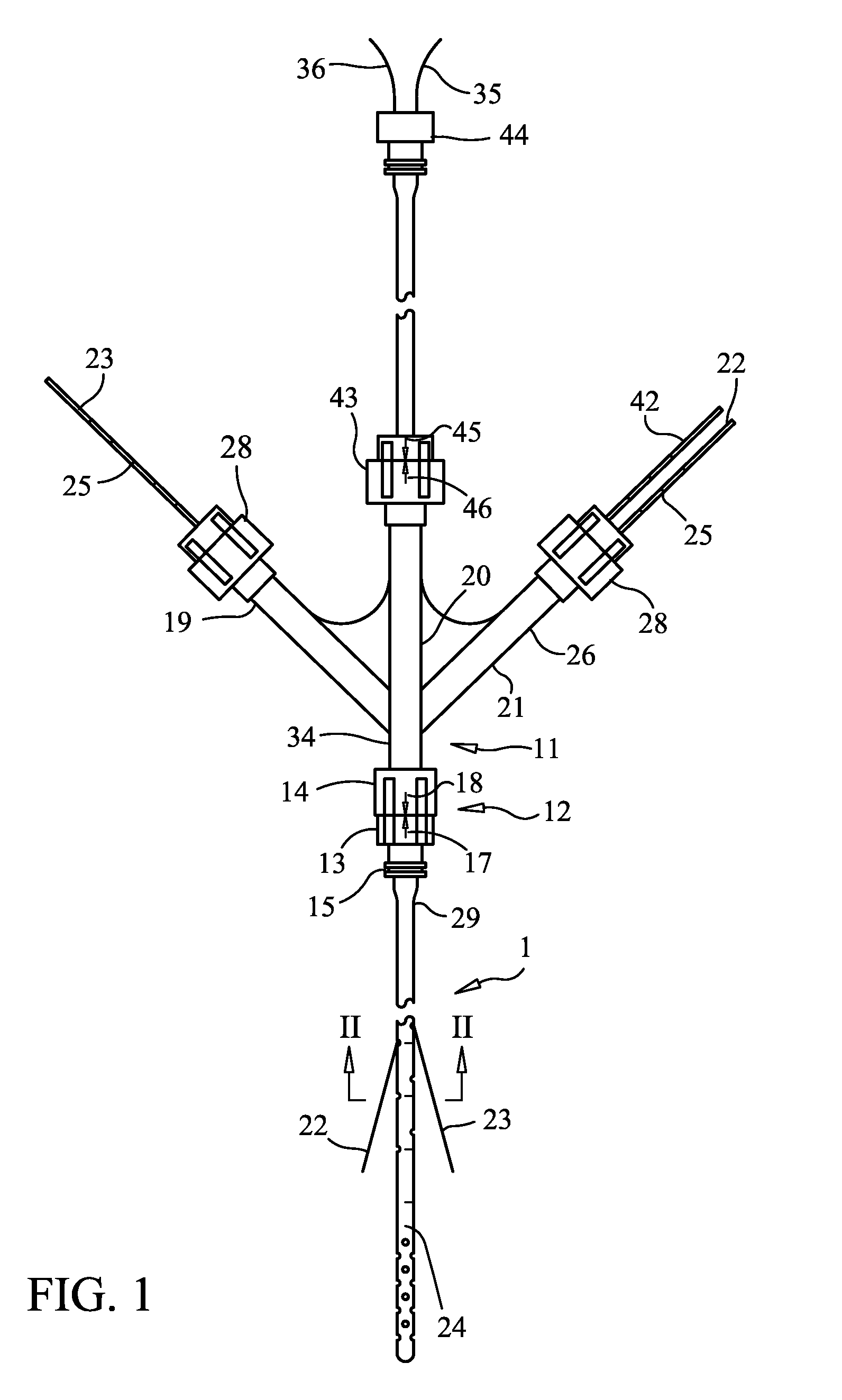
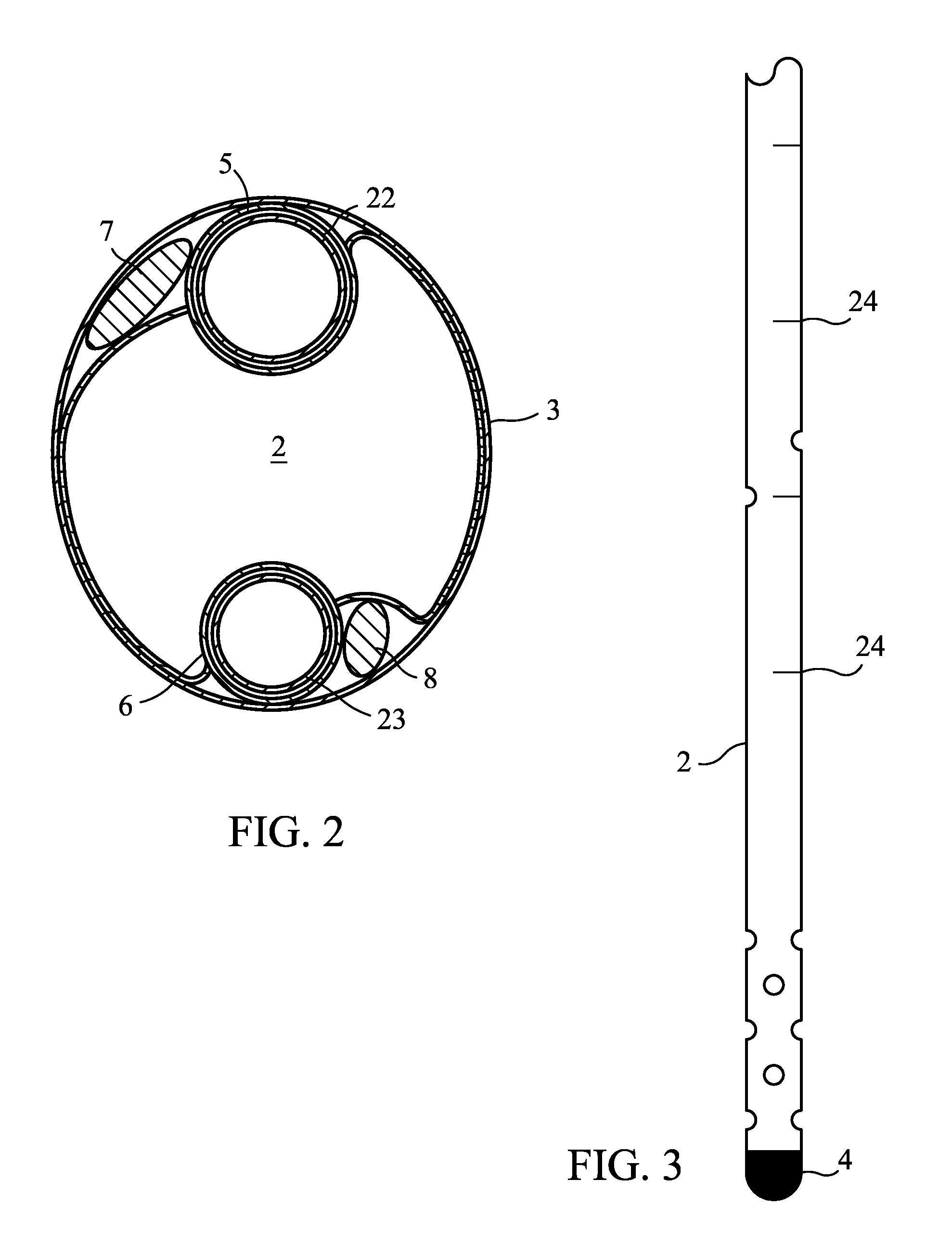
[0044]Referring now to the figures of the drawing in detail and first, particularly, to FIG. 1 thereof, there is seen a ventricular catheter 1 according to the invention. The ventricular catheter 1 includes an external ventricular drainage (EVD) catheter 2; see FIGS. 2-4. The EVD catheter 2 is a generally elongated tube formed by a wall 3. The EVD catheter 2 preferably has a length of twenty centimeters (20 cm). This length allow for the distal tip 4 to be inserted into the brain and the proximate end 29 to be tunneled subcutaneously along the skull. The outer diameter of the EVD catheter 2 is preferably between 3.2 and 3.8 mm. The inner diameter of the EVD catheter 2 is preferably between 1.8 and 2.0 mm. The distal tip 4 of the EVD catheter 2 is made of a radio-opaque material.

[0045]FIG. 2 shows a cross section of the ventricular catheter 1 taken along Line II-II in FIG. 1. The overall crossection of the ventricular catheter 1 is oval shaped. The wall 3 of the ventricular catheter ...


PUM


Abstract

A ventricular catheter includes an external ventricular drainage (EVD) catheter and has at least one conduit formed in the wall of the EVD catheter. The conduit opens at a side port of the wall at an angle to the EVD catheter. The side port is formed in the wall to allow a probe to be extended from the conduit at the angle into brain tissue that is not altered by the insertion of the EVD catheter. The ventricular catheter has an oval shape to minimize the total cross-sectional area while still allowing the necessary number of conduits.

Description

CROSS-REFERENCE TO RELATED APPLICATIONS[0001]Not ApplicableSTATEMENT REGARDING FEDERALLY SPONSORED RESEARCH OR DEVELOPMENT[0002]Not ApplicableTHE NAMES OF PARTIES TO A JOINT RESEARCH AGREEMENT[0003]Not ApplicableINCORPORATION-BY-REFERENCE OF MATERIAL SUBMITTED ON A COMPACT DISC[0004]Not ApplicableBACKGROUND OF THE INVENTION[0005]1. Field of the Invention[0006]The invention relates to ventricular catheters and more particular to multimodal catheters for focal brain monitoring and ventriculostomies.[0007]2. Description of the Related Art[0008]External ventricular drainage catheters are known in the art. External Ventricular Drainage (EVD) catheters are indicated for drainage of cerebrospinal fluid (CSF) from the lateral ventricles of the brain and for relieving intracranial pressure. A typical EVD catheter has a length of fifteen to thirty-five centimeters (15-35 cm) with an outer diameter between two and one half and three and three tenths millimeter (2.5-3.3 mm) and an inner diamete...

Claims


Application Information

Patent Timeline
Patent Type & Authority Applications(United States)
IPC IPC(8): A61M5/32
CPCA61B5/01A61B5/031A61B5/14539A61B5/14553A61B5/1459A61M2025/0078A61M25/007A61M25/0108A61M27/006A61M2025/0008A61M2025/0036A61M25/0032A61B5/4076
Inventor ZAUNER, ALOIS
Owner ZAUNER ALOIS
Why Eureka
Try Eureka