Device for spatial localization of a movable part of the body

a technology for movable parts and devices, applied in the field of devices for spatial localization of movable parts of the body, can solve the problems of damage (“invasive”) methods, their complexity, their expense and costs, and remain, and achieve the effects of greater measurement dynamics and/or faster, accurate measurement localization, and fast and easy calculation

Inactive Publication Date: 2008-08-07
SIEMENS SWITZERLAND
View PDF8 Cites 2 Cited by
  • Summary
  • Abstract
  • Description
  • Claims
  • Application Information

AI Technical Summary

Benefits of technology

[0009]The variable number 1, 2, 3 of the sound receiver inputs on the one hand allows the one-, two- or three-dimensional localization of the exciter in an absolute, measurement based manner in the desired coordinate system. With larger numbers of sound receiver inputs and / or sound sources, the measurement localization is undertaken more accurately, with greater measurement dynamics and / or more quickly, where necessary.
[0010]Thus, at least sound-related measurement signals emanating from the part of the body are determined by the sound receiver device of which the delay times, frequencies, phases and / or propagation amplitudes vary at each sound receiver input of the sound receiver device depending on the distance between the part of the body and the sound receiver input. By determining characteristics of measuring signals an evaluation of the distances between each sound receiver input and the birthplace of the second sound wave is undertaken by the exciter. The fact that the relative locations / distances between the origin of the second sound wave and the sound receiver inputs have been made known, and the fact that both or all sound receiver inputs in a known, spatial coordinate system are known in an absolute way enables the precise position of the origin of the second sound wave in the absolute coordinate system to be calculated quickly and easily. In this way, the invention allows a unique one-, two- or three-dimensional localization of a part of the body movable within a movement volume (=origin of the second sound wave) as well as a high-speed tracking of the movement of the associated part of the body.
[0011]The fact that what is referred to as an actual isocenter to be determined is able to be moved with the movement of the part of the body (=of the exciter), means that an output signal of a sound receiver input changes or a number of output signals from the sound receiver inputs change simultaneously. The deviations of the output signal formed and determined by this thus allow a spatially dynamic tracking of the part of the body (=of the exciter) with high accuracy, which occurs relative to a reference point (e.g., a required isocenter of a radiation system). An attempt is subsequently made to permanently determine newly occurring deviations relative to a previously localized actual isocenter (and to compensate for them, if necessary, by a triggered positioning means and to keep the actual isocenter from the detected exciter permanently known as the next required isocenter), so that the part of the body always remains detectable with the sound receiver device.
[0012]Naturally, more than one sound source or more than two spatially separated sound receiver inputs can be used. The accuracy and / or the speed of the localization / following are increased in this way. In this way, possible sound-absorbing locations (especially within the body) can be covered better / more intensively with sound waves. The intensity of the sound waves is tailored in the interests of minimally invasive characteristics such that a sick patient is not disturbed.

Problems solved by technology

A few methods (e.g., “in-vivo” imaging methods that use radiography with x-rays) are however classified as damaging (“invasive”) methods and should therefore only be used in a restricted-manner.
Even the few damaging “in-vivo” methods, such as, for example, an imaging method using magnetic resonance, in addition to their complexity, their expense and their costs, remain difficult to combine with and use at the same time as a radiation treatment.
However these methods lack accuracy and also reproducibility if physiological changes of the patient (e.g., gaining weight) or unavoidable (which are in the worst case unexpected) movements (e.g., caused by breathing) occur during treatments or if there is repositioning of the patient / parts of the body between treatments.
Furthermore, methods and apparatuses for holding patients or parts of a body on a support table, for example, during radiation therapy, or devices for measuring breathing exist; however, on the one hand these solutions reduce the “comfort” of the patient, or do not allow a 100%-accurate spatial determination / localization of a tumor in a known coordinate system.

Method used


Image

  • Device for spatial localization of a movable part of the body
  • Device for spatial localization of a movable part of the body
  • Device for spatial localization of a movable part of the body

Examples


Embodiment Construction

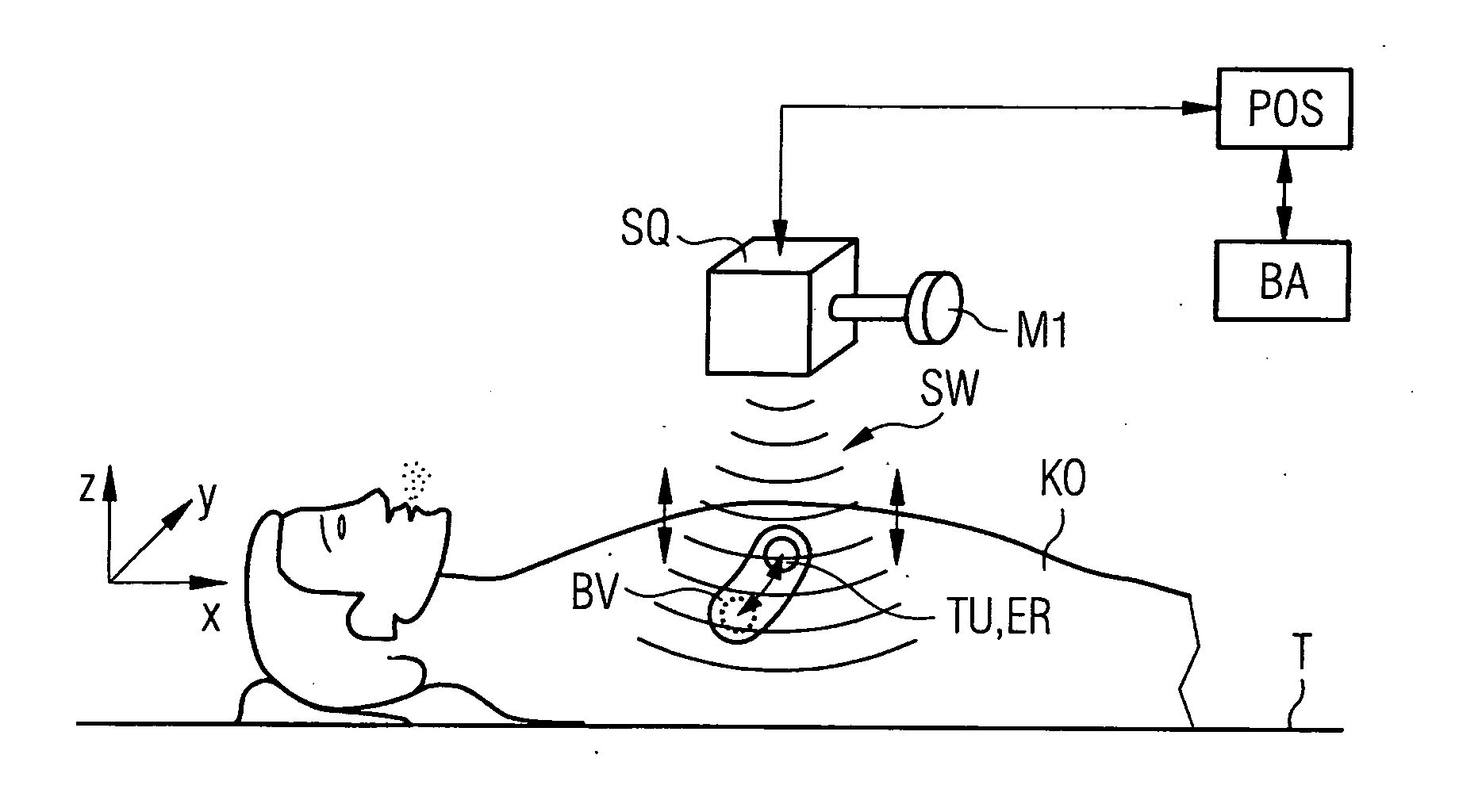
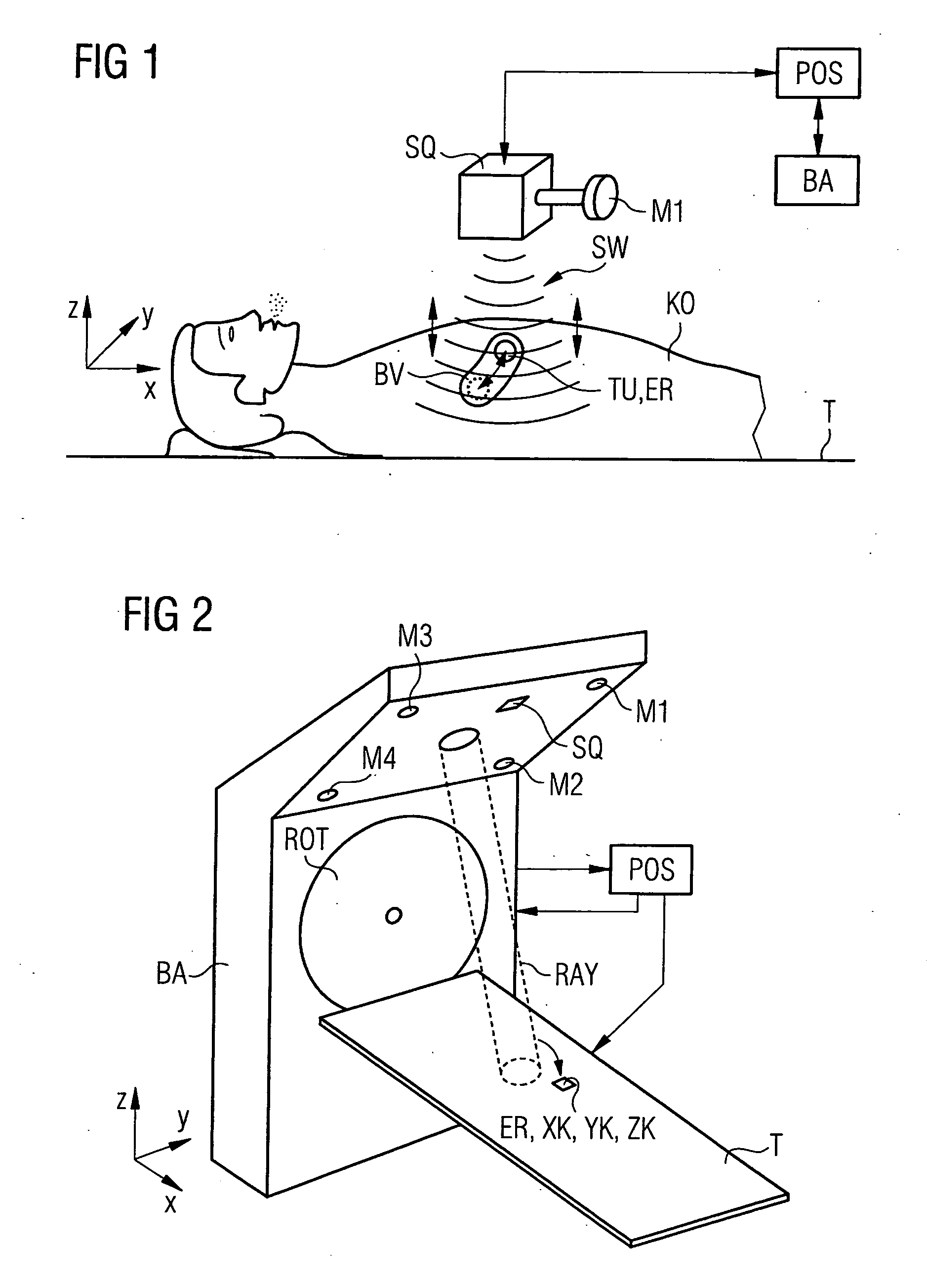
[0021]FIG. 1 shows a radiation system BA (only shown symbolically here) such as a linear accelerator for radio-therapeutic treatments, of which at least one beam output or axis is intended to target a tumor TU to be destroyed (here in the chest or lung area) of a person / patient KO lying on a table T. The breathing or the unexpected movement of the person inevitably causes the tumor TU to move relative to the pre-planned radiation target in the body KO and thus forms what is referred to as a movement volume BV which includes all positions of the tumor. Before a radiation therapy is performed it is usual to carry out a computer tomography in the chest or lung so that subsequently a repositioning of the inventive device is facilitated relative to the movement volume BV of the tumor determined thereby. After the computer tomography, the patient is positioned for regular radio-therapeutic treatments on the table T or another positionable table, if possible, in the same position as for th...


PUM


Abstract

A device for spatially localizing a movable part of a body, wherein movement of the body part is within a movement volume at surface parts and inside a living being, has a sound receiver device and a sound source. The sound receiver device is outside the living being and has spatially distinguishable sound receiver inputs located in an X-Y-Z coordinate system. The sound source is outside the living being, and configured to emit a first sound wave to propagate as far as the movement volume. The sound receiver device detects a second sound wave having a defined excitation wavelength or frequency, wherein the second sound wave is generated when the first sound wave causes a spectral exciter to oscillate. The sound receiver device is positioned such that the second sound wave encounters at least a maximum number of sound receiver inputs.

Description

CROSS-REFERENCE TO RELATED APPLICATIONS[0001]The present application claims priority to European Patent Application No. 07002475.7, filed on Feb. 6, 2007, which is incorporated herein by reference.BACKGROUND OF THE INVENTION[0002]The invention relates to a device for spatial localization of a movable part of a body, and further to a method of determining three-dimensional coordinates of a body part of a living being in real time relative to a known three-dimensional coordinate system.[0003]Numerous methods or devices exist for radiation therapy of a tumor, for example, by means of Gamma or proton radiation, or for imaging of a tumor within the body of a being. These methods and devices aim to ensure a most accurate and reproducible three-dimensional localization of the tumor, or more generally of a movable part of the body. A few methods (e.g., “in-vivo” imaging methods that use radiography with x-rays) are however classified as damaging (“invasive”) methods and should therefore onl...

Claims


Application Information

Patent Timeline
Patent Type & Authority Applications(United States)
IPC IPC(8): A61B7/00
CPCA61B2019/5429A61N2005/1051A61N5/1049A61B2090/3929
Inventor BROENNIMANN, THOMASCLOUTOT, LAURENT
Owner SIEMENS SWITZERLAND
Why Eureka
Try Eureka