Structural quantification of cartilage changes using statistical parametric mapping

a structural and parametric mapping technology, applied in image analysis, medical science, image enhancement, etc., can solve the problems of small percentage of cartilage tissue, insensitive to focal morphological changes, and large effort placed on accurate quantification of cartilage morphological parameters, so as to reduce total system variability, improve system ability to detect focal changes, and minimal cartilage morphology changes

Inactive Publication Date: 2008-08-21
VIRTUALSCOPICS
View PDF18 Cites 8 Cited by
  • Summary
  • Abstract
  • Description
  • Claims
  • Application Information

AI Technical Summary

Benefits of technology

[0007]It is another object to reduce the error in quantitative MRI measurements due to the nature of human-computer performance.
[0010]Quantification of structural changes using SPM is a very useful technique for the evaluation of focal changes in cartilage morphology. The technique was evaluated with synthetic data and reduced the total system variability and improved the system ability to detect focal changes. It also provided the location and size of newly formed regions. The application of this concept in nine healthy volunteer data sets demonstrated that there is minimal change in cartilage morphology over one year. Although no major changes were observed, the delta map approach improved the precision and the ability to detect smaller changes in cartilage morphology. The improved precision was archived by the ability to map the baseline segmentation into the follow-up segmentation and look for changes on all the points that shared a common supporting area. The use of a common area reduces the errors in the definition of the extent of the cartilage surface. Furthermore, precision gains are achieved by the ability to compensate for magnet distortions. The distortions are compensated by adjusting the global cartilage shape to mimic one time point to the other time point. The ability to correct and map all the cartilage points to each other allows for the measurement of the thickness error and the estimation of areas with significant changes. The precision of those significant changes were better that the precision of the raw changes; hence the ability to detect changes is improved. On the other hand, the additional extracted information from the SPM was used to test differences among the distribution of two cartilage plates. The test in change among two distributions can be used to test the difference among two different groups, which is important when evaluating drug efficacy in newly designed disease modifying osteoarthritis drugs (DMOAD).

Problems solved by technology

Therefore, a great deal of effort has been placed in the accurate quantification of cartilage morphological parameters such as volume, average thickness, and surface area.
The problem is related to the focal nature of OA changes, which affect a small percentage of the cartilage tissue.
However, they are not very sensitive to the detection of focal morphological changes in cartilage structure.
Even standard evaluation of cartilage volume depends on human intervention, which tends to introduce error in the analysis.
To minimize the requirements of an expert observer for the evaluation of cartilage changes, researchers have proposed automation; even still, full cartilage analysis can not be achieved with the desired precision to detect focal changes.
Although SPM has been commonly used for the evaluation of activation regions in functional MRI, little effort has been made towards the application of SPM in the detection of structural changes in human anatomy.

Method used


Image

  • Structural quantification of cartilage changes using statistical parametric mapping
  • Structural quantification of cartilage changes using statistical parametric mapping
  • Structural quantification of cartilage changes using statistical parametric mapping

Examples


Embodiment Construction

[0022]A preferred embodiment of the present invention will be set forth in detail with reference to the drawings, in which like reference numerals refer to like elements or steps throughout.

[0023]The data set used for the evaluation of the method consisted of a pair of scans from nine healthy volunteers between 31 and 71 years old (mean age of 44), five female and four male, with no clinical evidence of OA. One knee of each of the nine volunteers was scanned using a GE Sigma 1.5 T scanner (GE, Milwaukee, Wis.) at two time points: baseline scan and the one year follow-up. All volunteers consented to the study protocol which was previously approved by the Institutional Research Subjects Review Board. Two sets of images were acquired for each knee: first a sagittal 3D SPGR fat saturated sequence with a TR of 39, TE of 7.0 ms, Nex 1, Flip angle of 20°, and a 256 by 256 matrix with a 14.0 cm field of view. Second, a sagittal 3D GRE image with a TR of 29, TE of 15.0 sec, Nex 1, Flip angle...


PUM


Abstract

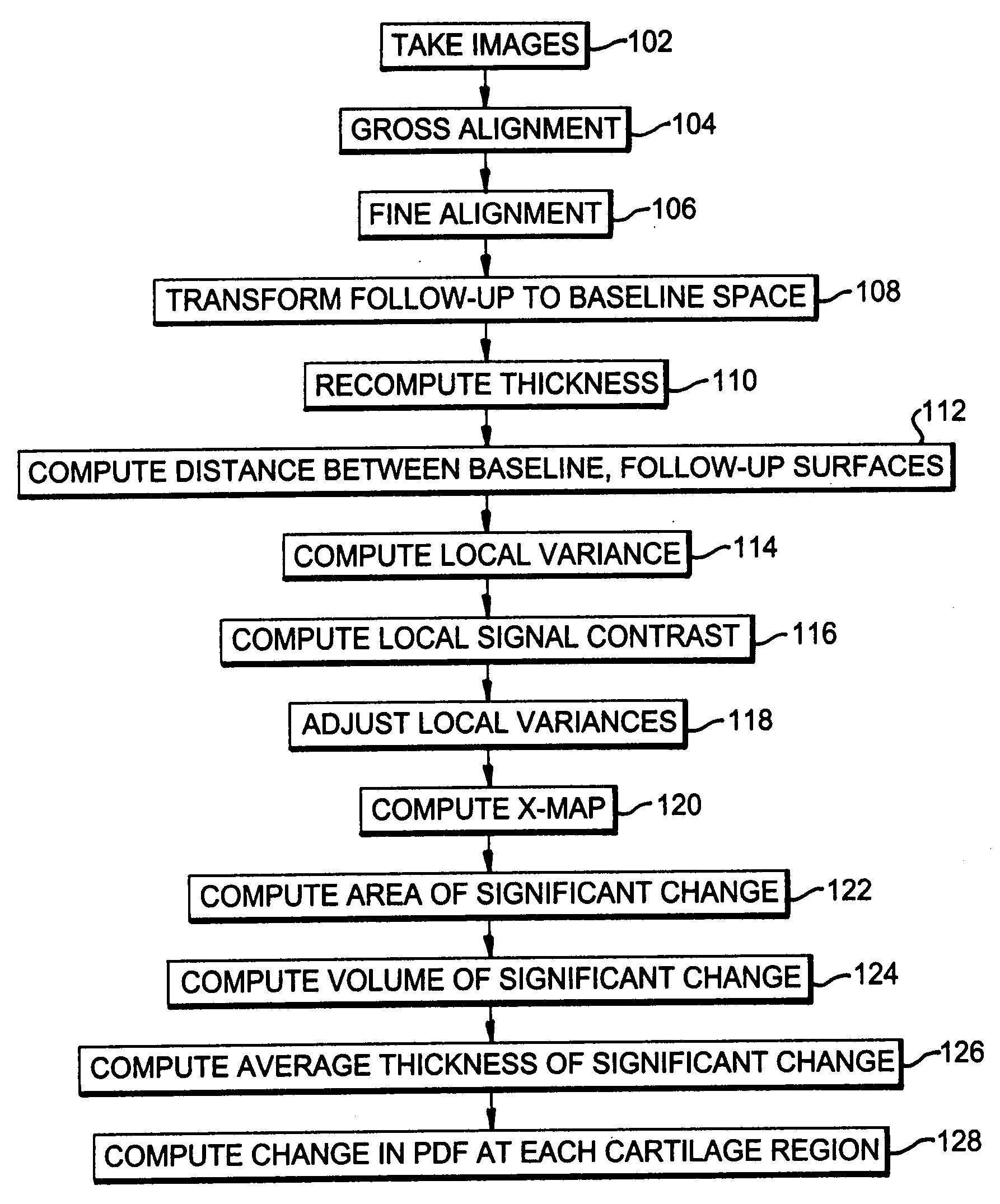
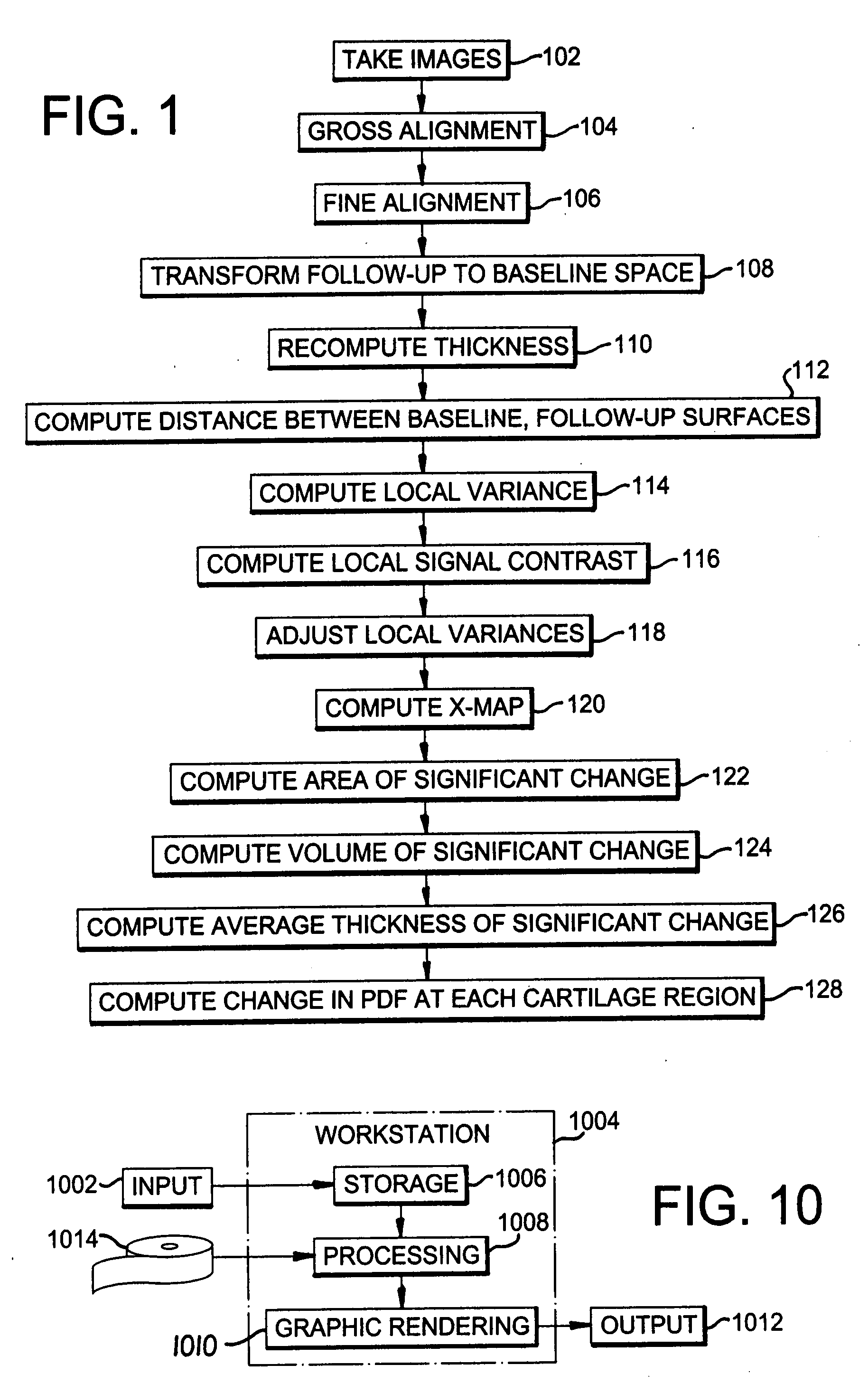
The analysis of the focal changes in the morphology of a tissue such as cartilage is completed through statistical parametric mapping by first detecting the amount of thickness changes; followed by the point by point estimation of the variance in the thickness delta estimation. Once the change and the variance are estimated, the z-map is computed. The z-map is used to compute single change parameters. i.e: volume significant change, area of significant change, average thickness of the significant changes, and D values from the probability distributions. That can be used for treatment decisions.

Description

FIELD OF THE INVENTION[0001]The present invention is directed to the evaluation of osteoarthritis progression and more particularly to such evaluation using statistical parametric mapping to evaluate cartilage degradation.DESCRIPTION OF RELATED ART[0002]The detection of structural changes is an important task in the evaluation of osteoarthritis progression. The cartilage tissue is one of the primary affected structures in OA which is a debilitating disease. The early detection of OA treatment efficacy requires monitoring of small changes in cartilage morphology.[0003]It has been reported that 70% of women and 60% of men aged 65 years or older suffer from OA. Therefore, a great deal of effort has been placed in the accurate quantification of cartilage morphological parameters such as volume, average thickness, and surface area. Cartilage tissue in normal subjects has very similar quantitative properties to OA subjects, and the evolution of those changes is very small. The problem is ...

Claims


Application Information

Patent Timeline
Patent Type & Authority Applications(United States)
IPC IPC(8): A61B5/107
CPCA61B5/055A61B5/4514G06T7/0012G06T2207/30008G06T7/60G06T2207/10088G06T7/0022G06T7/97
Inventor TAMEZ-PENA, JOSE GERARADOBARBU-MCINNIS, MONICATOTTERMAN, SAARA
Owner VIRTUALSCOPICS
Why Eureka
Try Eureka